

Search Count: 10
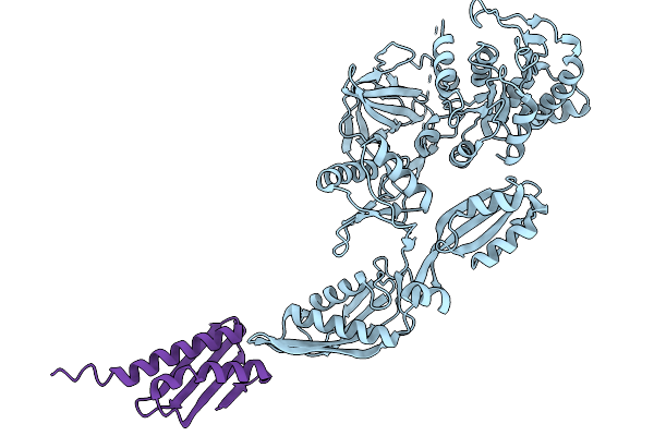 |
Cryo-Em Structure Of Ef-G And Raia Simultaneously Bound To An E. Coli Ribosome Imaged In A Cell Lysate
Organism: Escherichia coli k-12
Method: ELECTRON MICROSCOPY Release Date: 2026-02-18 Classification: RIBOSOMAL PROTEIN |
Organism: Escherichia coli k-12
Method: ELECTRON MICROSCOPY
Release Date: 2026-02-18
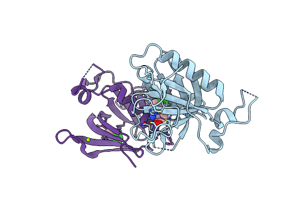 |
Crystal Structure Of 5-Chloroindoline-2,3-Dione Covalently Bound To The Ph Domain Of Bruton'S Tyrosine Kinase Mutant R28C
Organism: Homo sapiens
Method: X-RAY DIFFRACTION Resolution:1.93 Å Release Date: 2021-05-12 Classification: TRANSFERASE Ligands: 3IS, ZN, MG |
Organism: Homo sapiens
Method: X-RAY DIFFRACTION
Release Date: 2021-05-12
Ligands: 3IS, ZN, MG
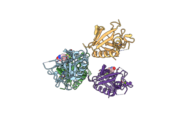 |
Crystal Structure Of 5-(Trifluoromethoxy)Indoline-2,3-Dione Covalently Bound To The Ph Domain Of Bruton'S Tyrosine Kinase Mutant R28C
Organism: Homo sapiens
Method: X-RAY DIFFRACTION Resolution:1.95 Å Release Date: 2021-05-12 Classification: TRANSFERASE Ligands: Q1B, MG, ZN |
Organism: Homo sapiens
Method: X-RAY DIFFRACTION
Release Date: 2021-05-12
Ligands: Q1B, MG, ZN
 |
Crystal Structure Of 1,5-Dimethylindoline-2,3-Dione Covalently Bound To The Ph Domain Of Bruton'S Tyrosine Kinase Mutant R28C
Organism: Homo sapiens
Method: X-RAY DIFFRACTION Resolution:2.04 Å Release Date: 2021-05-12 Classification: TRANSFERASE Ligands: ZN, MG, IS7 |
Organism: Homo sapiens
Method: X-RAY DIFFRACTION
Release Date: 2021-05-12
Ligands: ZN, MG, IS7
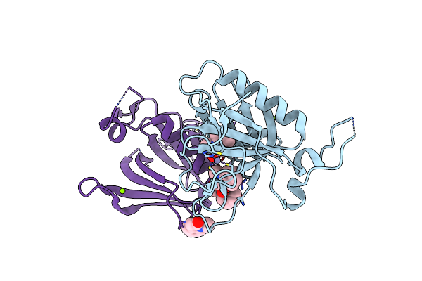 |
Crystal Structure Of 1-Methylindoline-2,3-Dione Covalently Bound To The Ph Domain Of Bruton'S Tyrosine Kinase Mutant R28C
Organism: Homo sapiens
Method: X-RAY DIFFRACTION Resolution:1.41 Å Release Date: 2020-11-25 Classification: TRANSFERASE Ligands: MG, 72V, ZN |
Organism: Homo sapiens
Method: X-RAY DIFFRACTION
Release Date: 2020-11-25
Ligands: MG, 72V, ZN
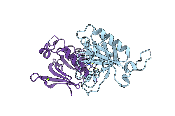 |
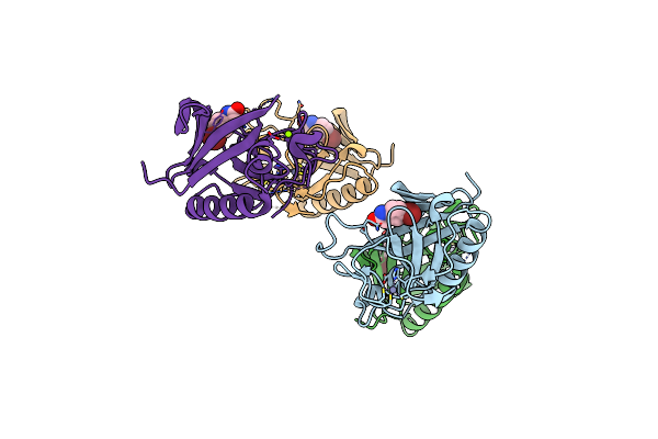
The Ph Domain Of Bruton'S Tyrosine Kinase Mutant R28C
Organism: Homo sapiens
Method: X-RAY DIFFRACTION Resolution:1.36 Å Release Date: 2020-11-25 Classification: TRANSFERASE Ligands: MG, ZN |
Organism: Homo sapiens
Method: X-RAY DIFFRACTION
Release Date: 2020-11-25
Ligands: MG, ZN
 |
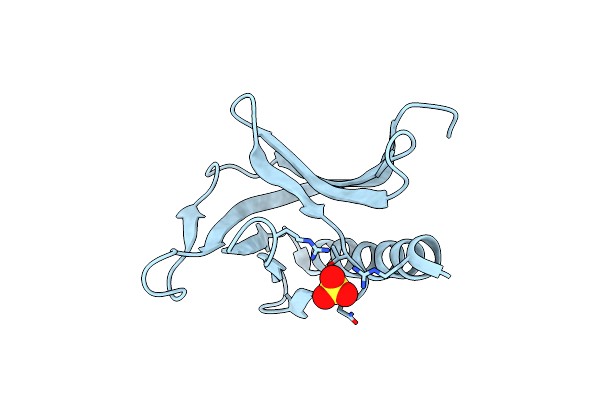
Crystal Structure Of 5-Bromoindoline-2,3-Dione Covalently Bound To The Ph Domain Of Bruton'S Tyrosine Kinase
Organism: Homo sapiens
Method: X-RAY DIFFRACTION Resolution:2.31 Å Release Date: 2020-11-25 Classification: TRANSFERASE Ligands: NYQ, ZN, MG |
Organism: Homo sapiens
Method: X-RAY DIFFRACTION
Release Date: 2020-11-25
Ligands: NYQ, ZN, MG
 |
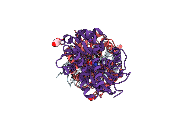
Crystal Structure Of Human Homer3 Evh1 Domain
Organism: Homo sapiens
Method: X-RAY DIFFRACTION Resolution:1.85 Å Release Date: 2007-11-20 Classification: SIGNALING PROTEIN Ligands: SO4 |
Organism: Homo sapiens
Method: X-RAY DIFFRACTION
Release Date: 2007-11-20
Ligands: SO4
 |
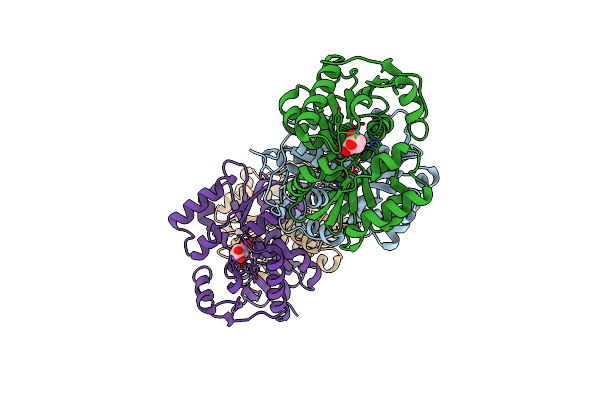
Crystal Structure Of Glutamate Racemase Isoform Race1 From Bacillus Anthracis
Organism: Bacillus anthracis
Method: X-RAY DIFFRACTION Resolution:1.60 Å Release Date: 2007-06-19 Classification: ISOMERASE Ligands: K, DGL, GOL |
Organism: Bacillus anthracis
Method: X-RAY DIFFRACTION
Release Date: 2007-06-19
Ligands: K, DGL, GOL
 |
Crystal Structure Of The Glutamate Racemase From Bacillus Anthracis
Organism: Bacillus anthracis
Method: X-RAY DIFFRACTION Resolution:1.99 Å Release Date: 2007-05-29 Classification: ISOMERASE Ligands: DGL |
Organism: Bacillus anthracis
Method: X-RAY DIFFRACTION
Release Date: 2007-05-29
Ligands: DGL