

Search Count: 20
 |
Crystal Structure Of Endoplasmic Reticulum Aminopeptidase 2 (Erap2) In Complex With A Diaminobenzoic Acid Derivative Ligand.
Organism: Homo sapiens
Method: X-RAY DIFFRACTION Resolution:2.90 Å Release Date: 2017-03-29 Classification: HYDROLASE Ligands: ZN, 6PX, NAG |
Organism: Homo sapiens
Method: X-RAY DIFFRACTION
Release Date: 2017-03-29
Ligands: ZN, 6PX, NAG
 |
Crystal Structure Of Endoplasmic Reticulum Aminopeptidase 2 (Erap2) In Complex With A Hydroxamic Derivative Ligand
Organism: Homo sapiens
Method: X-RAY DIFFRACTION Resolution:2.80 Å Release Date: 2017-03-15 Classification: HYDROLASE Ligands: ZN, NAG, 6GA |
Organism: Homo sapiens
Method: X-RAY DIFFRACTION
Release Date: 2017-03-15
Ligands: ZN, NAG, 6GA
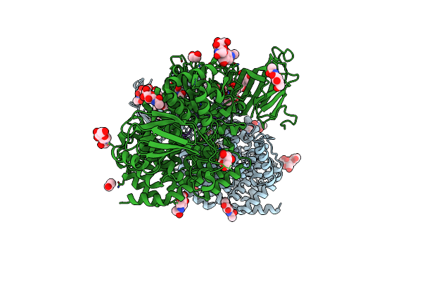 |
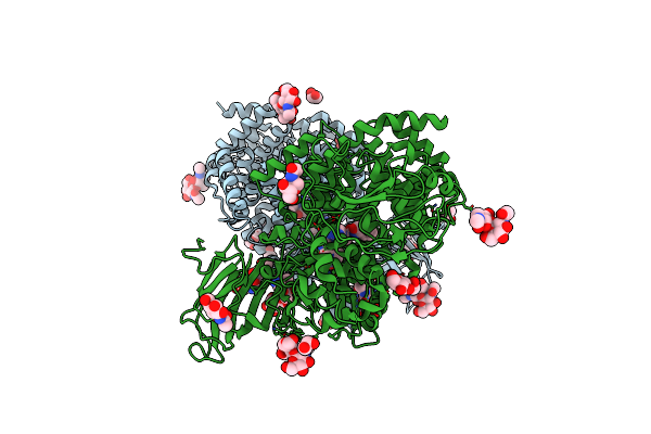
Crystal Structure Of Aminopeptidase Erap2 With Ligand
Organism: Homo sapiens, Synthetic construct
Method: X-RAY DIFFRACTION Resolution:2.50 Å Release Date: 2015-09-30 Classification: HYDROLASE Ligands: ZN, NAG, EDO, MES |
Organism: Homo sapiens, Synthetic construct
Method: X-RAY DIFFRACTION
Release Date: 2015-09-30
Ligands: ZN, NAG, EDO, MES
 |
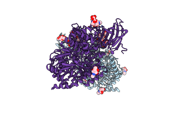
Crystal Structure Of Aminopeptidase Erap2 With Ligand
Organism: Homo sapiens, Synthetic construct
Method: X-RAY DIFFRACTION Resolution:2.73 Å Release Date: 2015-09-30 Classification: HYDROLASE Ligands: NAG, ZN, EDO |
Organism: Homo sapiens, Synthetic construct
Method: X-RAY DIFFRACTION
Release Date: 2015-09-30
Ligands: NAG, ZN, EDO
 |
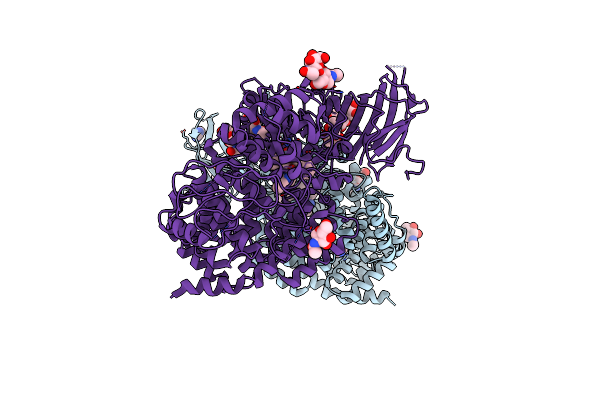
Crystal Structure Of Erap2 Without Catalytic Zn(Ii) Atom
Organism: Homo sapiens
Method: X-RAY DIFFRACTION Resolution:3.02 Å Release Date: 2015-09-23 Classification: HYDROLASE Ligands: NAG |
Organism: Homo sapiens
Method: X-RAY DIFFRACTION
Release Date: 2015-09-23
Ligands: NAG
 |
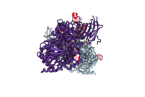
Crystal Structure Of The Human Endoplasmic Reticulum Aminopeptidase 2 In Complex With Phosphinic Pseudotripeptide Inhibitor.
Organism: Homo sapiens
Method: X-RAY DIFFRACTION Resolution:2.79 Å Release Date: 2013-12-11 Classification: Hydrolase/Hydrolase Inhibitor Ligands: NAG, ZN, P52, IMD |
Organism: Homo sapiens
Method: X-RAY DIFFRACTION
Release Date: 2013-12-11
Ligands: NAG, ZN, P52, IMD
 |
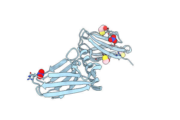
Crystal Structure Of The Human Endoplasmic Reticulum Aminopeptidase 2 Variant N392K
Organism: Homo sapiens
Method: X-RAY DIFFRACTION Resolution:3.22 Å Release Date: 2012-08-29 Classification: HYDROLASE Ligands: LYS, NAG, MES, ZN |
Organism: Homo sapiens
Method: X-RAY DIFFRACTION
Release Date: 2012-08-29
Ligands: LYS, NAG, MES, ZN
 |
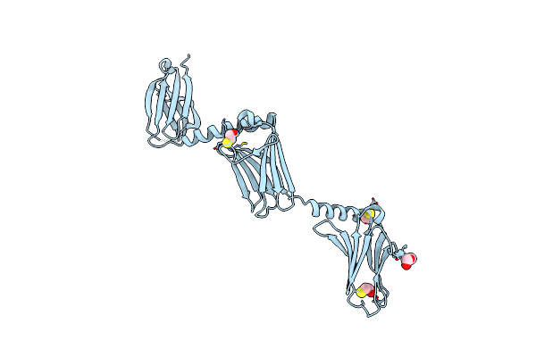
Crystal Structure Of The Myomesin Domains 10 And 11
Organism: Homo sapiens
Method: X-RAY DIFFRACTION Resolution:1.85 Å Release Date: 2012-03-07 Classification: CONTRACTILE PROTEIN Ligands: BME, NO3 |
Organism: Homo sapiens
Method: X-RAY DIFFRACTION
Release Date: 2012-03-07
Ligands: BME, NO3
 |
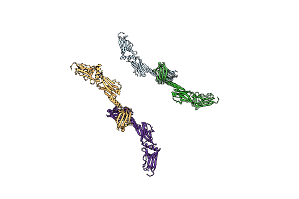
Crystal Structure Of The Myomesin Domains My9-My11
Organism: Homo sapiens
Method: X-RAY DIFFRACTION Resolution:2.50 Å Release Date: 2011-12-21 Classification: STRUCTURAL PROTEIN Ligands: BME, PG0 |
Organism: Homo sapiens
Method: X-RAY DIFFRACTION
Release Date: 2011-12-21
Ligands: BME, PG0
 |
Crystal Structure Of The Myomesin Domains My11-My13
Organism: Homo sapiens
Method: X-RAY DIFFRACTION Resolution:3.50 Å Release Date: 2011-12-21 Classification: STRUCTURAL PROTEIN |
Organism: Homo sapiens
Method: X-RAY DIFFRACTION
Release Date: 2011-12-21
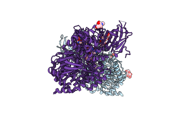 |
Crystal Structure Of The Human Endoplasmic Reticulum Aminopeptidase 2
Organism: Homo sapiens
Method: X-RAY DIFFRACTION Resolution:3.08 Å Release Date: 2011-12-21 Classification: HYDROLASE Ligands: ZN, LYS, NAG, MES |
Organism: Homo sapiens
Method: X-RAY DIFFRACTION
Release Date: 2011-12-21
Ligands: ZN, LYS, NAG, MES
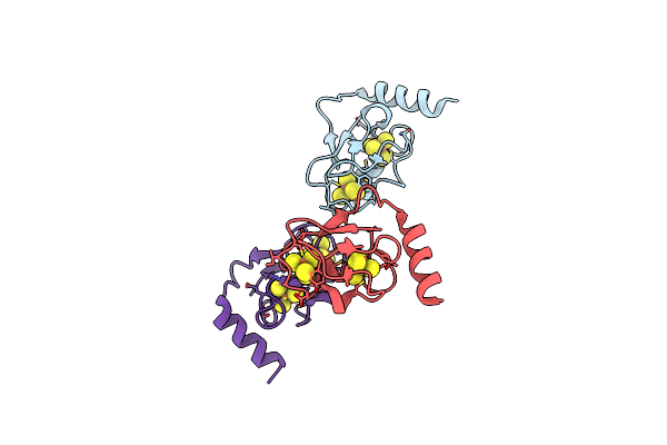 |
Crystal Structure Of The 2[4Fe-4S] Ferredoxin From Escherichia Coli
Organism: Escherichia coli
Method: X-RAY DIFFRACTION Resolution:1.65 Å Release Date: 2009-08-25 Classification: ELECTRON TRANSPORT Ligands: SF4 |
Organism: Escherichia coli
Method: X-RAY DIFFRACTION
Release Date: 2009-08-25
Ligands: SF4
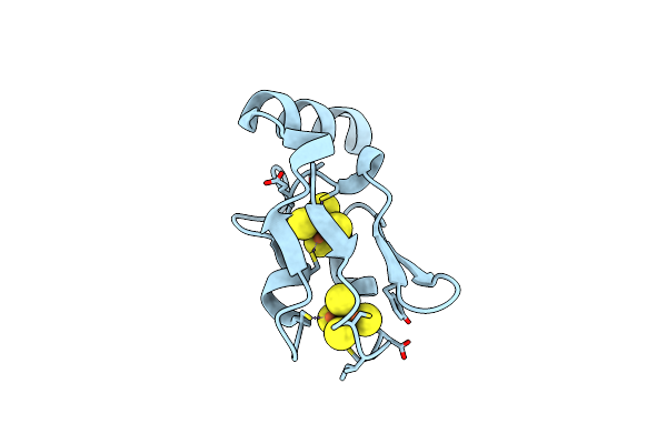 |
Crystal Structure Of The 2[4Fe-4S] C57A Ferredoxin Variant From Allochromatium Vinosum
Organism: Allochromatium vinosum
Method: X-RAY DIFFRACTION Resolution:1.05 Å Release Date: 2009-08-18 Classification: ELECTRON TRANSPORT Ligands: SF4 |
Organism: Allochromatium vinosum
Method: X-RAY DIFFRACTION
Release Date: 2009-08-18
Ligands: SF4
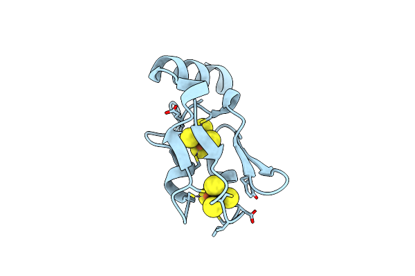 |
Crystal Structure Of The 2[4Fe-4S] Ferredoxin V13G Variant From Allochromatium Vinosum
Organism: Allochromatium vinosum
Method: X-RAY DIFFRACTION Resolution:1.48 Å Release Date: 2009-05-05 Classification: ELECTRON TRANSPORT Ligands: SF4 |
Organism: Allochromatium vinosum
Method: X-RAY DIFFRACTION
Release Date: 2009-05-05
Ligands: SF4
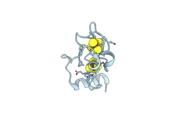 |
Structure Of The 2[4Fe-4S] Ferredoxin From Pseudomonas Aeruginosa
Organism: Pseudomonas aeruginosa
Method: X-RAY DIFFRACTION Resolution:1.32 Å Release Date: 2006-04-18 Classification: ELECTRON TRANSPORT Ligands: SF4 |
Organism: Pseudomonas aeruginosa
Method: X-RAY DIFFRACTION
Release Date: 2006-04-18
Ligands: SF4
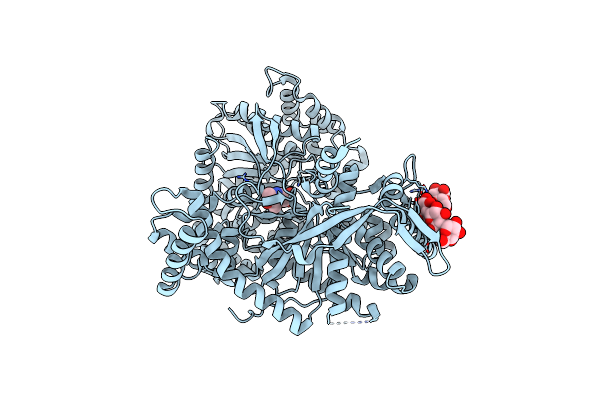 |
Crystal Structure Of Glycogen Phosphorylase B In Complex With Maltopentaose
Organism: Oryctolagus cuniculus
Method: X-RAY DIFFRACTION Resolution:2.20 Å Release Date: 2003-09-02 Classification: TRANSFERASE Ligands: PLP |
Organism: Oryctolagus cuniculus
Method: X-RAY DIFFRACTION
Release Date: 2003-09-02
Ligands: PLP
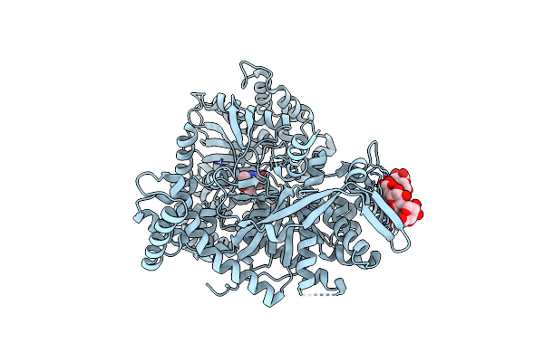 |
Crystal Structure Of Glycogen Phosphorylase B In Complex With Maltoheptaose
Organism: Oryctolagus cuniculus
Method: X-RAY DIFFRACTION Resolution:2.20 Å Release Date: 2003-09-02 Classification: TRANSFERASE Ligands: PLP |
Organism: Oryctolagus cuniculus
Method: X-RAY DIFFRACTION
Release Date: 2003-09-02
Ligands: PLP
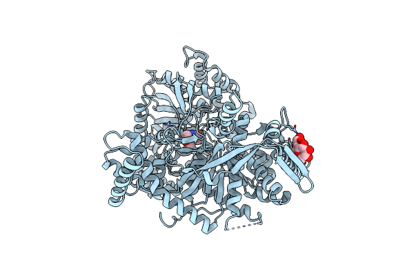 |
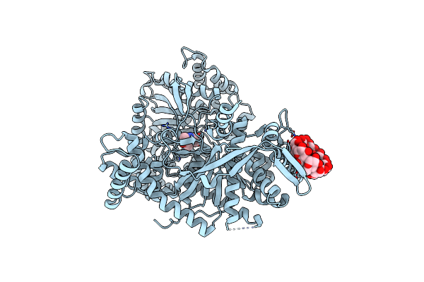
Crystal Structure Of Glycogen Phosphorylase B In Complex With Beta Cyclodextrin
Organism: Oryctolagus cuniculus
Method: X-RAY DIFFRACTION Resolution:1.94 Å Release Date: 2003-09-02 Classification: TRANSFERASE Ligands: PLP |
Organism: Oryctolagus cuniculus
Method: X-RAY DIFFRACTION
Release Date: 2003-09-02
Ligands: PLP
 |
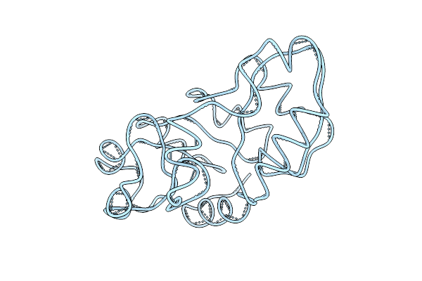
Crystal Structure Of Glycogen Phosphorylase B In Complex With Gamma Cyclodextrin
Organism: Oryctolagus cuniculus
Method: X-RAY DIFFRACTION Resolution:2.30 Å Release Date: 2003-09-02 Classification: TRANSFERASE Ligands: PLP |
Organism: Oryctolagus cuniculus
Method: X-RAY DIFFRACTION
Release Date: 2003-09-02
Ligands: PLP
 |
Structure Of 2-Keto-3-Deoxy-6-Phosphogluconate Aldolase At 2.8 Angstroms Resolution
Organism: Pseudomonas putida
Method: X-RAY DIFFRACTION Resolution:3.50 Å Release Date: 1978-10-13 Classification: LYASE |
Organism: Pseudomonas putida
Method: X-RAY DIFFRACTION
Release Date: 1978-10-13