

Search Count: 34
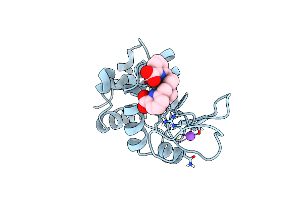 |
Crystal Structure Of Hen Egg White Lysozyme Co-Crystallized With 10 Mm Tbxo4
Organism: Gallus gallus
Method: X-RAY DIFFRACTION Resolution:1.70 Å Release Date: 2024-07-17 Classification: HYDROLASE Ligands: TB, 7MT, CL, NA |
Organism: Gallus gallus
Method: X-RAY DIFFRACTION
Release Date: 2024-07-17
Ligands: TB, 7MT, CL, NA
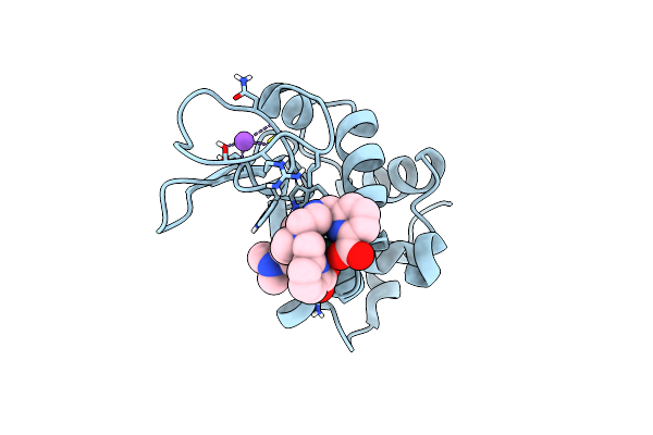 |
Crystal Structure Of Hen Egg White Lysozyme Co-Crystallized With 10 Mm Tbxo4-Nmet2
Organism: Gallus gallus
Method: X-RAY DIFFRACTION Resolution:1.72 Å Release Date: 2024-07-10 Classification: HYDROLASE Ligands: TB, ZHT, CL, NA |
Organism: Gallus gallus
Method: X-RAY DIFFRACTION
Release Date: 2024-07-10
Ligands: TB, ZHT, CL, NA
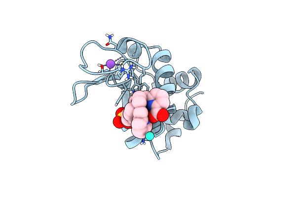 |
Crystal Structure Of Hen Egg White Lysozyme Co-Crystallized With 10 Mm Tbxo4-So3
Organism: Gallus gallus
Method: X-RAY DIFFRACTION Resolution:1.74 Å Release Date: 2024-07-10 Classification: HYDROLASE Ligands: TB, ZW0, CL, NA |
Organism: Gallus gallus
Method: X-RAY DIFFRACTION
Release Date: 2024-07-10
Ligands: TB, ZW0, CL, NA
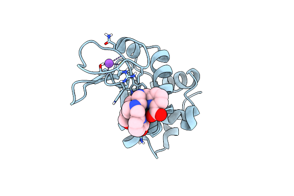 |
Crystal Structure Of Hen Egg White Lysozyme Co-Crystallized With 10 Mm Tbxo4-Oh
Organism: Gallus gallus
Method: X-RAY DIFFRACTION Resolution:1.69 Å Release Date: 2024-06-05 Classification: HYDROLASE Ligands: WRT, CL, NA |
Organism: Gallus gallus
Method: X-RAY DIFFRACTION
Release Date: 2024-06-05
Ligands: WRT, CL, NA
 |
Mosquitocidal Cry11Aa Determined At Ph 7 From Naturally-Occurring Nanocrystals By Serial Femtosecond Crystallography
Organism: Bacillus thuringiensis serovar israelensis
Method: X-RAY DIFFRACTION Resolution:2.60 Å Release Date: 2022-07-27 Classification: TOXIN |
Organism: Bacillus thuringiensis serovar israelensis
Method: X-RAY DIFFRACTION
Release Date: 2022-07-27
 |
Mosquitocidal Cry11Aa-Y449F Determined At Ph 7 From Naturally-Occurring Nanocrystals By Serial Femtosecond Crystallography
Organism: Bacillus thuringiensis serovar israelensis
Method: X-RAY DIFFRACTION Resolution:3.10 Å Release Date: 2022-07-27 Classification: TOXIN |
Organism: Bacillus thuringiensis serovar israelensis
Method: X-RAY DIFFRACTION
Release Date: 2022-07-27
 |
Mosquitocidal Cry11Aa-E583Q Determined At Ph 7 From Naturally-Occurring Nanocrystals By Serial Femtosecond Crystallography
Organism: Bacillus thuringiensis serovar israelensis
Method: X-RAY DIFFRACTION Resolution:3.30 Å Release Date: 2022-07-27 Classification: TOXIN |
Organism: Bacillus thuringiensis serovar israelensis
Method: X-RAY DIFFRACTION
Release Date: 2022-07-27
 |
Mosquitocidal Cry11Aa-F17Y Determined At Ph 7 From Naturally-Occurring Nanocrystals By Serial Femtosecond Crystallography
Organism: Bacillus thuringiensis serovar israelensis
Method: X-RAY DIFFRACTION Resolution:3.40 Å Release Date: 2022-07-27 Classification: TOXIN |
Organism: Bacillus thuringiensis serovar israelensis
Method: X-RAY DIFFRACTION
Release Date: 2022-07-27
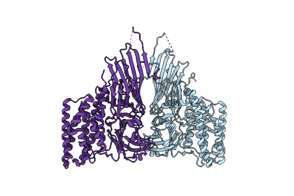 |
Mosquitocidal Cry11Ba Determined At Ph 6.5 From Naturally-Occurring Nanocrystals By Serial Femtosecond Crystallography
Organism: Bacillus thuringiensis serovar jegathesan
Method: X-RAY DIFFRACTION Resolution:2.40 Å Release Date: 2022-07-27 Classification: TOXIN |
Organism: Bacillus thuringiensis serovar jegathesan
Method: X-RAY DIFFRACTION
Release Date: 2022-07-27
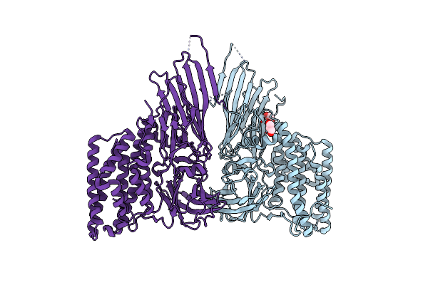 |
Mosquitocidal Cry11Ba Determined At Ph 10.4 From Naturally-Occurring Nanocrystals By Serial Femtosecond Crystallography
Organism: Bacillus thuringiensis serovar jegathesan
Method: X-RAY DIFFRACTION Resolution:2.65 Å Release Date: 2022-07-27 Classification: TOXIN Ligands: GOL |
Organism: Bacillus thuringiensis serovar jegathesan
Method: X-RAY DIFFRACTION
Release Date: 2022-07-27
Ligands: GOL
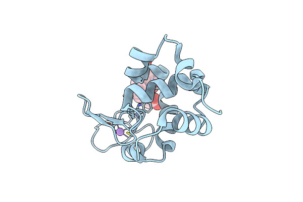 |
Structure Of Hen Egg White Lysozyme Crystallized In The Presence Of Tb-Xo4 Crystallophore In The Xtalcontroller Device
Organism: Gallus gallus
Method: X-RAY DIFFRACTION Resolution:1.51 Å Release Date: 2020-12-16 Classification: ANTIMICROBIAL PROTEIN |
Organism: Gallus gallus
Method: X-RAY DIFFRACTION
Release Date: 2020-12-16
 |
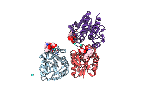
Structure Of A Psychrophilic Cca-Adding Enzyme Crystallized In The Xtalcontroller Device
Organism: Planococcus halocryophilus
Method: X-RAY DIFFRACTION Resolution:2.28 Å Release Date: 2020-12-16 Classification: RNA BINDING PROTEIN Ligands: PO4, GOL, ACT |
Organism: Planococcus halocryophilus
Method: X-RAY DIFFRACTION
Release Date: 2020-12-16
Ligands: PO4, GOL, ACT
 |
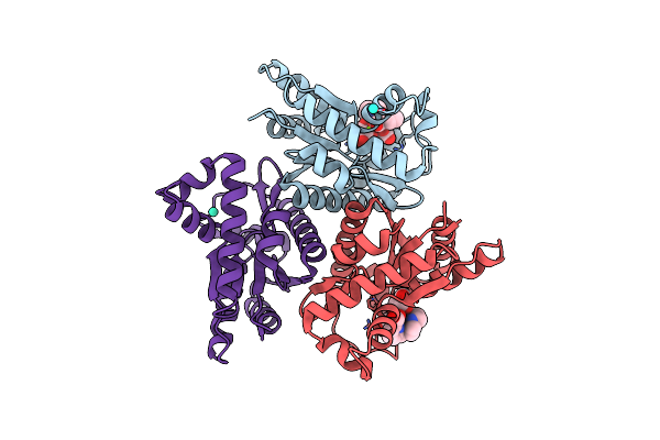
Crystal Structure Of The Protease 1 (E29A,E60A,E80A) From Pyrococcus Horikoshii Co-Crystallized With Tb-Xo4.
Organism: Pyrococcus horikoshii (strain atcc 700860 / dsm 12428 / jcm 9974 / nbrc 100139 / ot-3)
Method: X-RAY DIFFRACTION Resolution:2.00 Å Release Date: 2019-06-19 Classification: HYDROLASE Ligands: 7MT, MLI, TB |
Organism: Pyrococcus horikoshii (strain atcc 700860 / dsm 12428 / jcm 9974 / nbrc 100139 / ot-3)
Method: X-RAY DIFFRACTION
Release Date: 2019-06-19
Ligands: 7MT, MLI, TB
 |
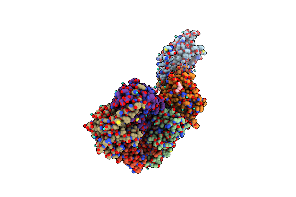
Crystal Structure Of The Adenylate Kinase From Methanothermococcus Thermolithotrophicus Co-Crystallized With Tb-Xo4
Organism: Methanothermococcus thermolithotrophicus
Method: X-RAY DIFFRACTION Resolution:1.96 Å Release Date: 2019-06-19 Classification: TRANSFERASE Ligands: 7MT, TB, MG, GOL |
Organism: Methanothermococcus thermolithotrophicus
Method: X-RAY DIFFRACTION
Release Date: 2019-06-19
Ligands: 7MT, TB, MG, GOL
 |
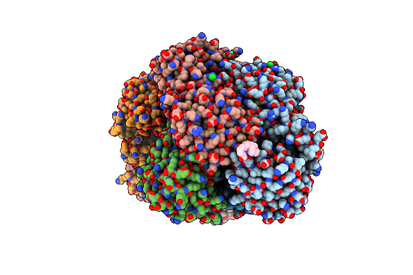
Crystal Structure Of The Thiazole Synthase From Methanothermococcus Thermolithotrophicus Co-Crystallized With Tb-Xo4
Organism: Methanothermococcus thermolithotrophicus
Method: X-RAY DIFFRACTION Resolution:2.55 Å Release Date: 2019-06-19 Classification: BIOSYNTHETIC PROTEIN Ligands: TB, 48F, GOL, PGE, PEG, NA |
Organism: Methanothermococcus thermolithotrophicus
Method: X-RAY DIFFRACTION
Release Date: 2019-06-19
Ligands: TB, 48F, GOL, PGE, PEG, NA
 |
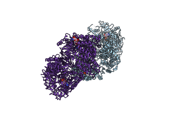
Crystal Structure Of Coenzyme F420H2 Oxidase (Fpra) Co-Crystallized With 10 Mm Tb-Xo4
Organism: Methanothermococcus thermolithotrophicus
Method: X-RAY DIFFRACTION Resolution:2.20 Å Release Date: 2018-10-31 Classification: OXIDOREDUCTASE Ligands: FMN, 7MT, TB, CL, FE |
Organism: Methanothermococcus thermolithotrophicus
Method: X-RAY DIFFRACTION
Release Date: 2018-10-31
Ligands: FMN, 7MT, TB, CL, FE
 |
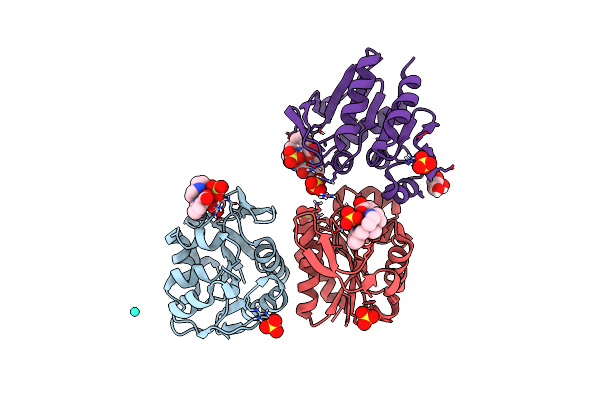
Tri-Functional Propionyl-Coa Synthase Of Erythrobacter Sp. Nap1 With Bound Nadp+ And Phosphomethylphosphonic Acid Adenylate Ester
Organism: Erythrobacter sp. nap1
Method: X-RAY DIFFRACTION Resolution:2.70 Å Release Date: 2018-10-24 Classification: OXIDOREDUCTASE Ligands: ACP, NAP |
Organism: Erythrobacter sp. nap1
Method: X-RAY DIFFRACTION
Release Date: 2018-10-24
Ligands: ACP, NAP
 |
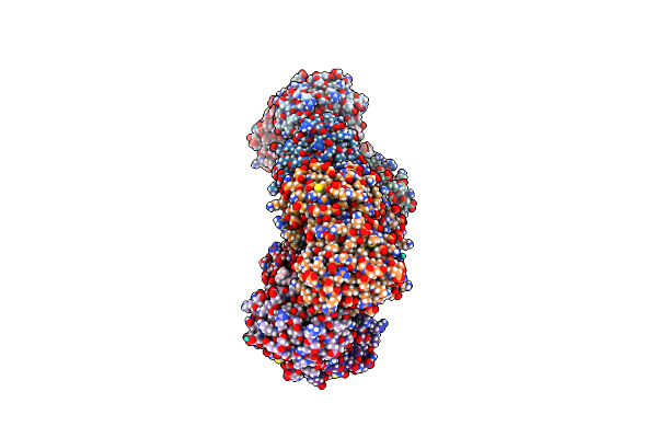
Crystal Structure Of Protease 1 From Pyrococcus Horikoshii Co-Cystallized In Presence Of 10 Mm Tb-Xo4 And Ammonium Sulfate.
Organism: Pyrococcus horikoshii
Method: X-RAY DIFFRACTION Resolution:1.65 Å Release Date: 2018-10-03 Classification: CELL CYCLE Ligands: 7MT, SO4, TB, 2HA |
Organism: Pyrococcus horikoshii
Method: X-RAY DIFFRACTION
Release Date: 2018-10-03
Ligands: 7MT, SO4, TB, 2HA
 |
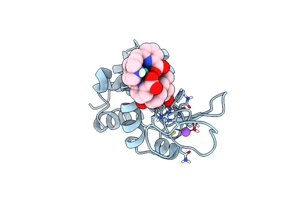
Structure Of Protease 1 From Pyrococcus Horikoshii Co-Crystallized In Presence Of 10 Mm Tb-Xo4 And Potassium Iodide.
Organism: Pyrococcus horikoshii
Method: X-RAY DIFFRACTION Resolution:2.19 Å Release Date: 2018-10-03 Classification: HYDROLASE Ligands: 7MT, IOD, TB |
Organism: Pyrococcus horikoshii
Method: X-RAY DIFFRACTION
Release Date: 2018-10-03
Ligands: 7MT, IOD, TB
 |
Crystal Structure Of Hen Egg-White Lysozyme Co-Crystallized In Presence Of 100 Mm Tb-Xo4
Organism: Gallus gallus
Method: X-RAY DIFFRACTION Resolution:1.20 Å Release Date: 2018-10-03 Classification: HYDROLASE Ligands: TB, 7MT, CL, NA, ACT |
Organism: Gallus gallus
Method: X-RAY DIFFRACTION
Release Date: 2018-10-03
Ligands: TB, 7MT, CL, NA, ACT