

Search Count: 19
 |
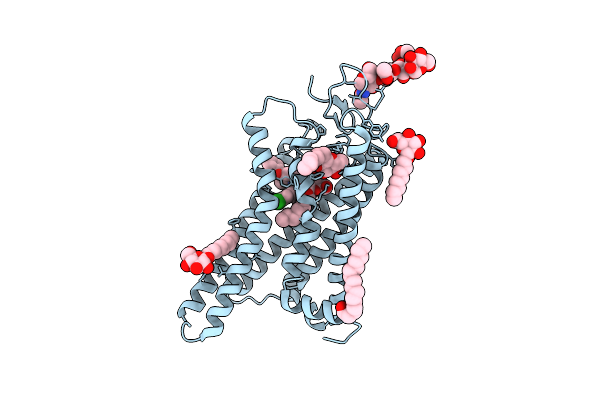
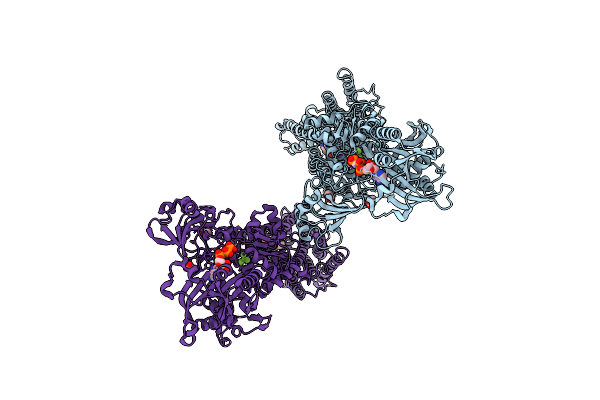
Structure Of The Human Cc Chemokine Receptor 7 In Complex With The Intracellular Allosteric Antagonist Cmp2105 And The Insertion Protein Sialidase Nana
Organism: Homo sapiens, Streptococcus pneumoniae
Method: X-RAY DIFFRACTION Resolution:2.10 Å Release Date: 2019-09-04 Classification: SIGNALING PROTEIN Ligands: PGE, JLW, NA, TAR |
Organism: Homo sapiens, Streptococcus pneumoniae
Method: X-RAY DIFFRACTION
Release Date: 2019-09-04
Ligands: PGE, JLW, NA, TAR
 |
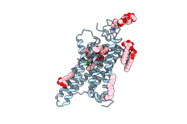
Crystal Structure Of N2C/D282C Stabilized Opsin Bound To Rs01
Organism: Bos taurus
Method: X-RAY DIFFRACTION Resolution:2.36 Å Release Date: 2018-04-04 Classification: MEMBRANE PROTEIN Ligands: PLM, BOG, DOK |
Organism: Bos taurus
Method: X-RAY DIFFRACTION
Release Date: 2018-04-04
Ligands: PLM, BOG, DOK
 |
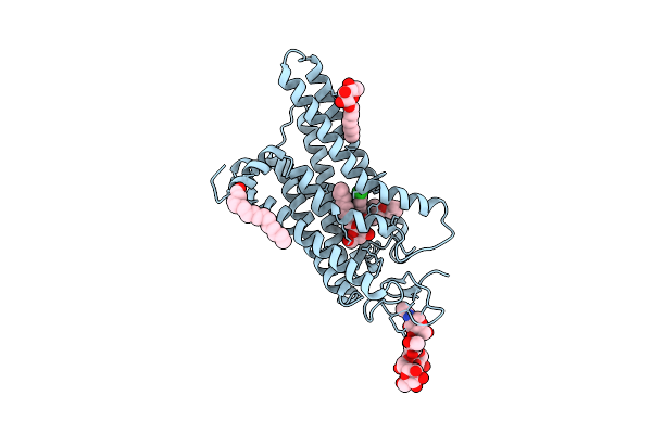
Crystal Structure Of N2C/D282C Stabilized Opsin Bound To Rs06
Organism: Bos taurus
Method: X-RAY DIFFRACTION Resolution:2.62 Å Release Date: 2018-04-04 Classification: MEMBRANE PROTEIN Ligands: PLM, BOG, DO5 |
Organism: Bos taurus
Method: X-RAY DIFFRACTION
Release Date: 2018-04-04
Ligands: PLM, BOG, DO5
 |
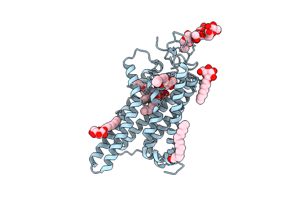
Crystal Structure Of N2C/D282C Stabilized Opsin Bound To Rs08
Organism: Bos taurus
Method: X-RAY DIFFRACTION Resolution:2.87 Å Release Date: 2018-04-04 Classification: MEMBRANE PROTEIN Ligands: PLM, DNZ, BOG |
Organism: Bos taurus
Method: X-RAY DIFFRACTION
Release Date: 2018-04-04
Ligands: PLM, DNZ, BOG
 |
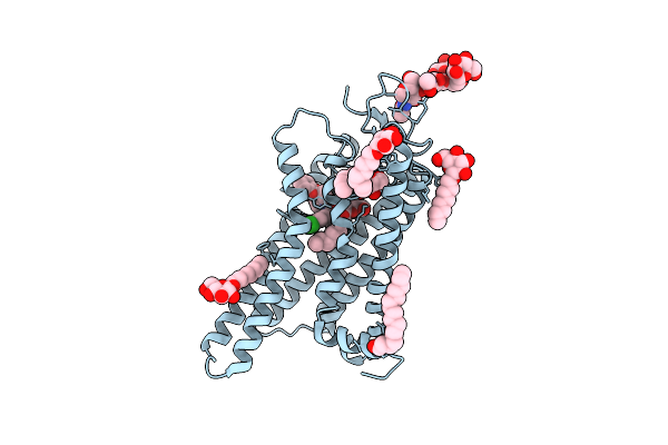
Crystal Structure Of N2C/D282C Stabilized Opsin Bound To Rs09
Organism: Bos taurus
Method: X-RAY DIFFRACTION Resolution:2.63 Å Release Date: 2018-04-04 Classification: MEMBRANE PROTEIN Ligands: PLM, BOG, DNK |
Organism: Bos taurus
Method: X-RAY DIFFRACTION
Release Date: 2018-04-04
Ligands: PLM, BOG, DNK
 |
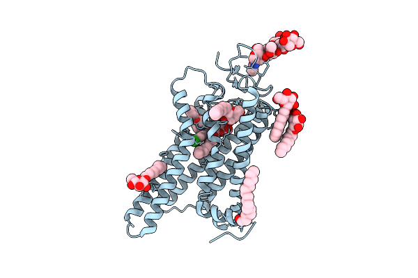
Crystal Structure Of N2C/D282C Stabilized Opsin Bound To Rs11
Organism: Bos taurus
Method: X-RAY DIFFRACTION Resolution:2.70 Å Release Date: 2018-04-04 Classification: MEMBRANE PROTEIN Ligands: PLM, BOG, DN5 |
Organism: Bos taurus
Method: X-RAY DIFFRACTION
Release Date: 2018-04-04
Ligands: PLM, BOG, DN5
 |
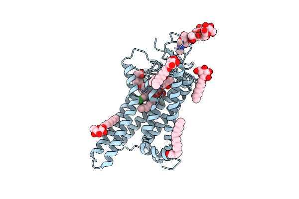
Crystal Structure Of N2C/D282C Stabilized Opsin Bound To Rs13
Organism: Bos taurus
Method: X-RAY DIFFRACTION Resolution:3.03 Å Release Date: 2018-04-04 Classification: MEMBRANE PROTEIN Ligands: PLM, NAG, BMA, MAN, BOG, DLH |
Organism: Bos taurus
Method: X-RAY DIFFRACTION
Release Date: 2018-04-04
Ligands: PLM, NAG, BMA, MAN, BOG, DLH
 |
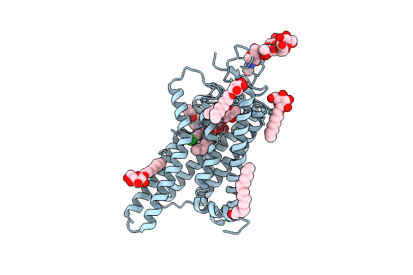
Crystal Structure Of N2C/D282C Stabilized Opsin Bound To Rs15
Organism: Bos taurus
Method: X-RAY DIFFRACTION Resolution:2.46 Å Release Date: 2018-04-04 Classification: MEMBRANE PROTEIN Ligands: PLM, DLB, BOG |
Organism: Bos taurus
Method: X-RAY DIFFRACTION
Release Date: 2018-04-04
Ligands: PLM, DLB, BOG
 |
Crystal Structure Of N2C/D282C Stabilized Opsin Bound To Rs16
Organism: Bos taurus
Method: X-RAY DIFFRACTION Resolution:2.49 Å Release Date: 2018-04-04 Classification: MEMBRANE PROTEIN Ligands: PLM, BOG, DL2 |
Organism: Bos taurus
Method: X-RAY DIFFRACTION
Release Date: 2018-04-04
Ligands: PLM, BOG, DL2
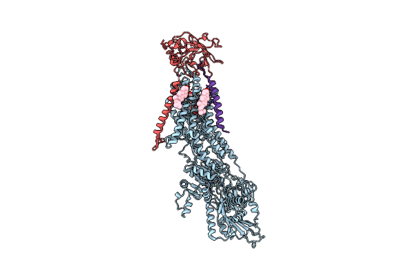 |
Crystal Structure Of The Na,K-Atpase From Bovine
Organism: Bos taurus
Method: X-RAY DIFFRACTION Resolution:3.90 Å Release Date: 2016-03-09 Classification: HYDROLASE Ligands: MG, OBN, CLR, BEF |
Organism: Bos taurus
Method: X-RAY DIFFRACTION
Release Date: 2016-03-09
Ligands: MG, OBN, CLR, BEF
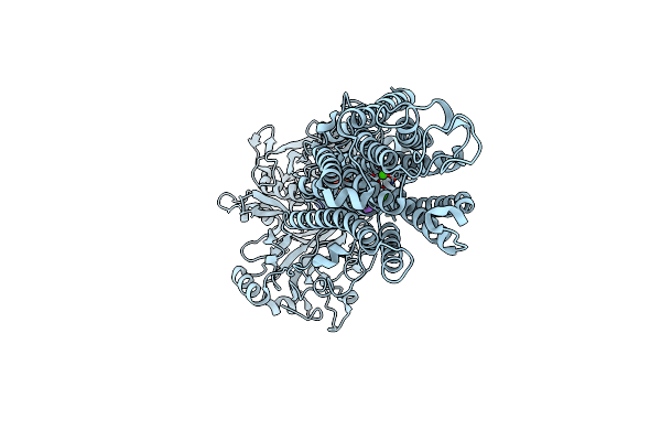 |
Crystal Structure Of The Sr Ca2+-Atpase In The Ca2-E1-Mgamppcp Form Determined By Serial Femtosecond Crystallography Using An X-Ray Free-Electron Laser.
Organism: Oryctolagus cuniculus
Method: X-RAY DIFFRACTION Resolution:2.80 Å Release Date: 2015-06-10 Classification: HYDROLASE Ligands: CA, K, ACP |
Organism: Oryctolagus cuniculus
Method: X-RAY DIFFRACTION
Release Date: 2015-06-10
Ligands: CA, K, ACP
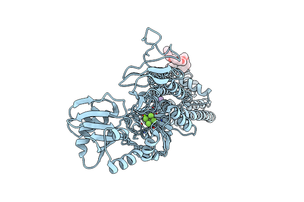 |
Atpase Crystal Structure
Organism: Legionella pneumophila subsp. pneumophila str. philadelphia 1
Method: X-RAY DIFFRACTION Resolution:2.85 Å Release Date: 2014-08-06 Classification: HYDROLASE Ligands: ALF, MG, K, 15P |
Organism: Legionella pneumophila subsp. pneumophila str. philadelphia 1
Method: X-RAY DIFFRACTION
Release Date: 2014-08-06
Ligands: ALF, MG, K, 15P
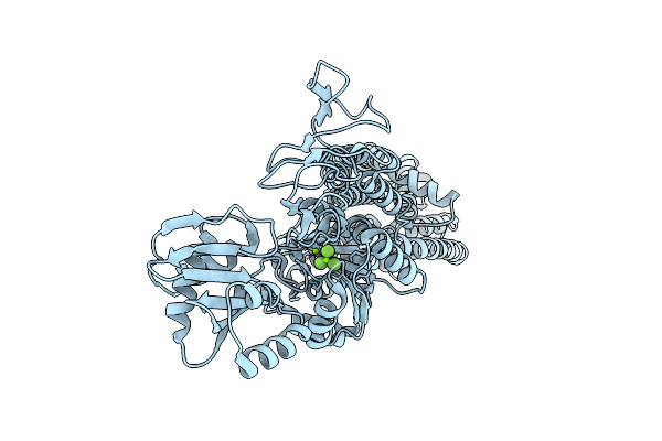 |
Atpase Crystal Structure With Bound Phosphate Analogue
Organism: Legionella pneumophila
Method: X-RAY DIFFRACTION Resolution:3.58 Å Release Date: 2014-04-02 Classification: HYDROLASE Ligands: MG, MGF |
Organism: Legionella pneumophila
Method: X-RAY DIFFRACTION
Release Date: 2014-04-02
Ligands: MG, MGF
 |
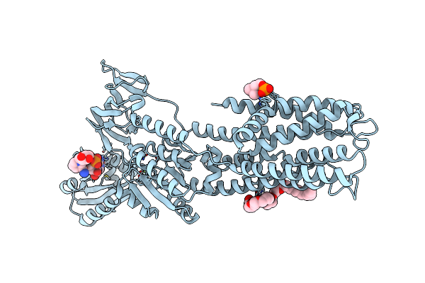
Serca Bound To Phosphate Analogue
Organism: Oryctolagus cuniculus
Method: X-RAY DIFFRACTION Resolution:2.50 Å Release Date: 2014-02-19 Classification: HYDROLASE Ligands: MG, K, CZA, MN, ACP, MGF, ACT |
Organism: Oryctolagus cuniculus
Method: X-RAY DIFFRACTION
Release Date: 2014-02-19
Ligands: MG, K, CZA, MN, ACP, MGF, ACT
 |
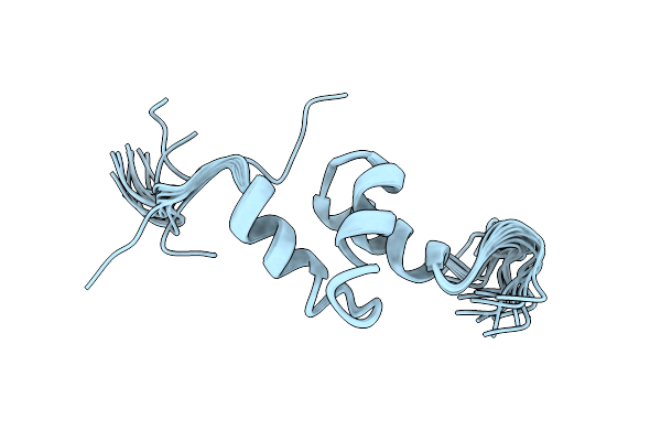
Copper-Transporting Pib-Atpase In Complex With Beryllium Fluoride Representing The E2P State
Organism: Legionella pneumophila subsp. pneumophila
Method: X-RAY DIFFRACTION Resolution:2.75 Å Release Date: 2013-12-11 Classification: HYDROLASE Ligands: MG, NAD, CE1, PC |
Organism: Legionella pneumophila subsp. pneumophila
Method: X-RAY DIFFRACTION
Release Date: 2013-12-11
Ligands: MG, NAD, CE1, PC
 |
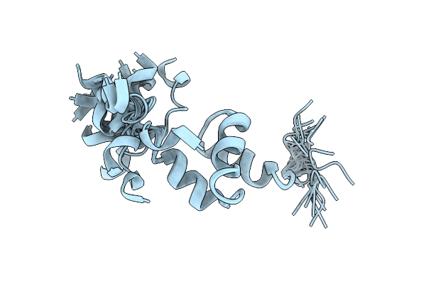
Nmr Structure Of The Uba Domain Of S. Cerevisiae Dcn1
Organism: Saccharomyces cerevisiae
Method: SOLUTION NMR Release Date: 2011-10-05 Classification: PROTEIN BINDING |
Organism: Saccharomyces cerevisiae
Method: SOLUTION NMR
Release Date: 2011-10-05
 |
Nmr Structure Of The Uba Domain Of S. Cerevisiae Dcn1 Bound To Ubiquitin
Organism: Saccharomyces cerevisiae
Method: SOLUTION NMR Release Date: 2011-10-05 Classification: PROTEIN BINDING |
Organism: Saccharomyces cerevisiae
Method: SOLUTION NMR
Release Date: 2011-10-05
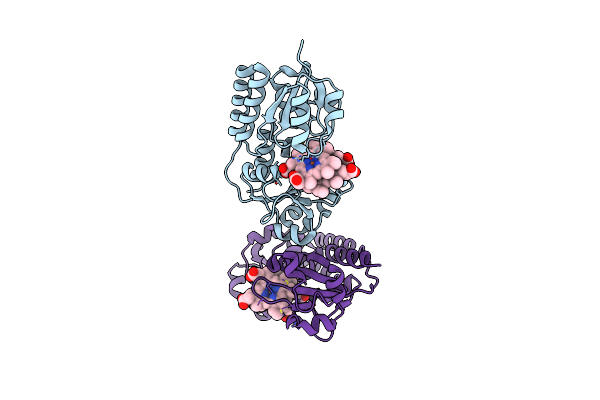 |
Structure Of Holo Form Of A Periplasmic Heme Binding Protein
Organism: Yersinia pestis
Method: X-RAY DIFFRACTION Resolution:2.50 Å Release Date: 2010-10-13 Classification: METAL TRANSPORT Ligands: HEM |
Organism: Yersinia pestis
Method: X-RAY DIFFRACTION
Release Date: 2010-10-13
Ligands: HEM
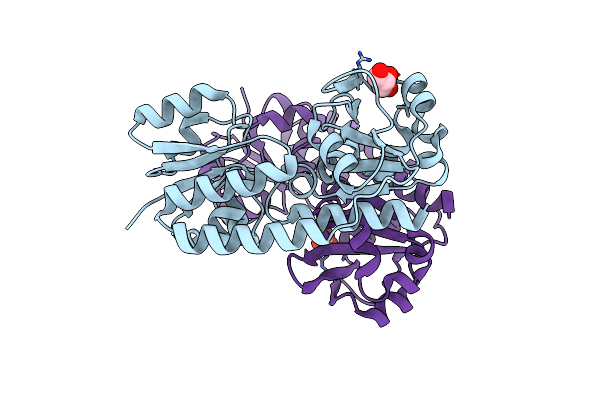 |
Structure Of Apo Form Of A Periplasmic Heme Binding Protein
Organism: Yersinia pestis
Method: X-RAY DIFFRACTION Resolution:1.50 Å Release Date: 2010-09-15 Classification: TRANSPORT PROTEIN Ligands: BR, GOL, SO4 |
Organism: Yersinia pestis
Method: X-RAY DIFFRACTION
Release Date: 2010-09-15
Ligands: BR, GOL, SO4