

Search Count: 34
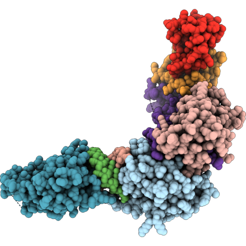 |
Divergent Interfaces And Convergent Outcome In The Structural Basis Of Hiv-2 Vif Antagonism Of Apobec3H
Organism: Pan troglodytes, Homo sapiens, Human immunodeficiency virus 2, Escherichia coli bl21(de3)
Method: ELECTRON MICROSCOPY Resolution:3.10 Å Release Date: 2026-10-14 Classification: VIRAL PROTEIN Ligands: ZN |
Organism: Pan troglodytes, Homo sapiens, Human immunodeficiency virus 2, Escherichia coli bl21(de3)
Method: ELECTRON MICROSCOPY
Release Date: 2026-10-14
Ligands: ZN
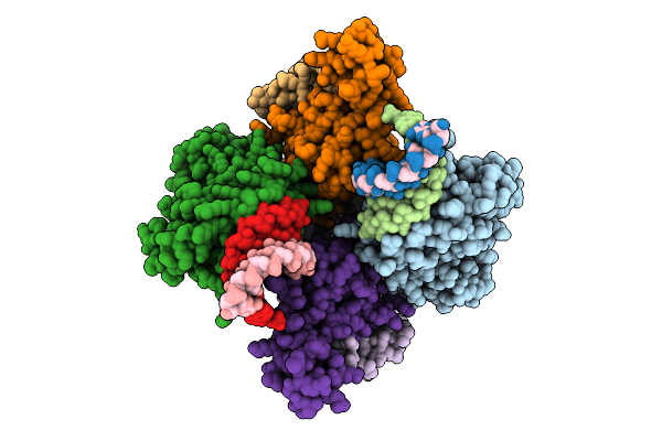 |
Structural Insights Into Hiv-1 Vif-Mediated Ubiquitination And Degradation Of Apobec3H
Organism: Pan troglodytes, Homo sapiens, Human immunodeficiency virus 1, Escherichia coli bl21(de3)
Method: ELECTRON MICROSCOPY Release Date: 2026-03-11 Classification: VIRAL PROTEIN/RNA Ligands: ZN |
Organism: Pan troglodytes, Homo sapiens, Human immunodeficiency virus 1, Escherichia coli bl21(de3)
Method: ELECTRON MICROSCOPY
Release Date: 2026-03-11
Ligands: ZN
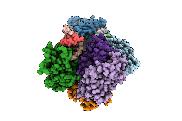 |
Structural Insights Into Hiv-1 Vif-Mediated Ubiquitination And Degradation Of Apobec3H
Organism: Pan troglodytes, Homo sapiens, Human immunodeficiency virus 1, Escherichia coli bl21(de3)
Method: ELECTRON MICROSCOPY Release Date: 2026-03-11 Classification: VIRAL PROTEIN/RNA Ligands: ZN |
Organism: Pan troglodytes, Homo sapiens, Human immunodeficiency virus 1, Escherichia coli bl21(de3)
Method: ELECTRON MICROSCOPY
Release Date: 2026-03-11
Ligands: ZN
 |
Cryo-Em Structure Of Human Glut9 Bound To Urate
Organism: Homo sapiens
Method: ELECTRON MICROSCOPY Resolution:3.39 Å Release Date: 2025-04-16 Classification: TRANSPORT PROTEIN Ligands: URC |
Organism: Homo sapiens
Method: ELECTRON MICROSCOPY
Release Date: 2025-04-16
Ligands: URC
 |
Cryo-Em Structure Of Human Glut9 Apo State
Organism: Homo sapiens
Method: ELECTRON MICROSCOPY Resolution:3.06 Å Release Date: 2025-04-16 Classification: TRANSPORT PROTEIN |
Organism: Homo sapiens
Method: ELECTRON MICROSCOPY
Release Date: 2025-04-16
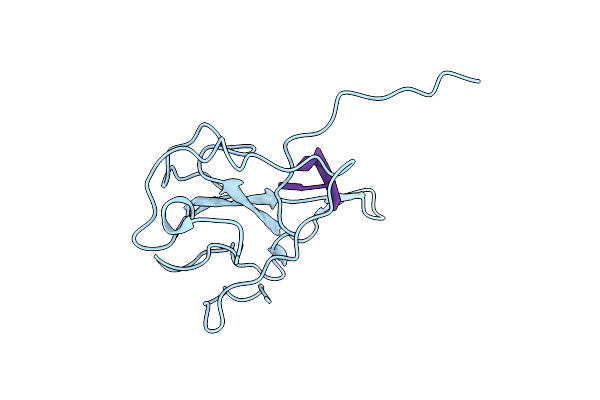 |
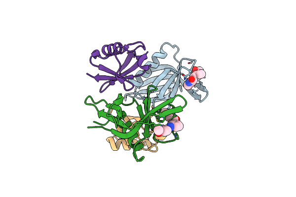
Crystal Structure Of Sars-Cov-2 N-Ntd With Part Of N-Arm Complex With Ssdna.
Organism: Severe acute respiratory syndrome coronavirus 2, Synthetic construct
Method: X-RAY DIFFRACTION Resolution:1.55 Å Release Date: 2024-12-11 Classification: VIRAL PROTEIN/DNA |
Organism: Severe acute respiratory syndrome coronavirus 2, Synthetic construct
Method: X-RAY DIFFRACTION
Release Date: 2024-12-11
 |
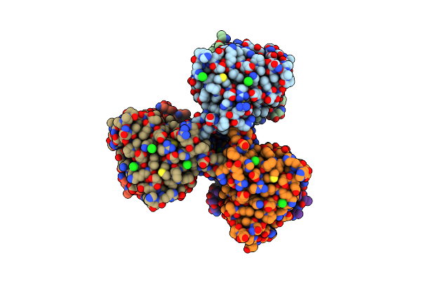
Crystal Structure Of Hrpn13 Pru Domain In Complex With Ubiquitin And Xl44
Organism: Homo sapiens
Method: X-RAY DIFFRACTION Resolution:2.10 Å Release Date: 2024-03-27 Classification: PROTEIN BINDING Ligands: ZTO |
Organism: Homo sapiens
Method: X-RAY DIFFRACTION
Release Date: 2024-03-27
Ligands: ZTO
 |
Crystal Structure Of The Small Ultra-Red Fluorescent Protein (Smurfp)
Organism: Synthetic construct
Method: X-RAY DIFFRACTION Resolution:2.80 Å Release Date: 2023-07-19 Classification: FLUORESCENT PROTEIN Ligands: CL, NA |
Organism: Synthetic construct
Method: X-RAY DIFFRACTION
Release Date: 2023-07-19
Ligands: CL, NA
 |
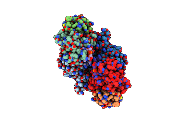
Cryo-Em Structure Of Apobec3G-Vif Complex
Organism: Homo sapiens, Human immunodeficiency virus 1, Synthetic construct
Method: ELECTRON MICROSCOPY Release Date: 2023-07-19 Classification: ANTIVIRAL PROTEIN Ligands: ZN, CL |
Organism: Homo sapiens, Human immunodeficiency virus 1, Synthetic construct
Method: ELECTRON MICROSCOPY
Release Date: 2023-07-19
Ligands: ZN, CL
 |
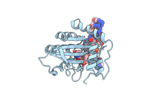
Cryo-Em Structure Of Apobec3G-Vif Complex
Organism: Homo sapiens, Human immunodeficiency virus 1, Synthetic construct
Method: ELECTRON MICROSCOPY Release Date: 2023-07-19 Classification: ANTIVIRAL PROTEIN Ligands: ZN |
Organism: Homo sapiens, Human immunodeficiency virus 1, Synthetic construct
Method: ELECTRON MICROSCOPY
Release Date: 2023-07-19
Ligands: ZN
 |
Crystal Structure Of Apobec3G Catalytic Domain Complex With Ssdna Containing 2'-Deoxy Zebularine.
Organism: Homo sapiens, Synthetic construct
Method: X-RAY DIFFRACTION Resolution:1.50 Å Release Date: 2022-12-14 Classification: HYDROLASE/DNA Ligands: ZN |
Organism: Homo sapiens, Synthetic construct
Method: X-RAY DIFFRACTION
Release Date: 2022-12-14
Ligands: ZN
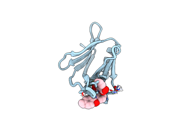 |
Structure Of Xl5-Ligated Hrpn13 Pru Domain
Organism: Homo sapiens
Method: SOLUTION NMR Release Date: 2021-12-15 Classification: PROTEIN BINDING Ligands: XAY |
Organism: Homo sapiens
Method: SOLUTION NMR
Release Date: 2021-12-15
Ligands: XAY
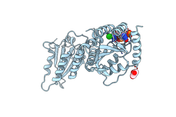 |
Crystal Structure Of A Soluble Variant Of Full-Length Human Apobec3G (Ph 7.6)
Organism: Homo sapiens, Escherichia coli
Method: X-RAY DIFFRACTION Resolution:2.50 Å Release Date: 2020-12-23 Classification: HYDROLASE/DNA Ligands: ZN, CL, GOL |
Organism: Homo sapiens, Escherichia coli
Method: X-RAY DIFFRACTION
Release Date: 2020-12-23
Ligands: ZN, CL, GOL
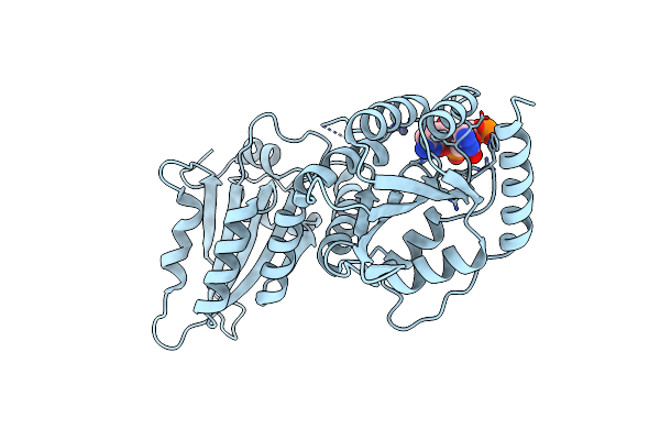 |
Crystal Structure Of A Soluble Variant Of Full-Length Human Apobec3G (Ph 8.0)
Organism: Homo sapiens, Escherichia coli
Method: X-RAY DIFFRACTION Resolution:3.02 Å Release Date: 2020-12-23 Classification: HYDROLASE/DNA Ligands: ZN |
Organism: Homo sapiens, Escherichia coli
Method: X-RAY DIFFRACTION
Release Date: 2020-12-23
Ligands: ZN
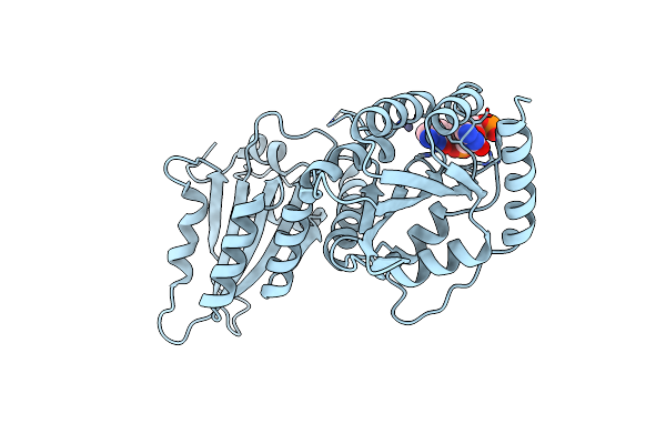 |
Crystal Structure Of A Soluble Variant Of Full-Length Human Apobec3G (Ph 9.0)
Organism: Homo sapiens, Escherichia coli
Method: X-RAY DIFFRACTION Resolution:3.49 Å Release Date: 2020-12-23 Classification: HYDROLASE/DNA Ligands: ZN |
Organism: Homo sapiens, Escherichia coli
Method: X-RAY DIFFRACTION
Release Date: 2020-12-23
Ligands: ZN
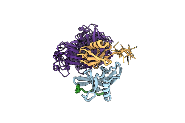 |
Hrpn13:Hrpn2:K48-Diubiquitin
Organism: Homo sapiens
Method: SOLUTION NMR Release Date: 2020-03-11 Classification: PROTEIN BINDING |
Organism: Homo sapiens
Method: SOLUTION NMR
Release Date: 2020-03-11
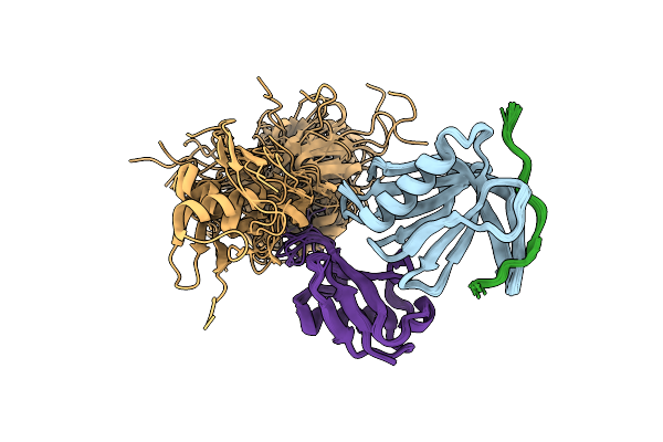 |
Hrpn13:Hrpn2:K48-Diubiquitin
Organism: Homo sapiens
Method: SOLUTION NMR Release Date: 2020-03-11 Classification: PROTEIN BINDING |
Organism: Homo sapiens
Method: SOLUTION NMR
Release Date: 2020-03-11
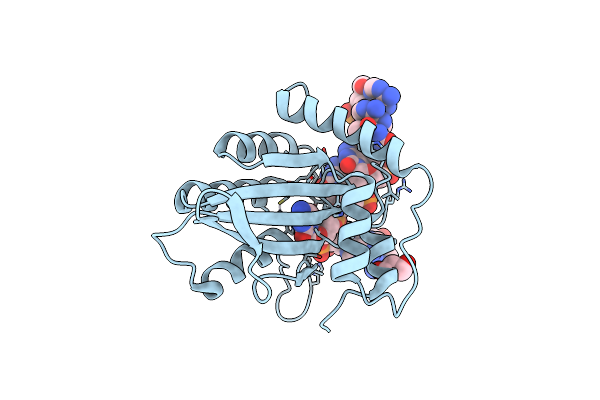 |
Crystal Structure Of Apobec3G Catalytic Domain Complex With Substrate Ssdna
Organism: Homo sapiens, Synthetic construct
Method: X-RAY DIFFRACTION Resolution:1.86 Å Release Date: 2018-07-18 Classification: HYDROLASE/DNA Ligands: ZN, GOL |
Organism: Homo sapiens, Synthetic construct
Method: X-RAY DIFFRACTION
Release Date: 2018-07-18
Ligands: ZN, GOL
 |
Crystal Structure Of Apobec3A In Complex With A Single-Stranded Dna
Organism: Homo sapiens, Synthetic construct
Method: X-RAY DIFFRACTION Resolution:2.20 Å Release Date: 2017-05-10 Classification: HYDROLASE/DNA Ligands: ZN, CA, CL |
Organism: Homo sapiens, Synthetic construct
Method: X-RAY DIFFRACTION
Release Date: 2017-05-10
Ligands: ZN, CA, CL
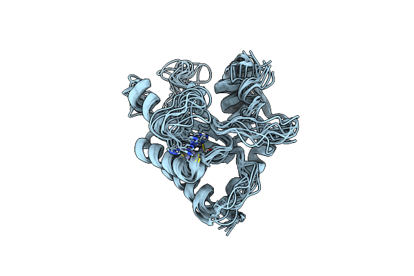 |
Nmr Structure Of Apobec3G Ntd Variant, Sntd
Organism: Artificial gene
Method: SOLUTION NMR Release Date: 2015-05-13 Classification: Hydrolase, Antiviral protein Ligands: ZN |
Organism: Artificial gene
Method: SOLUTION NMR
Release Date: 2015-05-13
Ligands: ZN