

Search Count: 32
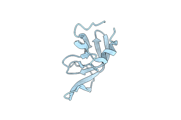 |
Crystal Structure Of A Multi-Drug Resistant Clinical Isolate-769 Hiv-1 Protease Variant That Is Resistant To The Dimerization Inhibitory Activity Of Tlf-Paff
Organism: Human immunodeficiency virus 1
Method: X-RAY DIFFRACTION Resolution:1.80 Å Release Date: 2014-07-09 Classification: HYDROLASE |
Organism: Human immunodeficiency virus 1
Method: X-RAY DIFFRACTION
Release Date: 2014-07-09
 |
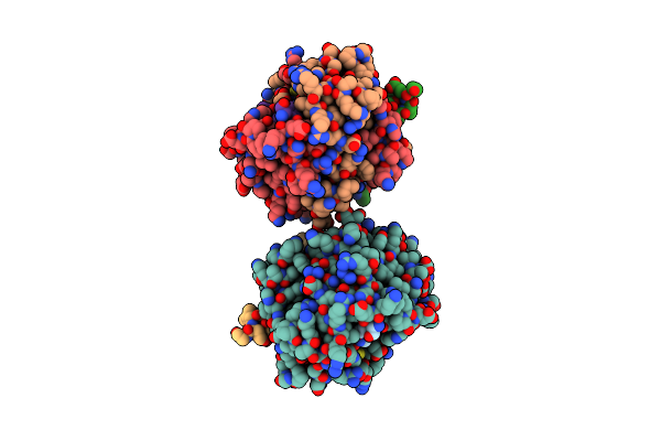
Crystal Structure Of The Aquifex Reactor Complex Formed By Dihydroorotase (H180A, H232A) With Dihydroorotate And Aspartate Transcarbamoylase With N-(Phosphonacetyl)-L-Aspartate (Pala)
Organism: Aquifex aeolicus
Method: X-RAY DIFFRACTION Resolution:2.20 Å Release Date: 2013-12-18 Classification: HYDROLASE/TRANSFERASE Ligands: ZN, DOR, PAL, PO4, BA, EDO |
Organism: Aquifex aeolicus
Method: X-RAY DIFFRACTION
Release Date: 2013-12-18
Ligands: ZN, DOR, PAL, PO4, BA, EDO
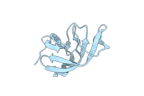 |
Crystal Structures Of Multidrug-Resistant Clinical Isolate 769 Hiv-1 Protease Variants
Organism: Human immunodeficiency virus 1
Method: X-RAY DIFFRACTION Resolution:1.71 Å Release Date: 2011-04-06 Classification: HYDROLASE |
Organism: Human immunodeficiency virus 1
Method: X-RAY DIFFRACTION
Release Date: 2011-04-06
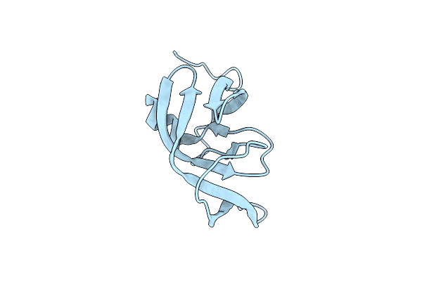 |
Crystal Structures Of Multidrug-Resistant Clinical Isolate 769 Hiv-1 Protease Variants
Organism: Human immunodeficiency virus 1
Method: X-RAY DIFFRACTION Resolution:2.25 Å Release Date: 2011-04-06 Classification: HYDROLASE |
Organism: Human immunodeficiency virus 1
Method: X-RAY DIFFRACTION
Release Date: 2011-04-06
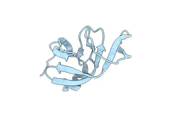 |
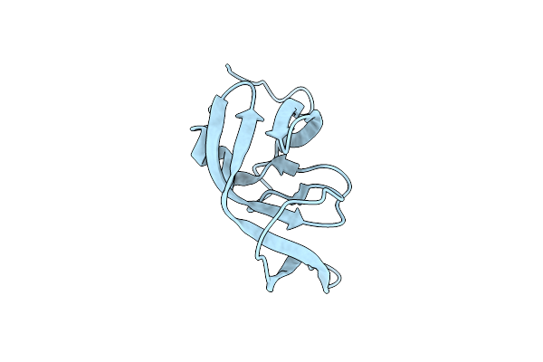
Crystal Structures Of Multidrug-Resistant Clinical Isolate 769 Hiv-1 Protease Variants
Organism: Human immunodeficiency virus 1
Method: X-RAY DIFFRACTION Resolution:1.71 Å Release Date: 2011-04-06 Classification: HYDROLASE |
Organism: Human immunodeficiency virus 1
Method: X-RAY DIFFRACTION
Release Date: 2011-04-06
 |
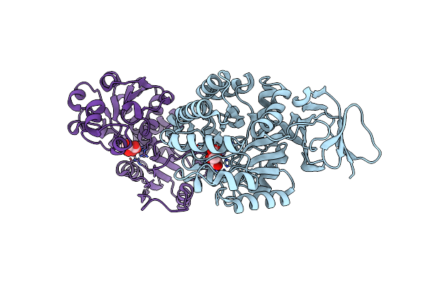
Crystal Structures Of Multidrug-Resistant Clinical Isolate 769 Hiv-1 Protease Variants
Organism: Human immunodeficiency virus 1
Method: X-RAY DIFFRACTION Resolution:2.25 Å Release Date: 2011-04-06 Classification: HYDROLASE |
Organism: Human immunodeficiency virus 1
Method: X-RAY DIFFRACTION
Release Date: 2011-04-06
 |
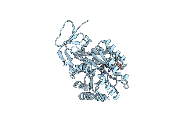
Crystal Structure Of Aquifex Dihydroorotase Activated By Aspartate Transcarbamoylase
Organism: Aquifex aeolicus
Method: X-RAY DIFFRACTION Resolution:2.30 Å Release Date: 2009-01-20 Classification: HYDROLASE/TRANSFERASE Ligands: ZN, FLC |
Organism: Aquifex aeolicus
Method: X-RAY DIFFRACTION
Release Date: 2009-01-20
Ligands: ZN, FLC
 |
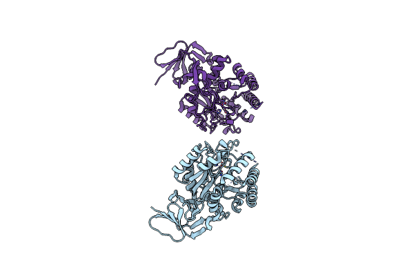
The Crystal Structure Of A Novel, Latent Dihydroorotase From Aquifex Aeolicus At 1.7 A Resolution
Organism: Aquifex aeolicus
Method: X-RAY DIFFRACTION Resolution:1.65 Å Release Date: 2005-07-05 Classification: HYDROLASE Ligands: ZN, SO4 |
Organism: Aquifex aeolicus
Method: X-RAY DIFFRACTION
Release Date: 2005-07-05
Ligands: ZN, SO4
 |
The Crystal Structure Of A Novel, Latent Dihydroorotase From Aquifex Aeolicus At 1.7 A Resolution
Organism: Aquifex aeolicus
Method: X-RAY DIFFRACTION Resolution:1.61 Å Release Date: 2005-07-05 Classification: HYDROLASE Ligands: ZN |
Organism: Aquifex aeolicus
Method: X-RAY DIFFRACTION
Release Date: 2005-07-05
Ligands: ZN
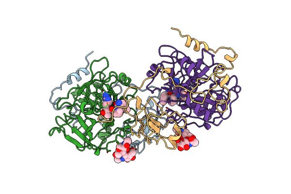 |
The X-Ray Crystal Structure Of Ppack-Meizothrombin Desf1: Kringle/Thrombin And Carbohydrate/Kringle/Thrombin Interactions And Location Of The Linker Chain
Organism: Bos taurus
Method: X-RAY DIFFRACTION Resolution:3.20 Å Release Date: 1998-06-17 Classification: HYDROLASE/HYDROLASE INHIBITOR Ligands: 0G6 |
Organism: Bos taurus
Method: X-RAY DIFFRACTION
Release Date: 1998-06-17
Ligands: 0G6
 |
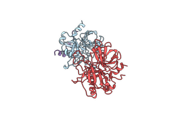
The Crystal Structure Of Fibrinogen-Aa Peptide 1-23 (F8Y) Bound To Bovine Thrombin Explains Why The Mutation Of Phe-8 To Tyrosine Strongly Inhibits Normal Cleavage At Arginine-16
Organism: Bos taurus
Method: X-RAY DIFFRACTION Resolution:2.50 Å Release Date: 1998-05-06 Classification: HYDROLASE/HYDROLASE SUBSTRATE |
Organism: Bos taurus
Method: X-RAY DIFFRACTION
Release Date: 1998-05-06
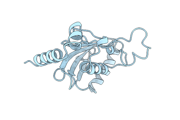 |
Ubiquitin Conjugating Enzyme (Ubc7) From Saccharomyces Cerevisiae
Organism: Saccharomyces cerevisiae
Method: X-RAY DIFFRACTION Resolution:2.93 Å Release Date: 1998-03-18 Classification: UBIQUITIN CONJUGATION |
Organism: Saccharomyces cerevisiae
Method: X-RAY DIFFRACTION
Release Date: 1998-03-18
 |
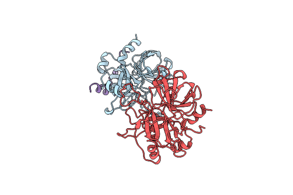
The Co-Crystal Structure Of Unliganded Bovine Alpha-Thrombin And Prethrombin-2: Movement Of The Yppw Segment And Active Site Residues Upon Ligand Binding
Organism: Bos taurus
Method: X-RAY DIFFRACTION Resolution:2.30 Å Release Date: 1997-07-07 Classification: COMPLEX (BLOOD COAGULATION/PROENZYME) |
Organism: Bos taurus
Method: X-RAY DIFFRACTION
Release Date: 1997-07-07
 |
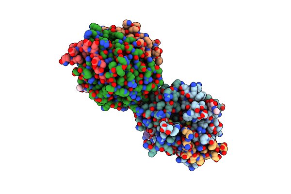
The Co-Crystal Structure Of Unliganded Bovine Alpha-Thrombin And Prethrombin-2: Movement Of The Yppw Segment And Active Site Residues Upon Ligand Binding
Organism: Bos taurus
Method: X-RAY DIFFRACTION Resolution:2.20 Å Release Date: 1997-07-07 Classification: COMPLEX (BLOOD COAGULATION/PROENZYME) |
Organism: Bos taurus
Method: X-RAY DIFFRACTION
Release Date: 1997-07-07
 |
Thrombin:Hirudin 51-65 Complex
Organism: Bos taurus
Method: X-RAY DIFFRACTION Resolution:3.20 Å Release Date: 1997-04-21 Classification: COMPLEX (SERINE PROTEASE/INHIBITOR) Ligands: NAG |
Organism: Bos taurus
Method: X-RAY DIFFRACTION
Release Date: 1997-04-21
Ligands: NAG
 |
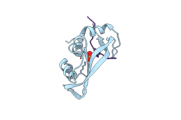
Thrombin Complexed With Fibrinopeptide A Alpha (Residues 7-19). Three Complexes, One With Epsilon-Thrombin And Two With Alpha-Thrombin
Organism: Bos taurus
Method: X-RAY DIFFRACTION Resolution:2.20 Å Release Date: 1997-02-12 Classification: COMPLEX (SERINE PROTEASE/COAGULATION) |
Organism: Bos taurus
Method: X-RAY DIFFRACTION
Release Date: 1997-02-12
 |
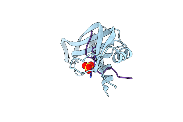
The 1.6 Angstroms Structure Of A Semisynthetic Ribonuclease Crystallized From Aqueous Ethanol. Comparison With Crystals From Salt Solutions And With Rnase A From Aqueous Alcohol Solutions
Organism: Bos taurus
Method: X-RAY DIFFRACTION Resolution:2.00 Å Release Date: 1995-01-26 Classification: ENDONUCLEASE Ligands: PO4 |
Organism: Bos taurus
Method: X-RAY DIFFRACTION
Release Date: 1995-01-26
Ligands: PO4
 |
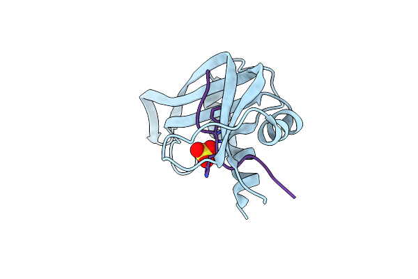
Structural Changes That Accompany The Reduced Catalytic Efficiency Of Two Semisynthetic Ribonuclease Analogs
Organism: Bos taurus
Method: X-RAY DIFFRACTION Resolution:2.00 Å Release Date: 1994-12-20 Classification: HYDROLASE(NUCLEIC ACID,RNA) Ligands: SO4 |
Organism: Bos taurus
Method: X-RAY DIFFRACTION
Release Date: 1994-12-20
Ligands: SO4
 |
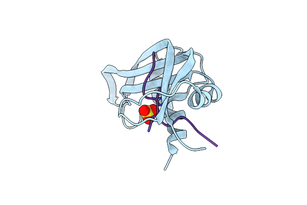
Structural Changes That Accompany The Reduced Catalytic Efficiency Of Two Semisynthetic Ribonuclease Analogs
Organism: Bos taurus
Method: X-RAY DIFFRACTION Resolution:2.00 Å Release Date: 1994-12-20 Classification: HYDROLASE(NUCLEIC ACID,RNA) Ligands: SO4 |
Organism: Bos taurus
Method: X-RAY DIFFRACTION
Release Date: 1994-12-20
Ligands: SO4
 |
A Structural Investigation Of Catalytically Modified F12Ol And F12Oy Semisynthetic Ribonucleases
Organism: Bos taurus
Method: X-RAY DIFFRACTION Resolution:2.00 Å Release Date: 1994-09-30 Classification: HYDROLASE(ENDORIBONUCLEASE) Ligands: SO4 |
Organism: Bos taurus
Method: X-RAY DIFFRACTION
Release Date: 1994-09-30
Ligands: SO4