

Search Count: 47
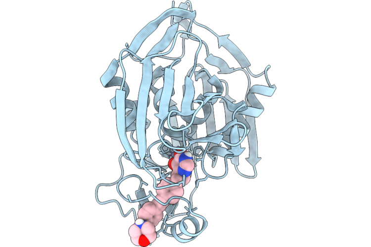 |
Crystal Structure Of Palpxc Complexed With Compound 6
Organism: Pseudomonas aeruginosa
Method: X-RAY DIFFRACTION Resolution:2.20 Å Release Date: 2026-07-29 Classification: ANTIBIOTIC Ligands: ZN, A1J1T |
Organism: Pseudomonas aeruginosa
Method: X-RAY DIFFRACTION
Release Date: 2026-07-29
Ligands: ZN, A1J1T
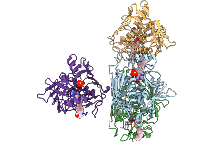 |
Crystal Structure Of Eclpxc Complexed With Compound 7A
Organism: Escherichia coli
Method: X-RAY DIFFRACTION Resolution:2.62 Å Release Date: 2026-07-29 Classification: ANTIBIOTIC Ligands: ZN, A1J1S, SO4 |
Organism: Escherichia coli
Method: X-RAY DIFFRACTION
Release Date: 2026-07-29
Ligands: ZN, A1J1S, SO4
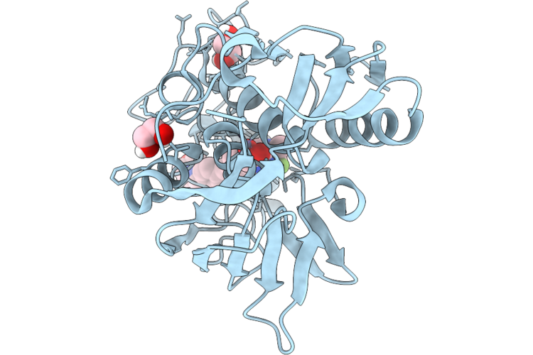 |
Crystal Structure Of Palpxc Complexed With Compound 8K
Organism: Pseudomonas aeruginosa
Method: X-RAY DIFFRACTION Resolution:1.50 Å Release Date: 2026-07-29 Classification: ANTIBIOTIC Ligands: ZN, A1J1V, EDO |
Organism: Pseudomonas aeruginosa
Method: X-RAY DIFFRACTION
Release Date: 2026-07-29
Ligands: ZN, A1J1V, EDO
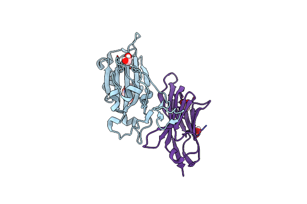 |
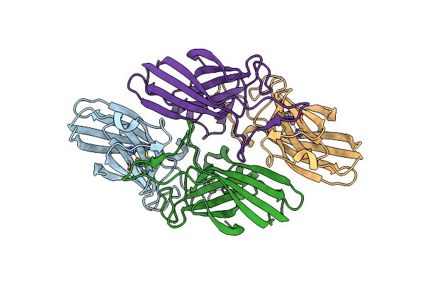
The Crystal Structure Of The Sars-Cov-2 Receptor Binding Domain In Complex With The Neutralizing Nanobody 4.
Organism: Severe acute respiratory syndrome coronavirus 2, Camelus dromedarius
Method: X-RAY DIFFRACTION Resolution:1.21 Å Release Date: 2025-06-11 Classification: ANTIVIRAL PROTEIN Ligands: NAG, EDO, ACT |
Organism: Severe acute respiratory syndrome coronavirus 2, Camelus dromedarius
Method: X-RAY DIFFRACTION
Release Date: 2025-06-11
Ligands: NAG, EDO, ACT
 |
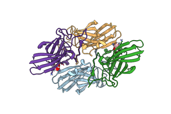
The Crystal Structure Of Brucella Abortus Ftra
Organism: Brucella abortus
Method: X-RAY DIFFRACTION Resolution:1.84 Å Release Date: 2025-02-05 Classification: METAL BINDING PROTEIN Ligands: CU |
Organism: Brucella abortus
Method: X-RAY DIFFRACTION
Release Date: 2025-02-05
Ligands: CU
 |
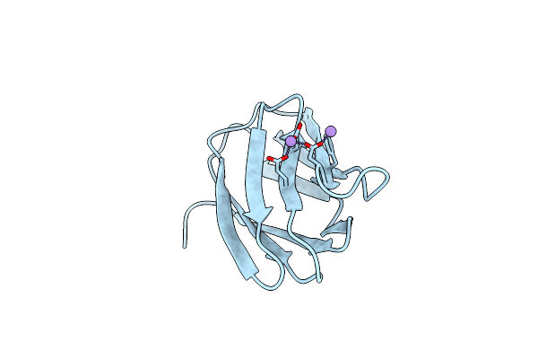
The Crystal Structure Of Brucella Abortus Ftra-H151A Mutant
Organism: Brucella abortus
Method: X-RAY DIFFRACTION Resolution:1.55 Å Release Date: 2025-02-05 Classification: METAL BINDING PROTEIN Ligands: GOL |
Organism: Brucella abortus
Method: X-RAY DIFFRACTION
Release Date: 2025-02-05
Ligands: GOL
 |
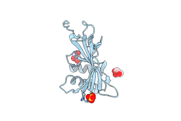
The Crystal Structure Of Brucella Abortus Ftrb At 1.3 Angstrom Resolution
Organism: Brucella abortus
Method: X-RAY DIFFRACTION Resolution:1.30 Å Release Date: 2025-01-22 Classification: METAL BINDING PROTEIN Ligands: MN |
Organism: Brucella abortus
Method: X-RAY DIFFRACTION
Release Date: 2025-01-22
Ligands: MN
 |
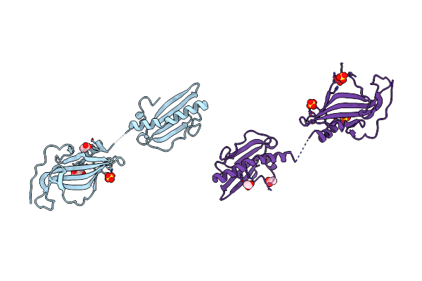
Crystal Structure Of Protein E From Non-Typeable Haemophilus Influenzae
Organism: Haemophilus influenzae
Method: X-RAY DIFFRACTION Resolution:1.92 Å Release Date: 2019-05-29 Classification: CELL ADHESION Ligands: SO4, GOL, CL |
Organism: Haemophilus influenzae
Method: X-RAY DIFFRACTION
Release Date: 2019-05-29
Ligands: SO4, GOL, CL
 |
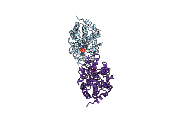
Crystal Structure Of Non-Typeable Haemophilus Influenzae Protein E And Pila Expressed As A Single-Chain Chimeric Protein
Organism: Haemophilus influenzae
Method: X-RAY DIFFRACTION Resolution:1.63 Å Release Date: 2019-05-29 Classification: CELL ADHESION Ligands: SO4, ACT, GOL |
Organism: Haemophilus influenzae
Method: X-RAY DIFFRACTION
Release Date: 2019-05-29
Ligands: SO4, ACT, GOL
 |
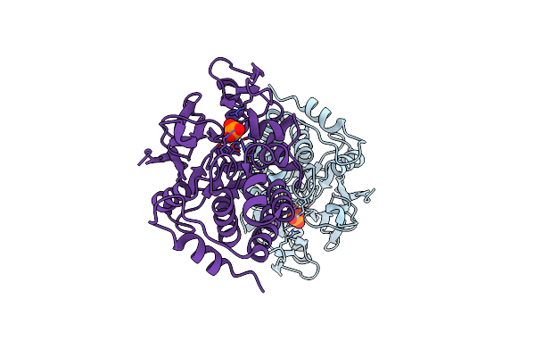
X-Ray Structure Of The Human Pp1 Gamma Catalytic Subunit Treated With Ascorbate
Organism: Homo sapiens
Method: X-RAY DIFFRACTION Resolution:1.96 Å Release Date: 2015-07-22 Classification: HYDROLASE Ligands: MN, PO4 |
Organism: Homo sapiens
Method: X-RAY DIFFRACTION
Release Date: 2015-07-22
Ligands: MN, PO4
 |
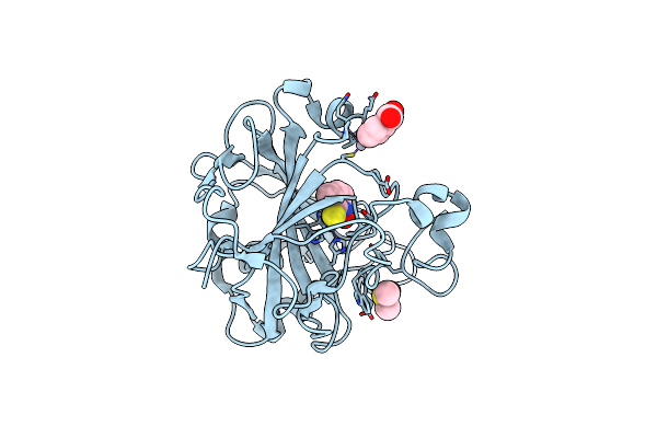
X-Ray Structure Of The Human Pp1 Gamma Catalytic Subunit Treated With Hydrogen Peroxide
Organism: Homo sapiens
Method: X-RAY DIFFRACTION Resolution:2.19 Å Release Date: 2015-07-22 Classification: HYDROLASE Ligands: MN, PO4 |
Organism: Homo sapiens
Method: X-RAY DIFFRACTION
Release Date: 2015-07-22
Ligands: MN, PO4
 |
Crystal Structure Of 1-Hydroxy-3-Methylpyridine-2(1H)-Thione Bound To Hcaii
Organism: Homo sapiens
Method: X-RAY DIFFRACTION Resolution:1.65 Å Release Date: 2015-03-11 Classification: LYASE Ligands: ZN, 3MH, MBO, DMS |
Organism: Homo sapiens
Method: X-RAY DIFFRACTION
Release Date: 2015-03-11
Ligands: ZN, 3MH, MBO, DMS
 |
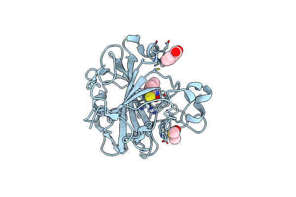
Crystal Structure Of 1-Hydroxy-4-Methylpyridine-2(1H)-Thione Bound To Human Carbonic Anhydrase Ii
Organism: Homo sapiens
Method: X-RAY DIFFRACTION Resolution:1.80 Å Release Date: 2015-03-11 Classification: LYASE Ligands: ZN, 2YU, MBO |
Organism: Homo sapiens
Method: X-RAY DIFFRACTION
Release Date: 2015-03-11
Ligands: ZN, 2YU, MBO
 |
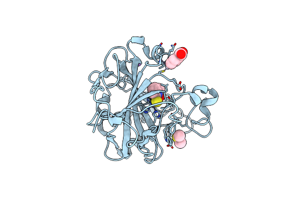
Crystal Structure Of 1-Hydroxy-5-Methylpyridine-2(1H)-Thione Bound To Human Carbonic Anhydrase Ii
Organism: Homo sapiens
Method: X-RAY DIFFRACTION Resolution:1.60 Å Release Date: 2015-03-11 Classification: LYASE Ligands: ZN, 5MH, MBO, DMS |
Organism: Homo sapiens
Method: X-RAY DIFFRACTION
Release Date: 2015-03-11
Ligands: ZN, 5MH, MBO, DMS
 |
Crystal Structure Of 1-Hydroxy-6-Methylpyridine-2(1H)-Thione Bound To Human Carbonic Anhydrase Ii
Organism: Homo sapiens
Method: X-RAY DIFFRACTION Resolution:1.45 Å Release Date: 2015-03-11 Classification: LYASE Ligands: ZN, 6MH, MBO, DMS, NA |
Organism: Homo sapiens
Method: X-RAY DIFFRACTION
Release Date: 2015-03-11
Ligands: ZN, 6MH, MBO, DMS, NA
 |
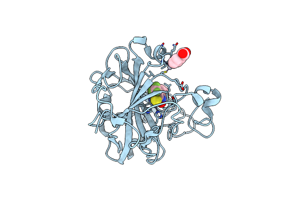
Crystal Structure Of 1-Hydroxy-4,6-Dimethylpyridine-2(1H)-Thione Bound To Human Carbonic Anhydrase Ii
Organism: Homo sapiens
Method: X-RAY DIFFRACTION Resolution:1.55 Å Release Date: 2015-03-11 Classification: LYASE Ligands: ZN, DMS, 7MH, MBO |
Organism: Homo sapiens
Method: X-RAY DIFFRACTION
Release Date: 2015-03-11
Ligands: ZN, DMS, 7MH, MBO
 |
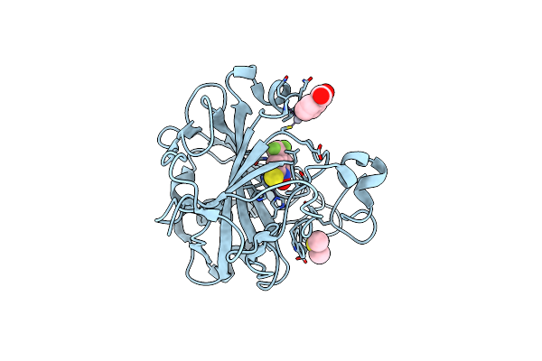
Crystal Structure Of 1-Hydroxy-3-(Trifluoromethyl)Pyridine-2(1H)-Thione Bound To Human Carbonic Anhydrase Ii
Organism: Homo sapiens
Method: X-RAY DIFFRACTION Resolution:1.55 Å Release Date: 2015-03-11 Classification: LYASE Ligands: ZN, 3FH, MBO |
Organism: Homo sapiens
Method: X-RAY DIFFRACTION
Release Date: 2015-03-11
Ligands: ZN, 3FH, MBO
 |
Crystal Structure Of 1-Hydroxy-4-(Trifluoromethyl)Pyridine-2(1H)-Thione Bound To Human Carbonic Anhydrase Ii
Organism: Homo sapiens
Method: X-RAY DIFFRACTION Resolution:1.55 Å Release Date: 2015-03-11 Classification: LYASE Ligands: ZN, MBO, 4FH, DMS |
Organism: Homo sapiens
Method: X-RAY DIFFRACTION
Release Date: 2015-03-11
Ligands: ZN, MBO, 4FH, DMS
 |
Crystal Structure Of 1-Hydroxy-5-(Trifluoromethyl)Pyridine-2(1H)-Thione Bound To Human Carbonic Anhydrase Ii
Organism: Homo sapiens
Method: X-RAY DIFFRACTION Resolution:1.55 Å Release Date: 2015-03-11 Classification: LYASE Ligands: ZN, MBO, DMS, 7FH |
Organism: Homo sapiens
Method: X-RAY DIFFRACTION
Release Date: 2015-03-11
Ligands: ZN, MBO, DMS, 7FH
 |
Crystal Structure Of 2-Hydroxyisoquinoline-1(2H)-Thione Bound To Human Carbonic Anhydrase Ii
Organism: Homo sapiens
Method: X-RAY DIFFRACTION Resolution:1.45 Å Release Date: 2015-03-11 Classification: LYASE Ligands: ZN, MBO, HQT |
Organism: Homo sapiens
Method: X-RAY DIFFRACTION
Release Date: 2015-03-11
Ligands: ZN, MBO, HQT