

Search Count: 101
 |
Crystal Structure Of Hdm2 In Complex With Caylin-1
Organism: Homo sapiens
Method: X-RAY DIFFRACTION Resolution:1.26 Å Release Date: 2022-10-05 Classification: LIGASE Ligands: CL, NA, SO4, AU0, DMS |
Organism: Homo sapiens
Method: X-RAY DIFFRACTION
Release Date: 2022-10-05
Ligands: CL, NA, SO4, AU0, DMS
 |
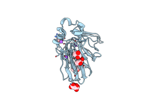
Structure Of Ribonucleotide Reductase R2 From Escherichia Coli Collected By Femtosecond Serial Crystallography On A Coc Membrane
Organism: Escherichia coli (strain k12)
Method: X-RAY DIFFRACTION Resolution:2.30 Å Release Date: 2022-01-12 Classification: OXIDOREDUCTASE Ligands: FE |
Organism: Escherichia coli (strain k12)
Method: X-RAY DIFFRACTION
Release Date: 2022-01-12
Ligands: FE
 |
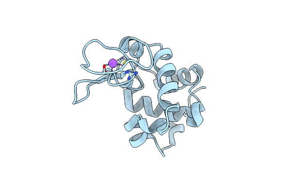
Structure Of Thaumatin Collected By Femtosecond Serial Crystallography On A Coc Membrane
Organism: Thaumatococcus daniellii
Method: X-RAY DIFFRACTION Resolution:1.46 Å Release Date: 2021-11-24 Classification: PLANT PROTEIN Ligands: TLA, PGR, NA |
Organism: Thaumatococcus daniellii
Method: X-RAY DIFFRACTION
Release Date: 2021-11-24
Ligands: TLA, PGR, NA
 |
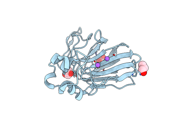
Structure Of Hen Egg White Lysozyme Collected By Rotation Serial Crystallography On A Coc Membrane At A Synchrotron Source
Organism: Gallus gallus
Method: X-RAY DIFFRACTION Resolution:1.51 Å Release Date: 2021-09-01 Classification: HYDROLASE Ligands: CL, NA |
Organism: Gallus gallus
Method: X-RAY DIFFRACTION
Release Date: 2021-09-01
Ligands: CL, NA
 |
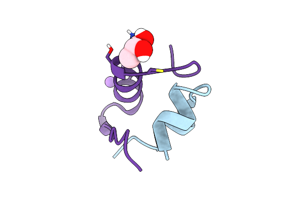
Structure Of Thaumatin Collected By Rotation Serial Crystallography On A Coc Membrane At A Synchrotron Source
Organism: Thaumatococcus daniellii
Method: X-RAY DIFFRACTION Resolution:1.65 Å Release Date: 2021-09-01 Classification: PLANT PROTEIN Ligands: TLA, PGO, NA |
Organism: Thaumatococcus daniellii
Method: X-RAY DIFFRACTION
Release Date: 2021-09-01
Ligands: TLA, PGO, NA
 |
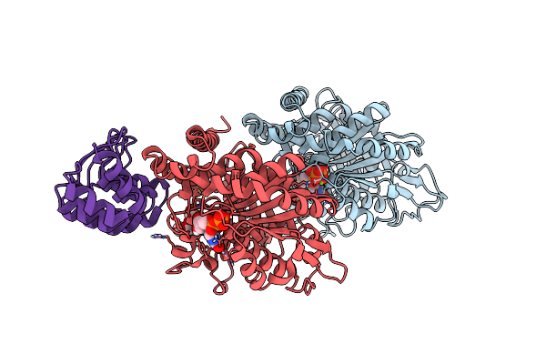
Structure Of Insulin Collected By Rotation Serial Crystallography On A Coc Membrane At A Synchrotron Source
Organism: Sus scrofa
Method: X-RAY DIFFRACTION Resolution:1.46 Å Release Date: 2021-09-01 Classification: HORMONE Ligands: PGR, NA |
Organism: Sus scrofa
Method: X-RAY DIFFRACTION
Release Date: 2021-09-01
Ligands: PGR, NA
 |
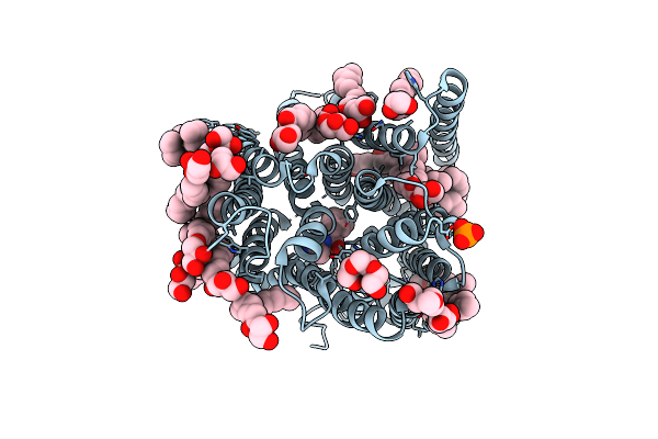
Structure Of Tubulin Darpin Complex 1 Collected By Rotation Serial Crystallography On A Coc Membrane At A Synchrotron Source
Organism: Bos taurus
Method: X-RAY DIFFRACTION Resolution:2.26 Å Release Date: 2021-09-01 Classification: CELL CYCLE Ligands: GTP, MG, GDP, PG0 |
Organism: Bos taurus
Method: X-RAY DIFFRACTION
Release Date: 2021-09-01
Ligands: GTP, MG, GDP, PG0
 |
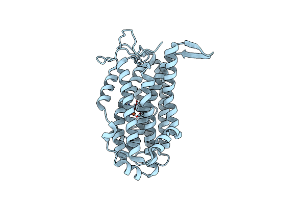
Structure Of Sponge-Phase Grown Peptst2 Collected By Rotation Serial Crystallography On A Coc Membrane At A Synchrotron Source
Organism: Streptococcus thermophilus (strain atcc baa-250 / lmg 18311)
Method: X-RAY DIFFRACTION Resolution:3.00 Å Release Date: 2021-09-01 Classification: TRANSPORT PROTEIN Ligands: PO4, 78M, PG0, PGE |
Organism: Streptococcus thermophilus (strain atcc baa-250 / lmg 18311)
Method: X-RAY DIFFRACTION
Release Date: 2021-09-01
Ligands: PO4, 78M, PG0, PGE
 |
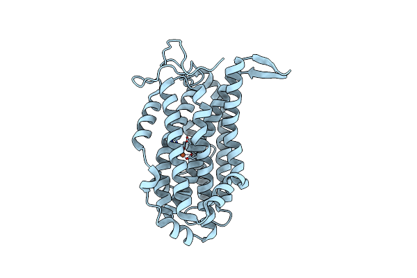
Structure Of Ribonucleotide Reductase R2 From Escherichia Coli Collected By Still Serial Crystallography On A Coc Membrane At A Synchrotron Source
Organism: Escherichia coli (strain k12)
Method: X-RAY DIFFRACTION Resolution:2.10 Å Release Date: 2021-09-01 Classification: OXIDOREDUCTASE Ligands: FE |
Organism: Escherichia coli (strain k12)
Method: X-RAY DIFFRACTION
Release Date: 2021-09-01
Ligands: FE
 |
Structure Of Ribonucleotide Reductase R2 From Escherichia Coli Collected By Rotation Serial Crystallography On A Coc Membrane At A Synchrotron Source
Organism: Escherichia coli (strain k12)
Method: X-RAY DIFFRACTION Resolution:2.00 Å Release Date: 2021-09-01 Classification: OXIDOREDUCTASE Ligands: FE |
Organism: Escherichia coli (strain k12)
Method: X-RAY DIFFRACTION
Release Date: 2021-09-01
Ligands: FE
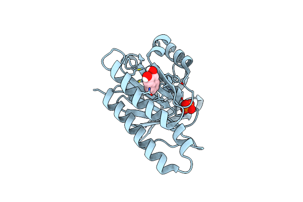 |
Humrada1.2 Soaked With Napht-2,3-Diol For 30 Seconds At 20 Mm
Organism: Pyrococcus furiosus
Method: X-RAY DIFFRACTION Resolution:1.35 Å Release Date: 2021-03-03 Classification: DNA BINDING PROTEIN Ligands: D48, PO4 |
Organism: Pyrococcus furiosus
Method: X-RAY DIFFRACTION
Release Date: 2021-03-03
Ligands: D48, PO4
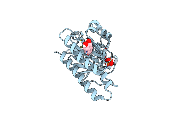 |
Humrada1.2 Soaked With Napht-2,3-Diol For 60 Seconds At 20 Mm
Organism: Pyrococcus furiosus
Method: X-RAY DIFFRACTION Resolution:1.43 Å Release Date: 2021-03-03 Classification: DNA BINDING PROTEIN Ligands: D48, PO4 |
Organism: Pyrococcus furiosus
Method: X-RAY DIFFRACTION
Release Date: 2021-03-03
Ligands: D48, PO4
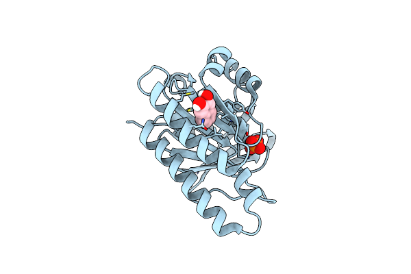 |
Humrada1.2 Soaked With Napht-2,3-Diol For 5 Minutes At 20 Mm
Organism: Pyrococcus furiosus
Method: X-RAY DIFFRACTION Resolution:1.30 Å Release Date: 2021-03-03 Classification: DNA BINDING PROTEIN Ligands: D48, PO4 |
Organism: Pyrococcus furiosus
Method: X-RAY DIFFRACTION
Release Date: 2021-03-03
Ligands: D48, PO4
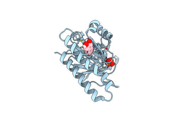 |
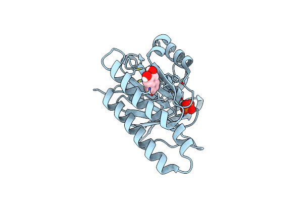
Humrada1.2 Soaked With Napht-2,3-Diol For 10 Minutes At 20 Mm
Organism: Pyrococcus furiosus
Method: X-RAY DIFFRACTION Resolution:1.30 Å Release Date: 2021-03-03 Classification: DNA BINDING PROTEIN Ligands: D48, PO4 |
Organism: Pyrococcus furiosus
Method: X-RAY DIFFRACTION
Release Date: 2021-03-03
Ligands: D48, PO4
 |
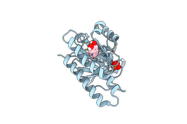
Humrada1.2 Soaked With Napht-2,3-Diol Overnight At 20 Mm
Organism: Pyrococcus furiosus
Method: X-RAY DIFFRACTION Resolution:1.35 Å Release Date: 2021-03-03 Classification: DNA BINDING PROTEIN Ligands: D48, PO4 |
Organism: Pyrococcus furiosus
Method: X-RAY DIFFRACTION
Release Date: 2021-03-03
Ligands: D48, PO4
 |
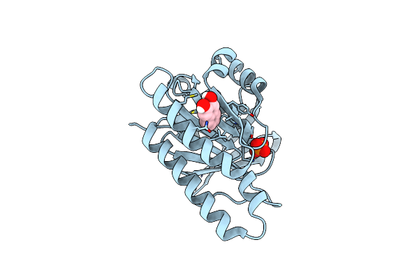
Humrada1.2 Soaked With Napht-2,3-Diol Overnight At 1 Mm
Organism: Pyrococcus furiosus
Method: X-RAY DIFFRACTION Resolution:1.48 Å Release Date: 2021-03-03 Classification: DNA BINDING PROTEIN Ligands: D48, PO4 |
Organism: Pyrococcus furiosus
Method: X-RAY DIFFRACTION
Release Date: 2021-03-03
Ligands: D48, PO4
 |
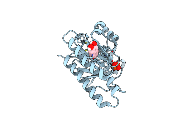
Humrada1.2 Soaked With Napht-2,3-Diol Overnight At 2 Mm
Organism: Pyrococcus furiosus
Method: X-RAY DIFFRACTION Resolution:1.38 Å Release Date: 2021-03-03 Classification: DNA BINDING PROTEIN Ligands: D48, PO4 |
Organism: Pyrococcus furiosus
Method: X-RAY DIFFRACTION
Release Date: 2021-03-03
Ligands: D48, PO4
 |
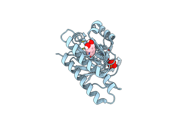
Humrada1.2 Soaked With Napht-2,3-Diol Overnight At 5 Mm
Organism: Pyrococcus furiosus
Method: X-RAY DIFFRACTION Resolution:1.40 Å Release Date: 2021-03-03 Classification: DNA BINDING PROTEIN Ligands: D48, PO4 |
Organism: Pyrococcus furiosus
Method: X-RAY DIFFRACTION
Release Date: 2021-03-03
Ligands: D48, PO4
 |
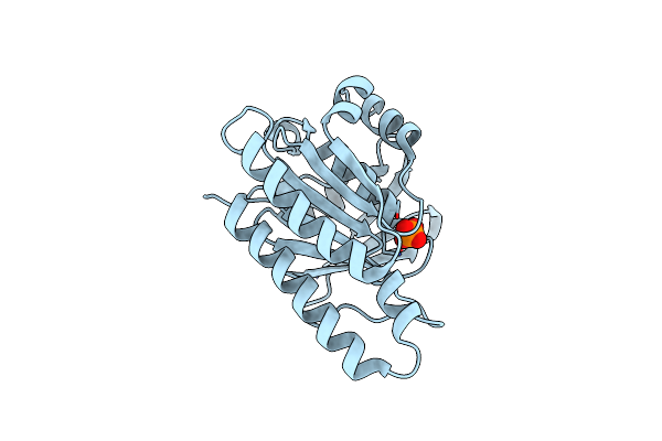
Humrada1.2 Soaked With Napht-2,3-Diol Overnight 10 Mm
Organism: Pyrococcus furiosus
Method: X-RAY DIFFRACTION Resolution:1.42 Å Release Date: 2021-03-03 Classification: DNA BINDING PROTEIN Ligands: D48, PO4 |
Organism: Pyrococcus furiosus
Method: X-RAY DIFFRACTION
Release Date: 2021-03-03
Ligands: D48, PO4
 |
Structure Of Unliganded Humrada1.2
Organism: Pyrococcus furiosus
Method: X-RAY DIFFRACTION Resolution:1.16 Å Release Date: 2021-03-03 Classification: DNA BINDING PROTEIN Ligands: PO4 |
Organism: Pyrococcus furiosus
Method: X-RAY DIFFRACTION
Release Date: 2021-03-03
Ligands: PO4