

Search Count: 34
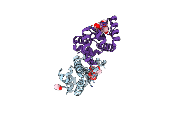 |
Structure Of Ms3494 From Mycobacterium Smegmatis Bound To Sucrose
Organism: Mycolicibacterium smegmatis mc2 155
Method: X-RAY DIFFRACTION Resolution:1.90 Å Release Date: 2023-08-09 Classification: UNKNOWN FUNCTION Ligands: ACT |
Organism: Mycolicibacterium smegmatis mc2 155
Method: X-RAY DIFFRACTION
Release Date: 2023-08-09
Ligands: ACT
 |
Structure Of Rv0455C From Mycobacterium Tuberculosis
Organism: Mycobacterium tuberculosis h37rv
Method: X-RAY DIFFRACTION Resolution:2.10 Å Release Date: 2023-08-02 Classification: UNKNOWN FUNCTION Ligands: CL |
Organism: Mycobacterium tuberculosis h37rv
Method: X-RAY DIFFRACTION
Release Date: 2023-08-02
Ligands: CL
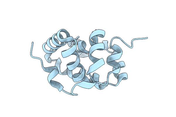 |
Structure Of Rv0455C From Mycobacterium Tuberculosis
Organism: Mycobacterium tuberculosis h37rv
Method: X-RAY DIFFRACTION Resolution:1.80 Å Release Date: 2023-08-02 Classification: UNKNOWN FUNCTION |
Organism: Mycobacterium tuberculosis h37rv
Method: X-RAY DIFFRACTION
Release Date: 2023-08-02
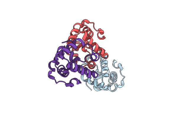 |
Structure Of Rv0455C From Mycobacterium Tuberculosis
Organism: Mycobacterium tuberculosis h37rv
Method: X-RAY DIFFRACTION Resolution:2.00 Å Release Date: 2023-08-02 Classification: UNKNOWN FUNCTION |
Organism: Mycobacterium tuberculosis h37rv
Method: X-RAY DIFFRACTION
Release Date: 2023-08-02
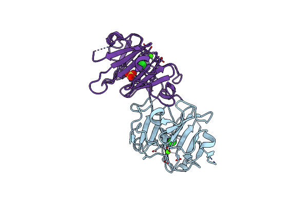 |
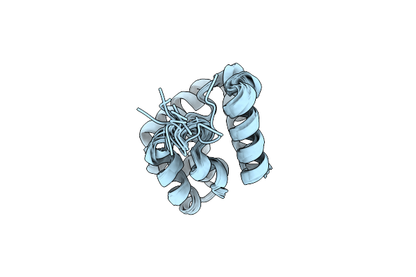
Crystal Structure Of The Vitronectin Hemopexin-Like Domain Binding Calcium
Organism: Homo sapiens
Method: X-RAY DIFFRACTION Resolution:1.70 Å Release Date: 2022-07-27 Classification: CELL ADHESION Ligands: CA, CL, NA, SO4 |
Organism: Homo sapiens
Method: X-RAY DIFFRACTION
Release Date: 2022-07-27
Ligands: CA, CL, NA, SO4
 |
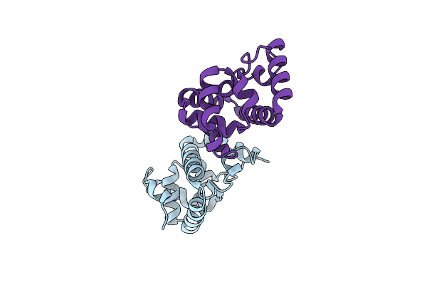
Structure Of Ms3494 From Mycobacterium Smegmatis Determined By Solution Nmr
Organism: Mycolicibacterium smegmatis
Method: SOLUTION NMR Release Date: 2021-10-06 Classification: UNKNOWN FUNCTION |
Organism: Mycolicibacterium smegmatis
Method: SOLUTION NMR
Release Date: 2021-10-06
 |
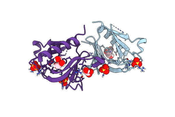
Structure Of Ms3494 From Mycobacterium Smegmatis
Organism: Mycolicibacterium smegmatis
Method: X-RAY DIFFRACTION Resolution:2.10 Å Release Date: 2021-08-18 Classification: UNKNOWN FUNCTION Ligands: BR |
Organism: Mycolicibacterium smegmatis
Method: X-RAY DIFFRACTION
Release Date: 2021-08-18
Ligands: BR
 |
Crystal Structure Of Plekha7 Ph Domain Biding So4
Organism: Homo sapiens
Method: X-RAY DIFFRACTION Resolution:1.45 Å Release Date: 2021-04-07 Classification: CELL ADHESION Ligands: SO4, GOL |
Organism: Homo sapiens
Method: X-RAY DIFFRACTION
Release Date: 2021-04-07
Ligands: SO4, GOL
 |
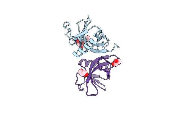
Crystal Structure Of Plekha7 Ph Domain Biding Inositol-Tetraphosphate
Organism: Homo sapiens
Method: X-RAY DIFFRACTION Resolution:2.43 Å Release Date: 2021-04-07 Classification: CELL ADHESION Ligands: 4IP, EDO |
Organism: Homo sapiens
Method: X-RAY DIFFRACTION
Release Date: 2021-04-07
Ligands: 4IP, EDO
 |
Crystal Structure Of Ligand-Free Plekha7 Ph Domain
Organism: Homo sapiens
Method: X-RAY DIFFRACTION Resolution:2.80 Å Release Date: 2021-04-07 Classification: CELL ADHESION Ligands: GOL, EDO |
Organism: Homo sapiens
Method: X-RAY DIFFRACTION
Release Date: 2021-04-07
Ligands: GOL, EDO
 |
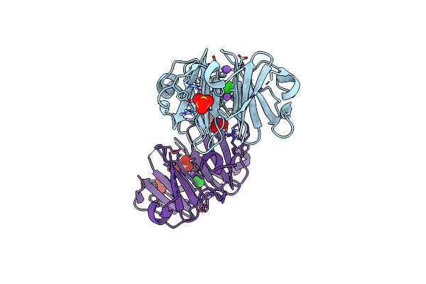
Structure Of The C-Terminal Domain Of Bcl-Xl In Membrane
Organism: Homo sapiens
Method: SOLUTION NMR, SOLID-STATE NMR Release Date: 2020-07-01 Classification: MEMBRANE PROTEIN |
Organism: Homo sapiens
Method: SOLUTION NMR, SOLID-STATE NMR
Release Date: 2020-07-01
 |
Crystal Structure Of The Vitronectin Hemopexin-Like Domain
Organism: Homo sapiens
Method: X-RAY DIFFRACTION Resolution:1.90 Å Release Date: 2019-09-18 Classification: CELL ADHESION |
Organism: Homo sapiens
Method: X-RAY DIFFRACTION
Release Date: 2019-09-18
 |
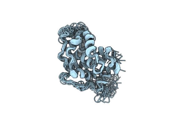
Structure Of Monomeric Interleukin-8 (1-66)
|
Organism: Homo sapiens
Method: SOLUTION NMR
Release Date: 2017-11-29
 |
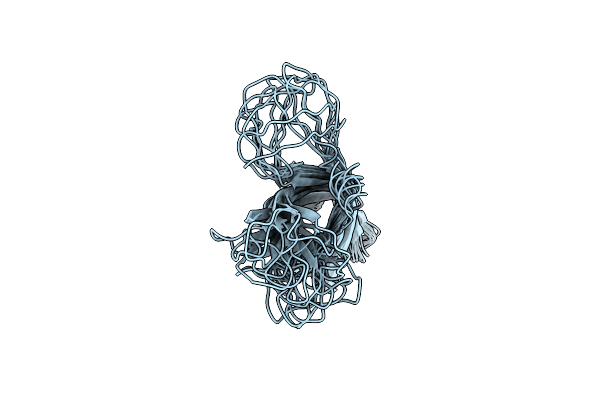
Solution Structure Of A Bcl-Xl S62E Mutant
|
Organism: Homo sapiens
Method: SOLUTION NMR
Release Date: 2017-11-08
 |
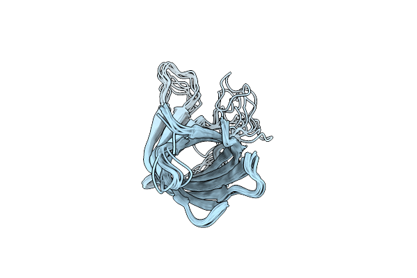
Backbone Structure Of The Yersinia Pestis Outer Membrane Protein Ail In Phospholipid Bilayer Nanodisc
Organism: Yersinia pestis
Method: SOLUTION NMR Release Date: 2017-05-17 Classification: MEMBRANE PROTEIN |
Organism: Yersinia pestis
Method: SOLUTION NMR
Release Date: 2017-05-17
 |
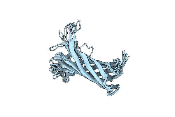
Nmr Structure Of Yersinia Pestis Ail (Attachment Invasion Locus) In Decylphosphocholine Micelles Calculated With Implicit Membrane Solvation
Organism: Yersinia pestis
Method: SOLUTION NMR Release Date: 2015-07-22 Classification: MEMBRANE PROTEIN |
Organism: Yersinia pestis
Method: SOLUTION NMR
Release Date: 2015-07-22
 |
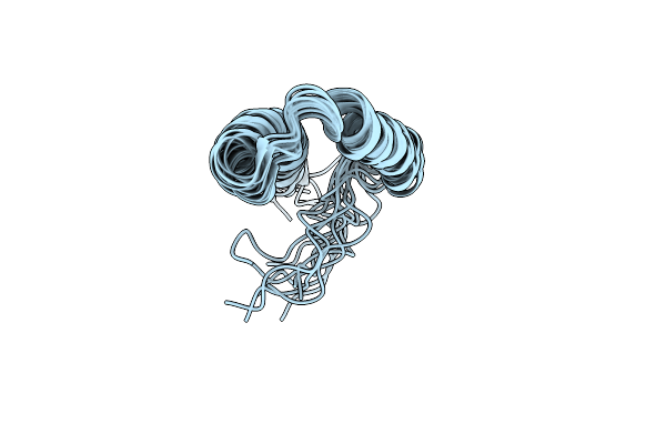
Nmr Structure Of Yersinia Pestis Ail (Attachment Invasion Locus) In Decylphosphocholine Micelles
Organism: Yersinia pestis
Method: SOLUTION NMR Release Date: 2015-07-22 Classification: MEMBRANE PROTEIN |
Organism: Yersinia pestis
Method: SOLUTION NMR
Release Date: 2015-07-22
 |
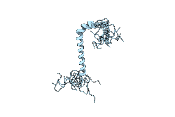
Structure Of The Membrane Protein Merf, A Bacterial Mercury Transporter, Improved By The Inclusion Of Chemical Shift Anisotropy Constraints
Organism: Morganella morganii
Method: SOLUTION NMR Release Date: 2014-07-30 Classification: MEMBRANE PROTEIN |
Organism: Morganella morganii
Method: SOLUTION NMR
Release Date: 2014-07-30
 |
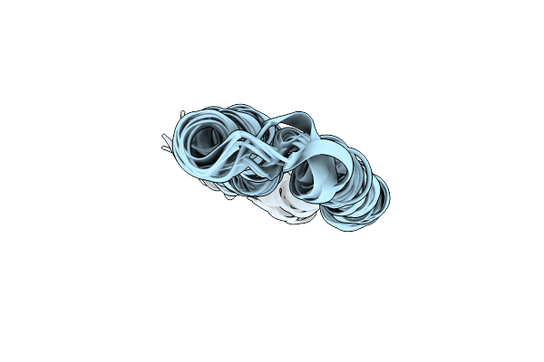
Structure Of The Na,K-Atpase Regulatory Protein Fxyd2B In Micelles
Organism: Homo sapiens
Method: SOLUTION NMR Release Date: 2014-05-14 Classification: TRANSPORT PROTEIN |
Organism: Homo sapiens
Method: SOLUTION NMR
Release Date: 2014-05-14
 |
Full-Length Mercury Transporter Protein Merf In Lipid Bilayer Membranes
Organism: Morganella morganii
Method: SOLID-STATE NMR Release Date: 2013-07-03 Classification: TRANSPORT PROTEIN |
Organism: Morganella morganii
Method: SOLID-STATE NMR
Release Date: 2013-07-03