

Search Count: 22
 |
Neuronal Rna Granules Are Ribosome Complexes Stalled At The Pre-Translocation State
Organism: Rattus norvegicus
Method: ELECTRON MICROSCOPY Release Date: 2022-10-26 Classification: RIBOSOME Ligands: MG, ZN |
Organism: Rattus norvegicus
Method: ELECTRON MICROSCOPY
Release Date: 2022-10-26
Ligands: MG, ZN
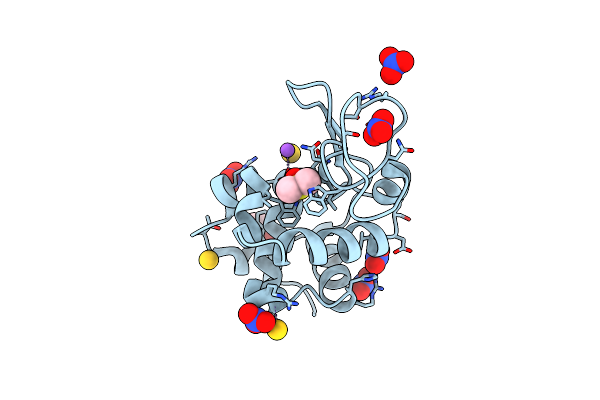 |
X-Ray Structure Of The Gold/Lysozyme Adduct Formed Upon 3 Days Exposure Of Protein Crystals To Compound 1
Organism: Gallus gallus
Method: X-RAY DIFFRACTION Resolution:1.90 Å Release Date: 2019-09-18 Classification: HYDROLASE Ligands: NO3, EDO, DMS, CL, AU, NA |
Organism: Gallus gallus
Method: X-RAY DIFFRACTION
Release Date: 2019-09-18
Ligands: NO3, EDO, DMS, CL, AU, NA
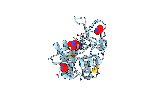 |
X-Ray Structure Of The Gold/Lysozyme Adduct Formed Upon 21H Exposure Of Protein Crystals To Compound 2
Organism: Gallus gallus
Method: X-RAY DIFFRACTION Resolution:1.95 Å Release Date: 2019-09-18 Classification: HYDROLASE Ligands: EDO, NO3, AU |
Organism: Gallus gallus
Method: X-RAY DIFFRACTION
Release Date: 2019-09-18
Ligands: EDO, NO3, AU
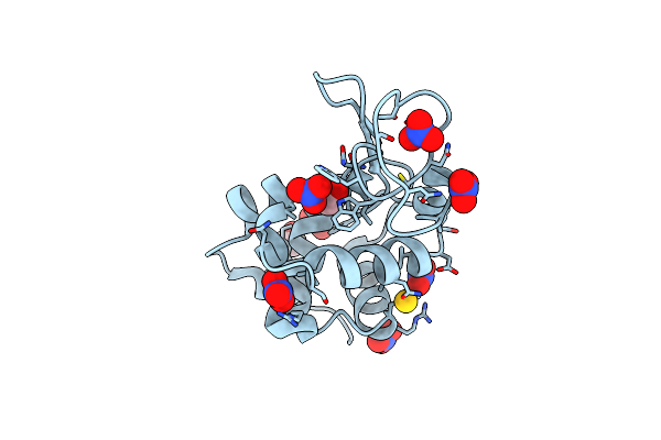 |
X-Ray Structure Of The Gold/Lysozyme Adduct Formed Upon 24H Exposure Of Protein Crystals To Compound 2
Organism: Gallus gallus
Method: X-RAY DIFFRACTION Resolution:2.12 Å Release Date: 2019-09-18 Classification: HYDROLASE Ligands: EDO, NO3, AU |
Organism: Gallus gallus
Method: X-RAY DIFFRACTION
Release Date: 2019-09-18
Ligands: EDO, NO3, AU
 |
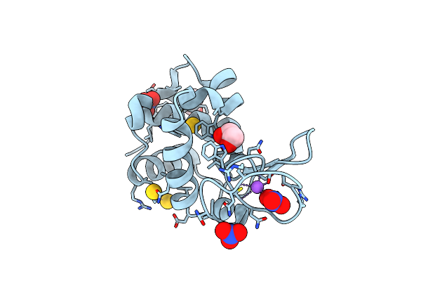
X-Ray Structure Of The Gold/Lysozyme Adduct Formed Upon 21H Exposure Of Protein Crystals To Compound 1
Organism: Gallus gallus
Method: X-RAY DIFFRACTION Resolution:1.78 Å Release Date: 2019-09-18 Classification: HYDROLASE Ligands: NO3, ACT, DMS, EDO, AU |
Organism: Gallus gallus
Method: X-RAY DIFFRACTION
Release Date: 2019-09-18
Ligands: NO3, ACT, DMS, EDO, AU
 |
X-Ray Structure Of The Gold/Lysozyme Adduct Formed Upon 24H Exposure Of Protein Crystals To Compound 1
Organism: Gallus gallus
Method: X-RAY DIFFRACTION Resolution:2.22 Å Release Date: 2019-09-18 Classification: HYDROLASE |
Organism: Gallus gallus
Method: X-RAY DIFFRACTION
Release Date: 2019-09-18
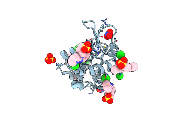 |
X-Ray Structure Of The Adduct Formed Upon Reaction Of Lysozyme With A Pd(Ii) Complex Bearing N,N-Pyridylbenzimidazole Derivative With An Alkylated Sulphonate Side Chain
Organism: Gallus gallus
Method: X-RAY DIFFRACTION Resolution:1.96 Å Release Date: 2018-07-25 Classification: HYDROLASE Ligands: PD, NO3, F6Q, SO4, CL |
Organism: Gallus gallus
Method: X-RAY DIFFRACTION
Release Date: 2018-07-25
Ligands: PD, NO3, F6Q, SO4, CL
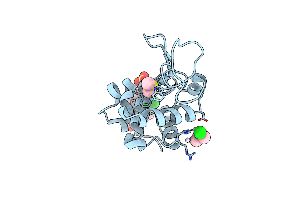 |
X-Ray Structure Of The Adduct Formed Upon Reaction Of Lysozyme With A Pt(Ii) Complex Bearing N,N-Pyridylbenzimidazole Derivative With An Alkylated Sulphonate Side Chain
Organism: Gallus gallus
Method: X-RAY DIFFRACTION Resolution:2.43 Å Release Date: 2018-07-25 Classification: HYDROLASE Ligands: DMS, PT, CL, F7T |
Organism: Gallus gallus
Method: X-RAY DIFFRACTION
Release Date: 2018-07-25
Ligands: DMS, PT, CL, F7T
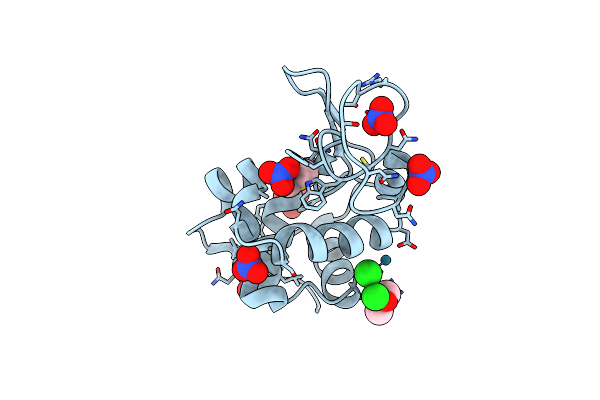 |
X-Ray Structure Of The Adduct Formed Upon Reaction Of Lysozyme With A Pd(Ii) Complex Bearing N,N-Pyridylbenzimidazole Derivative With An Alkylated Triphenylphosphonium Cation
Organism: Gallus gallus
Method: X-RAY DIFFRACTION Resolution:1.99 Å Release Date: 2018-07-25 Classification: HYDROLASE Ligands: NO3, PD, CL, DMS |
Organism: Gallus gallus
Method: X-RAY DIFFRACTION
Release Date: 2018-07-25
Ligands: NO3, PD, CL, DMS
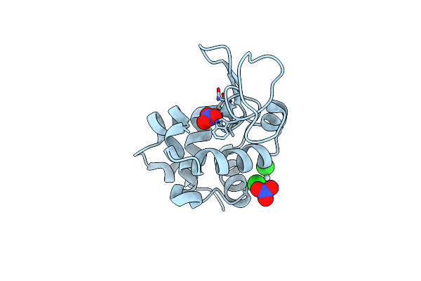 |
X-Ray Structure Of The Adduct Formed Upon Reaction Of Lysozyme With A Pt(Ii) Complex Bearing N,N-Pyridylbenzimidazole Derivative With An Alkylated Triphenylphosphonium Cation
Organism: Gallus gallus
Method: X-RAY DIFFRACTION Resolution:2.25 Å Release Date: 2018-07-25 Classification: HYDROLASE Ligands: PT, NO3, CL |
Organism: Gallus gallus
Method: X-RAY DIFFRACTION
Release Date: 2018-07-25
Ligands: PT, NO3, CL
 |
X-Ray Structure Of The Adduct Formed Upon Reaction Of Bovine Pancreatic Ribonuclease With A Pd(Ii) Complex Bearing N,N-Pyridylbenzimidazole Derivative With An Alkylated Sulphonate Side Chain
Organism: Bos taurus
Method: X-RAY DIFFRACTION Resolution:2.65 Å Release Date: 2018-07-25 Classification: HYDROLASE Ligands: PD, F6Q |
Organism: Bos taurus
Method: X-RAY DIFFRACTION
Release Date: 2018-07-25
Ligands: PD, F6Q
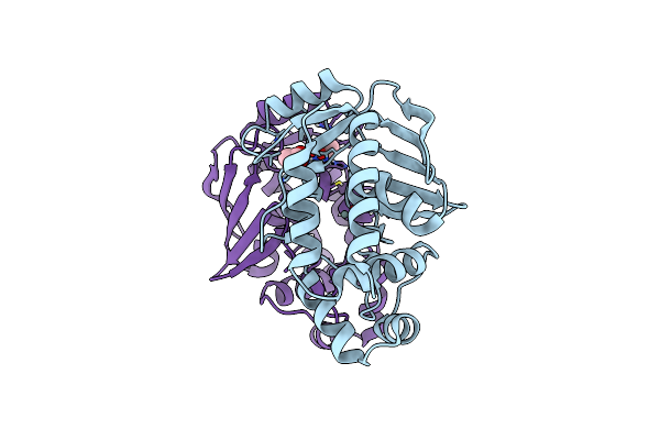 |
Crystal Structure Of A Complex Formed Between Merb And Dimethyltin
Organism: Escherichia coli
Method: X-RAY DIFFRACTION Resolution:1.60 Å Release Date: 2017-01-11 Classification: LYASE,METAL BINDING PROTEIN Ligands: ZN5, BR, ACT |
Organism: Escherichia coli
Method: X-RAY DIFFRACTION
Release Date: 2017-01-11
Ligands: ZN5, BR, ACT
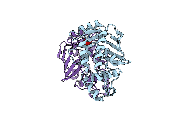 |
Crystal Structure Of A Complex Formed Between Merb And Dimethyltin
Organism: Escherichia coli
Method: X-RAY DIFFRACTION Resolution:1.53 Å Release Date: 2017-01-11 Classification: LYASE,METAL BINDING PROTEIN Ligands: ZN5, BR, PO4 |
Organism: Escherichia coli
Method: X-RAY DIFFRACTION
Release Date: 2017-01-11
Ligands: ZN5, BR, PO4
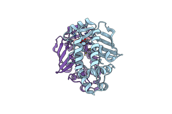 |
Crystal Structure Of A The Tin-Bound Form Of Merb Formed From Diethyltin.
Organism: Escherichia coli
Method: X-RAY DIFFRACTION Resolution:2.00 Å Release Date: 2017-01-11 Classification: LYASE,METAL BINDING PROTEIN Ligands: BR, ACT, ZN6 |
Organism: Escherichia coli
Method: X-RAY DIFFRACTION
Release Date: 2017-01-11
Ligands: BR, ACT, ZN6
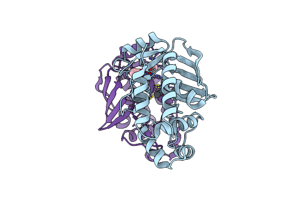 |
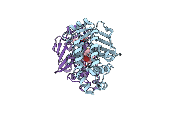
Crystal Structure Of The Lead-Bound Form Of Merb Formed From Diethyllead.
Organism: Escherichia coli
Method: X-RAY DIFFRACTION Resolution:1.75 Å Release Date: 2017-01-11 Classification: LYASE,METAL BINDING PROTEIN Ligands: ZN7, BR, ACT |
Organism: Escherichia coli
Method: X-RAY DIFFRACTION
Release Date: 2017-01-11
Ligands: ZN7, BR, ACT
 |
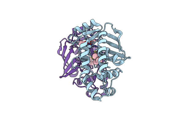
Crystal Structure Of A Merb-Triethyltin Complex
Organism: Escherichia coli
Method: X-RAY DIFFRACTION Resolution:1.85 Å Release Date: 2017-01-11 Classification: LYASE,METAL BINDING PROTEIN Ligands: ZN0, BR, ACT |
Organism: Escherichia coli
Method: X-RAY DIFFRACTION
Release Date: 2017-01-11
Ligands: ZN0, BR, ACT
 |
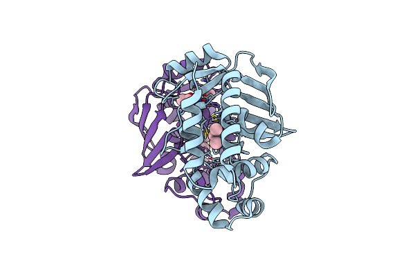
Crystal Structure Of A Merb-Trimethytin Complex.
Organism: Escherichia coli
Method: X-RAY DIFFRACTION Resolution:1.61 Å Release Date: 2017-01-11 Classification: LYASE,METAL BINDING PROTEIN Ligands: ZN8, ACT, BR |
Organism: Escherichia coli
Method: X-RAY DIFFRACTION
Release Date: 2017-01-11
Ligands: ZN8, ACT, BR
 |
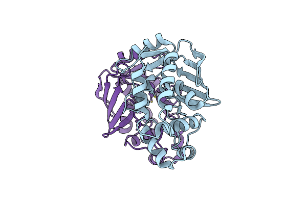
Crystal Structure Of A Merb-Triimethyllead Complex.
Organism: Escherichia coli
Method: X-RAY DIFFRACTION Resolution:1.80 Å Release Date: 2017-01-11 Classification: LYASE,METAL BINDING PROTEIN Ligands: ZN9, ACT |
Organism: Escherichia coli
Method: X-RAY DIFFRACTION
Release Date: 2017-01-11
Ligands: ZN9, ACT
 |
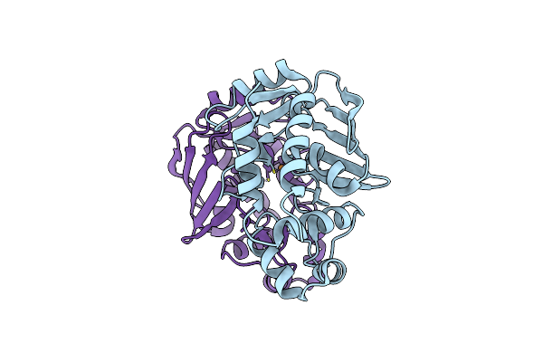
Crystal Structure Of The Mercury-Bound Form Of Merb Mutant D99S
Organism: Escherichia coli
Method: X-RAY DIFFRACTION Resolution:1.96 Å Release Date: 2016-02-03 Classification: LYASE,METAL BINDING PROTEIN Ligands: HG, BR |
Organism: Escherichia coli
Method: X-RAY DIFFRACTION
Release Date: 2016-02-03
Ligands: HG, BR
 |
Crystal Structure Of The Copper-Bound Form Of Merb Mutant D99S
Organism: Escherichia coli
Method: X-RAY DIFFRACTION Resolution:1.87 Å Release Date: 2016-02-03 Classification: LYASE,METAL BINDING PROTEIN Ligands: CU, BR |
Organism: Escherichia coli
Method: X-RAY DIFFRACTION
Release Date: 2016-02-03
Ligands: CU, BR