

Search Count: 52
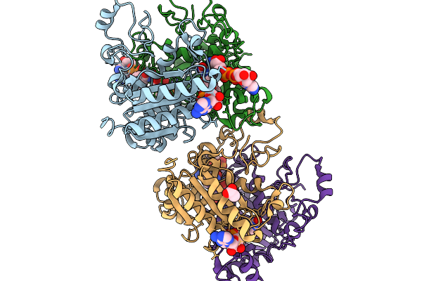 |
Xfel Structure Of Hnqo1 Mixed With Nadh In A Folded Orientation At 300 Ms
Organism: Homo sapiens
Method: X-RAY DIFFRACTION Resolution:2.51 Å Release Date: 2026-02-11 Classification: OXIDOREDUCTASE Ligands: NAD, FAD, ACT |
Organism: Homo sapiens
Method: X-RAY DIFFRACTION
Release Date: 2026-02-11
Ligands: NAD, FAD, ACT
 |
Xfel Structure Of Hnqo1 Mixed With Nadh In An Extended Orientation At 0.3 S
Organism: Homo sapiens
Method: X-RAY DIFFRACTION Resolution:2.51 Å Release Date: 2025-12-10 Classification: OXIDOREDUCTASE Ligands: NAD, FAD, ACT |
Organism: Homo sapiens
Method: X-RAY DIFFRACTION
Release Date: 2025-12-10
Ligands: NAD, FAD, ACT
 |
Room Temperature Structure Of Glycine Max Phya In Pfr/Pr Ensemble
Organism: Glycine max
Method: X-RAY DIFFRACTION Resolution:2.20 Å Release Date: 2025-06-04 Classification: PLANT PROTEIN Ligands: CYC |
Organism: Glycine max
Method: X-RAY DIFFRACTION
Release Date: 2025-06-04
Ligands: CYC
 |
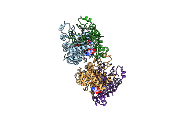
Xfel Structure Of The Free Hnqo1 Unmixed (P3083)
Organism: Homo sapiens
Method: X-RAY DIFFRACTION Resolution:2.50 Å Release Date: 2025-04-23 Classification: OXIDOREDUCTASE Ligands: FAD |
Organism: Homo sapiens
Method: X-RAY DIFFRACTION
Release Date: 2025-04-23
Ligands: FAD
 |
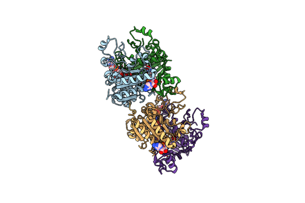
Xfel Structure Of Free Hnqo1 Unmixed (P4502)
Organism: Homo sapiens
Method: X-RAY DIFFRACTION Resolution:2.50 Å Release Date: 2025-04-23 Classification: OXIDOREDUCTASE Ligands: FAD |
Organism: Homo sapiens
Method: X-RAY DIFFRACTION
Release Date: 2025-04-23
Ligands: FAD
 |
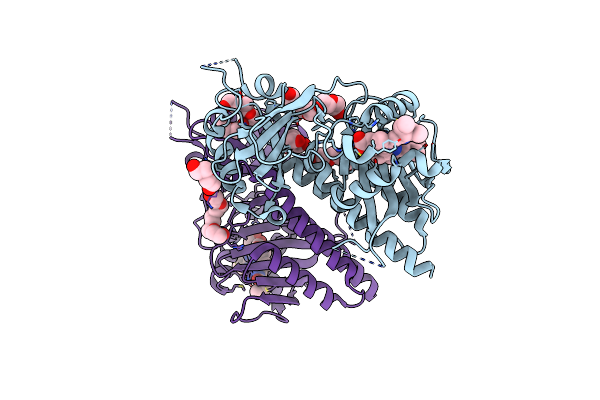
Xfel Structure Of Hnqo1 Mixed With Nadh For 1.2 S
Organism: Homo sapiens
Method: X-RAY DIFFRACTION Resolution:2.50 Å Release Date: 2025-04-23 Classification: OXIDOREDUCTASE Ligands: FAD, NAD |
Organism: Homo sapiens
Method: X-RAY DIFFRACTION
Release Date: 2025-04-23
Ligands: FAD, NAD
 |
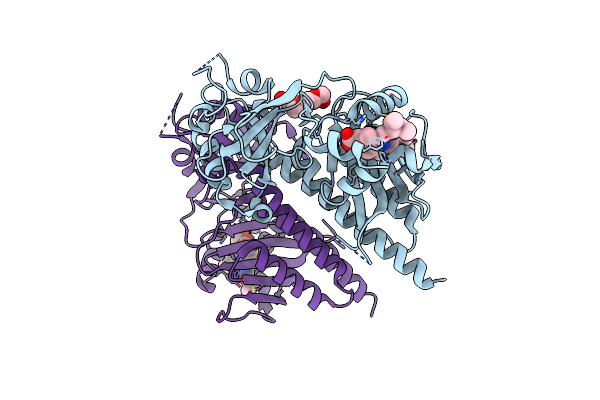
Pas-Gaf Bidomain Of Glycine Max Phytochrome A
Organism: Glycine max
Method: X-RAY DIFFRACTION Resolution:1.58 Å Release Date: 2025-01-08 Classification: PLANT PROTEIN Ligands: CYC, 1PE, PEG, PGE |
Organism: Glycine max
Method: X-RAY DIFFRACTION
Release Date: 2025-01-08
Ligands: CYC, 1PE, PEG, PGE
 |
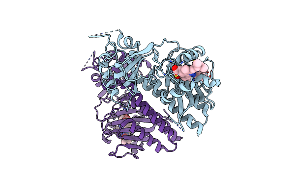
Phytochromobilin-Adducted Pas-Gaf Bidomain Of Glycine Max Phytochrome A
Organism: Glycine max
Method: X-RAY DIFFRACTION Resolution:1.86 Å Release Date: 2025-01-08 Classification: PLANT PROTEIN Ligands: O6E, PG4 |
Organism: Glycine max
Method: X-RAY DIFFRACTION
Release Date: 2025-01-08
Ligands: O6E, PG4
 |
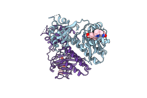
Room Temperature Structure Of Glycine Max Phya In Pr
Organism: Glycine max
Method: X-RAY DIFFRACTION Resolution:2.20 Å Release Date: 2025-01-08 Classification: PLANT PROTEIN Ligands: CYC |
Organism: Glycine max
Method: X-RAY DIFFRACTION
Release Date: 2025-01-08
Ligands: CYC
 |
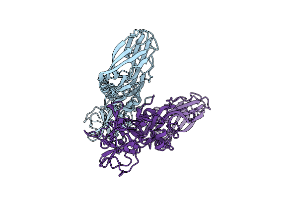
Room Temperature Structure Of Glycine Max Phya In Pfr
Organism: Glycine max
Method: X-RAY DIFFRACTION Resolution:2.20 Å Release Date: 2025-01-08 Classification: PLANT PROTEIN Ligands: CYC |
Organism: Glycine max
Method: X-RAY DIFFRACTION
Release Date: 2025-01-08
Ligands: CYC
 |
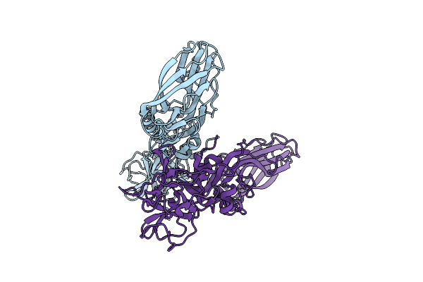
Structure Of The Lysinibacillus Sphaericus Tpp49Aa1 Pesticidal Protein At Ph 3
Organism: Lysinibacillus sphaericus
Method: X-RAY DIFFRACTION Resolution:1.78 Å Release Date: 2023-11-01 Classification: TOXIN |
Organism: Lysinibacillus sphaericus
Method: X-RAY DIFFRACTION
Release Date: 2023-11-01
 |
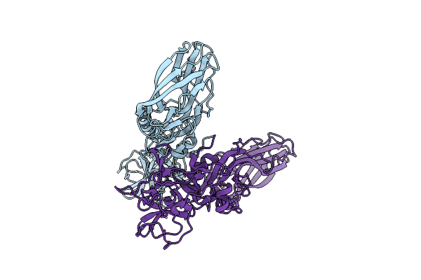
Structure Of The Lysinibacillus Sphaericus Tpp49Aa1 Pesticidal Protein At Ph 7
Organism: Lysinibacillus sphaericus
Method: X-RAY DIFFRACTION Resolution:1.62 Å Release Date: 2023-11-01 Classification: TOXIN |
Organism: Lysinibacillus sphaericus
Method: X-RAY DIFFRACTION
Release Date: 2023-11-01
 |
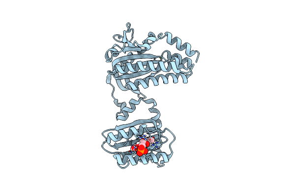
Structure Of The Lysinibacillus Sphaericus Tpp49Aa1 Pesticidal Protein At Ph 11
Organism: Lysinibacillus sphaericus
Method: X-RAY DIFFRACTION Resolution:1.75 Å Release Date: 2023-11-01 Classification: TOXIN |
Organism: Lysinibacillus sphaericus
Method: X-RAY DIFFRACTION
Release Date: 2023-11-01
 |
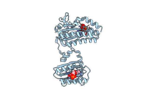
Cryogenic Crystal Structure Of The Photoactivated Adenylate Cyclase Oapac
Organism: Oscillatoria acuminata pcc 6304
Method: X-RAY DIFFRACTION Resolution:1.50 Å Release Date: 2023-11-01 Classification: LYASE Ligands: FMN, MG |
Organism: Oscillatoria acuminata pcc 6304
Method: X-RAY DIFFRACTION
Release Date: 2023-11-01
Ligands: FMN, MG
 |
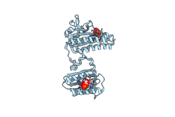
Cryogenic Crystal Structure Of The Photoactivated Adenylate Cyclase Oapac With Atp Bound
Organism: Oscillatoria acuminata pcc 6304
Method: X-RAY DIFFRACTION Resolution:2.10 Å Release Date: 2023-11-01 Classification: LYASE Ligands: FMN, ATP, MG |
Organism: Oscillatoria acuminata pcc 6304
Method: X-RAY DIFFRACTION
Release Date: 2023-11-01
Ligands: FMN, ATP, MG
 |
Cryogenic Crystal Structure Of The Photoactivated Adenylate Cyclase Oapac After 5 Seconds Of Blue Light Illumination
Organism: Oscillatoria acuminata pcc 6304
Method: X-RAY DIFFRACTION Resolution:1.70 Å Release Date: 2023-11-01 Classification: LYASE Ligands: FMN, ATP, MG |
Organism: Oscillatoria acuminata pcc 6304
Method: X-RAY DIFFRACTION
Release Date: 2023-11-01
Ligands: FMN, ATP, MG
 |
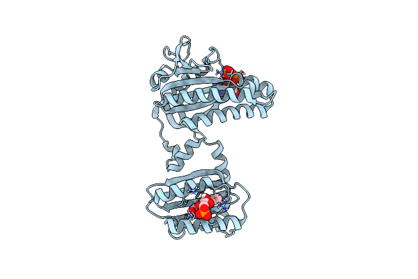
Room Temperature Crystal Structure Of The Photoactivated Adenylate Cyclase Oapac With Atp Bound
Organism: Oscillatoria acuminata pcc 6304
Method: X-RAY DIFFRACTION Resolution:1.80 Å Release Date: 2023-11-01 Classification: LYASE Ligands: FMN, ATP, MG |
Organism: Oscillatoria acuminata pcc 6304
Method: X-RAY DIFFRACTION
Release Date: 2023-11-01
Ligands: FMN, ATP, MG
 |
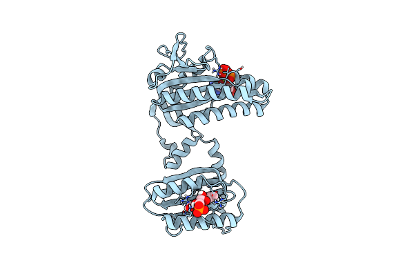
Room Temperature Crystal Structure Of The Photoactivated Adenylate Cyclase Oapac After Blue Light Excitation At 1.8 Us Delay
Organism: Oscillatoria acuminata pcc 6304
Method: X-RAY DIFFRACTION Resolution:1.90 Å Release Date: 2023-11-01 Classification: LYASE Ligands: FMN, ATP, MG |
Organism: Oscillatoria acuminata pcc 6304
Method: X-RAY DIFFRACTION
Release Date: 2023-11-01
Ligands: FMN, ATP, MG
 |
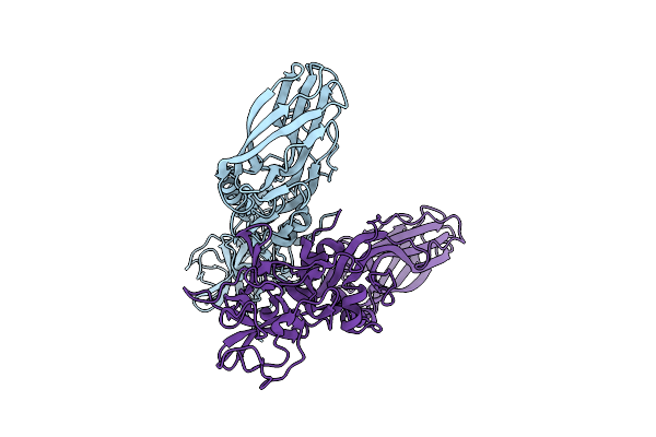
Room Temperature Crystal Structure Of The Photoactivated Adenylate Cyclase Oapac After Blue Light Excitation At 2.3 Us Delay
Organism: Oscillatoria acuminata pcc 6304
Method: X-RAY DIFFRACTION Resolution:1.90 Å Release Date: 2023-11-01 Classification: LYASE Ligands: FMN, ATP, MG |
Organism: Oscillatoria acuminata pcc 6304
Method: X-RAY DIFFRACTION
Release Date: 2023-11-01
Ligands: FMN, ATP, MG
 |
The Structure Of Natural Crystals Of The Lysinibacillus Sphaericus Tpp49Aa1 Pesticidal Protein Elucidated Using Serial Femtosecond Crystallography At An X-Ray Free Electron Laser
Organism: Lysinibacillus sphaericus
Method: X-RAY DIFFRACTION Resolution:2.20 Å Release Date: 2023-05-17 Classification: TOXIN |
Organism: Lysinibacillus sphaericus
Method: X-RAY DIFFRACTION
Release Date: 2023-05-17