

Search Count: 8
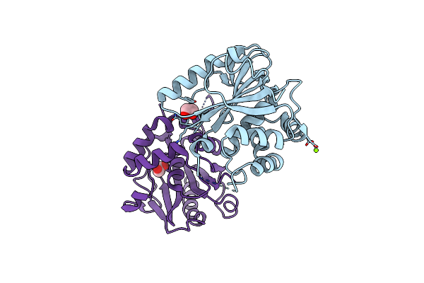 |
Crystal Structure Of The Catalytic Domain Of Uchl5, A Proteasome-Associated Human Deubiquitinating Enzyme, Reveals An Unproductive Form Of The Enzyme
Organism: Homo sapiens
Method: X-RAY DIFFRACTION Resolution:2.00 Å Release Date: 2011-11-09 Classification: HYDROLASE Ligands: EDO, MG |
Organism: Homo sapiens
Method: X-RAY DIFFRACTION
Release Date: 2011-11-09
Ligands: EDO, MG
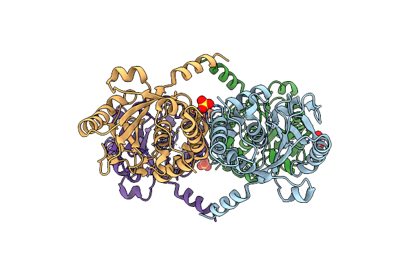 |
Crystal Structure Of The Catalytic Domain Of Uchl5, A Proteasome-Associated Human Deubiquitinating Enzyme, Reveals An Unproductive Form Of The Enzyme
Organism: Homo sapiens
Method: X-RAY DIFFRACTION Resolution:2.40 Å Release Date: 2011-11-09 Classification: HYDROLASE Ligands: SO4, GOL |
Organism: Homo sapiens
Method: X-RAY DIFFRACTION
Release Date: 2011-11-09
Ligands: SO4, GOL
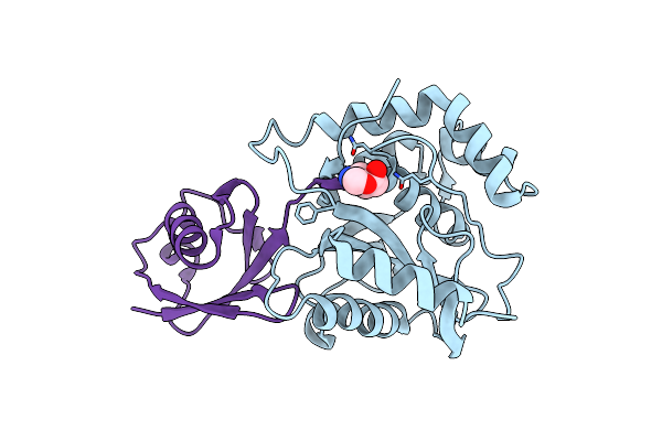 |
Crystal Structure Of The S18Y Variant Of Ubiquitin Carboxy Terminal Hydrolase L1 Bound To Ubiquitin Vinylmethylester.
Organism: Homo sapiens
Method: X-RAY DIFFRACTION Resolution:2.40 Å Release Date: 2010-06-16 Classification: hydrolase/hydrolase inhibitor Ligands: GVE |
Organism: Homo sapiens
Method: X-RAY DIFFRACTION
Release Date: 2010-06-16
Ligands: GVE
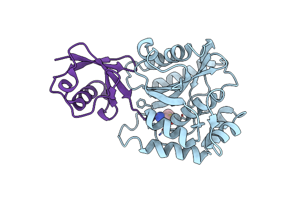 |
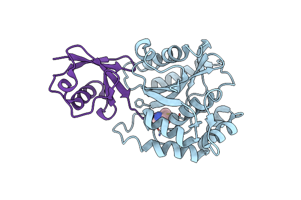
Crystal Structure Of The I93M Mutant Of Ubiquitin Carboxy Terminal Hydrolase L1 Bound To Ubiquitin Vinylmethylester
Organism: Homo sapiens
Method: X-RAY DIFFRACTION Resolution:2.80 Å Release Date: 2010-06-16 Classification: HYDROLASE/SIGNALING PROTEIN Ligands: GVE |
Organism: Homo sapiens
Method: X-RAY DIFFRACTION
Release Date: 2010-06-16
Ligands: GVE
 |
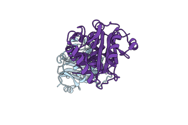
Crystal Structure Of Ubiquitin Carboxy Terminal Hydrolase L1 Bound To Ubiquitin Vinylmethylester
Organism: Homo sapiens
Method: X-RAY DIFFRACTION Resolution:2.83 Å Release Date: 2010-06-16 Classification: HYDROLASE/SIGNALING PROTEIN Ligands: GVE |
Organism: Homo sapiens
Method: X-RAY DIFFRACTION
Release Date: 2010-06-16
Ligands: GVE
 |
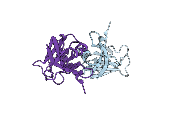
Crystal Structure Of The I93M Mutant Of Ubiquitin Carboxy-Terminal Hydrolase L1
Organism: Homo sapiens
Method: X-RAY DIFFRACTION Resolution:2.80 Å Release Date: 2010-06-09 Classification: HYDROLASE, LIGASE Ligands: CL |
Organism: Homo sapiens
Method: X-RAY DIFFRACTION
Release Date: 2010-06-09
Ligands: CL
 |
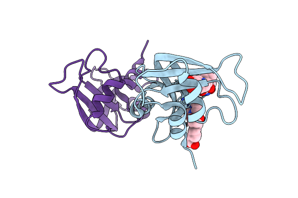
The Crystal Structure Of Rnase A From Monoclinic Crystals At 100 K
Organism: Bos taurus
Method: X-RAY DIFFRACTION Resolution:1.50 Å Release Date: 2006-08-15 Classification: HYDROLASE |
Organism: Bos taurus
Method: X-RAY DIFFRACTION
Release Date: 2006-08-15
 |
The Crystal Structure Of The Rnase A- 3-N-Piperidine-4-Carboxyl-3-Deoxy-Ara-Uridine Complex
Organism: Bos taurus
Method: X-RAY DIFFRACTION Resolution:1.70 Å Release Date: 2006-08-15 Classification: HYDROLASE Ligands: N3E |
Organism: Bos taurus
Method: X-RAY DIFFRACTION
Release Date: 2006-08-15
Ligands: N3E