

Search Count: 13
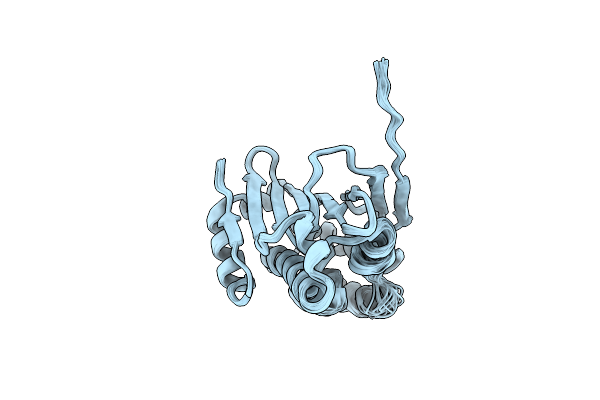 |
Human Hsp90 Alpha Atp Binding Domain, Atp-Lid Open Conformation, R60A
|
Organism: Homo sapiens
Method: SOLUTION NMR
Release Date: 2022-11-16
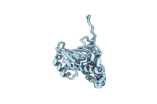 |
Human Hsp90 Alpha Atp Binding Domain, Atp-Lid Closed Conformation, R46A
|
Organism: Homo sapiens
Method: SOLUTION NMR
Release Date: 2022-11-16
 |
Crystal Structure Of Rahu Protein From Pseudomonas Aeruginosa
Organism: Pseudomonas aeruginosa pao1
Method: X-RAY DIFFRACTION Resolution:1.27 Å Release Date: 2021-04-07 Classification: TOXIN |
Organism: Pseudomonas aeruginosa pao1
Method: X-RAY DIFFRACTION
Release Date: 2021-04-07
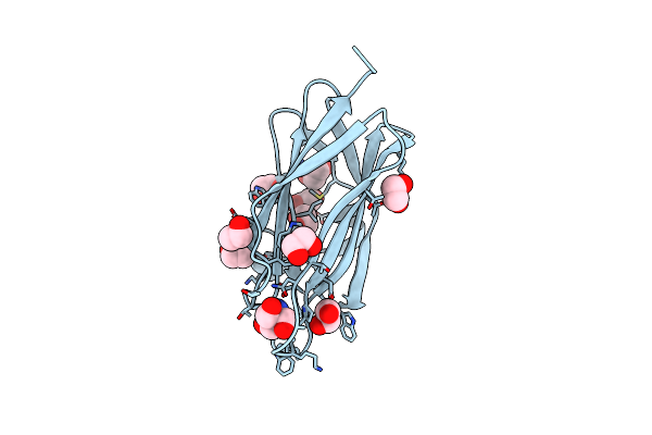 |
Crystal Structure Of Rahu Protein In Complex With Tris Molecule
Organism: Pseudomonas aeruginosa pao1
Method: X-RAY DIFFRACTION Resolution:1.13 Å Release Date: 2021-04-07 Classification: TOXIN Ligands: TRS, EDO |
Organism: Pseudomonas aeruginosa pao1
Method: X-RAY DIFFRACTION
Release Date: 2021-04-07
Ligands: TRS, EDO
 |
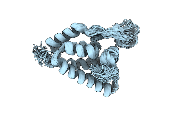
Structure Of The Self-Processing Module Of Iron-Regulated Frpc Of N. Meningitidis With Calcium Ions
Organism: Neisseria meningitidis nm95
Method: SOLUTION NMR Release Date: 2020-02-26 Classification: METAL BINDING PROTEIN Ligands: CA |
Organism: Neisseria meningitidis nm95
Method: SOLUTION NMR
Release Date: 2020-02-26
Ligands: CA
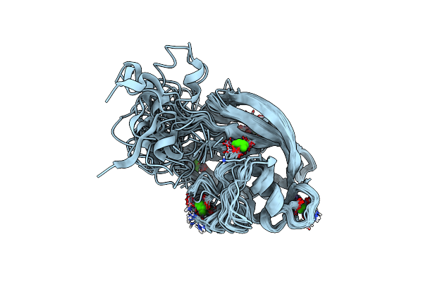 |
Precursor Structure Of The Self-Processing Module Of Iron-Regulated Frpc Of N. Meningitidis With Calcium Ions
Organism: Neisseria meningitidis serogroup c
Method: SOLUTION NMR Release Date: 2020-02-26 Classification: METAL BINDING PROTEIN Ligands: CA |
Organism: Neisseria meningitidis serogroup c
Method: SOLUTION NMR
Release Date: 2020-02-26
Ligands: CA
 |
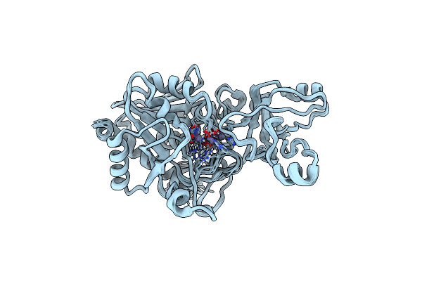
Structure Determination Of The Tetrahedral Aminopeptidase Tet2 From P. Horikoshii By Use Of Combined Solid-State Nmr, Solution-State Nmr And Em Data 4.1 A, Followed By Real_Space_Refinement At 4.1 A
Organism: Pyrococcus horikoshii ot3
Method: ELECTRON MICROSCOPY, SOLUTION NMR Release Date: 2019-08-14 Classification: PEPTIDE BINDING PROTEIN Ligands: ZN |
Organism: Pyrococcus horikoshii ot3
Method: ELECTRON MICROSCOPY, SOLUTION NMR
Release Date: 2019-08-14
Ligands: ZN
 |
Combined Solid-State Nmr, Solution-State Nmr And Em Data For Structure Determination Of The Tetrahedral Aminopeptidase Tet2 From P. Horikoshii
Organism: Pyrococcus horikoshii (strain atcc 700860 / dsm 12428 / jcm 9974 / nbrc 100139 / ot-3)
Method: ELECTRON MICROSCOPY, SOLUTION NMR, SOLID-STATE NMR Release Date: 2018-03-14 Classification: PEPTIDE BINDING PROTEIN Ligands: ZN |
Organism: Pyrococcus horikoshii (strain atcc 700860 / dsm 12428 / jcm 9974 / nbrc 100139 / ot-3)
Method: ELECTRON MICROSCOPY, SOLUTION NMR, SOLID-STATE NMR
Release Date: 2018-03-14
Ligands: ZN
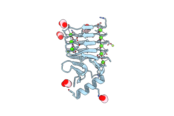 |
Crystal Structure Of Rtx Domain Block V Of Adenylate Cyclase Toxin From Bordetella Pertussis
Organism: Bordetella pertussis (strain atcc 9797 / dsm 5571 / nctc 10739 / 18323)
Method: X-RAY DIFFRACTION Resolution:1.25 Å Release Date: 2015-09-09 Classification: TOXIN Ligands: CA, EDO, GOL, MG |
Organism: Bordetella pertussis (strain atcc 9797 / dsm 5571 / nctc 10739 / 18323)
Method: X-RAY DIFFRACTION
Release Date: 2015-09-09
Ligands: CA, EDO, GOL, MG
 |
Crystal Structure Of Rtx Domain Block V Of Adenylate Cyclase Toxin From Bordetella Pertussis
Organism: Bordetella pertussis (strain tohama i / atcc baa-589 / nctc 13251)
Method: X-RAY DIFFRACTION Resolution:1.45 Å Release Date: 2015-09-02 Classification: TOXIN Ligands: CA, NO3 |
Organism: Bordetella pertussis (strain tohama i / atcc baa-589 / nctc 13251)
Method: X-RAY DIFFRACTION
Release Date: 2015-09-02
Ligands: CA, NO3
 |
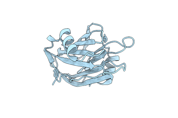
N-Terminal Domain Of Capsid Protein From The Mason-Pfizer Monkey Virus
Organism: Mason-pfizer monkey virus
Method: SOLUTION NMR Release Date: 2009-08-11 Classification: VIRAL PROTEIN |
Organism: Mason-pfizer monkey virus
Method: SOLUTION NMR
Release Date: 2009-08-11
 |
Crystal Structure Of The Equinatoxin Ii 8-69 Double Cysteine Mutant
Organism: Actinia equina
Method: X-RAY DIFFRACTION Resolution:2.30 Å Release Date: 2004-09-28 Classification: TOXIN |
Organism: Actinia equina
Method: X-RAY DIFFRACTION
Release Date: 2004-09-28
 |
Equinatoxin Ii
Organism: Actinia equina
Method: X-RAY DIFFRACTION Resolution:1.90 Å Release Date: 2001-04-04 Classification: TOXIN Ligands: SO4 |
Organism: Actinia equina
Method: X-RAY DIFFRACTION
Release Date: 2001-04-04
Ligands: SO4