

Search Count: 50
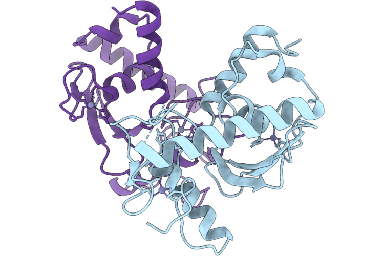 |
Mekk1 Swim And Ring Domains
Organism: Homo sapiens
Method: X-RAY DIFFRACTION Resolution:3.05 Å Release Date: 2026-06-24 Classification: SIGNALING PROTEIN Ligands: ZN |
Organism: Homo sapiens
Method: X-RAY DIFFRACTION
Release Date: 2026-06-24
Ligands: ZN
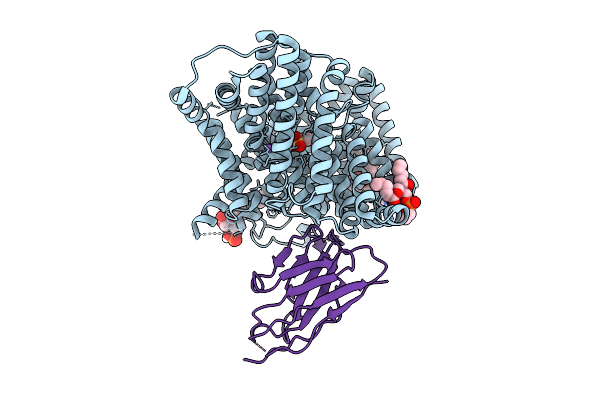 |
Cryo-Em Structure Of The Isethionate Trap Transporter Iseqm From Oleidesulfovibrio Alaskensis With Bound Isethionate
Organism: Oleidesulfovibrio alaskensis g20, Helicobacter pylori
Method: ELECTRON MICROSCOPY Release Date: 2025-11-12 Classification: TRANSPORT PROTEIN |
Organism: Oleidesulfovibrio alaskensis g20, Helicobacter pylori
Method: ELECTRON MICROSCOPY
Release Date: 2025-11-12
 |
Structure Of The Human Ddd-Ube2E2 Complex
Organism: Homo sapiens
Method: ELECTRON MICROSCOPY Release Date: 2025-03-05 Classification: PROTEIN BINDING |
Organism: Homo sapiens
Method: ELECTRON MICROSCOPY
Release Date: 2025-03-05
 |
Crystal Structure Of A Mes Bound Substrate Binding Protein (Isep) From An Isethionate Trap Transporter
Organism: Oleidesulfovibrio alaskensis g20
Method: X-RAY DIFFRACTION Resolution:1.65 Å Release Date: 2024-08-28 Classification: TRANSPORT PROTEIN Ligands: MES |
Organism: Oleidesulfovibrio alaskensis g20
Method: X-RAY DIFFRACTION
Release Date: 2024-08-28
Ligands: MES
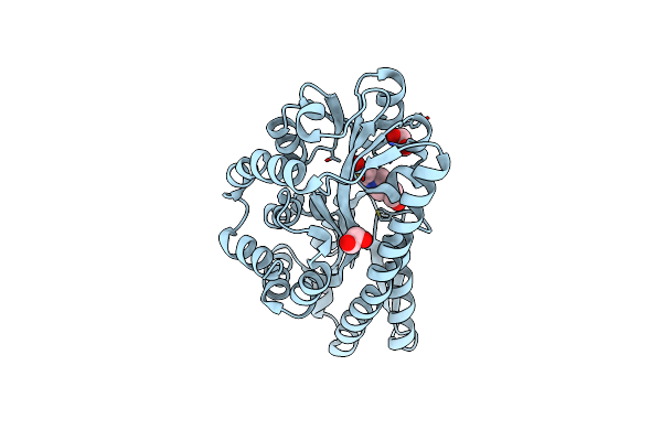 |
Structure Of A Hepes Bound Trap Transporter Substrate Binding Protein.
Organism: Oleidesulfovibrio alaskensis g20
Method: X-RAY DIFFRACTION Resolution:1.89 Å Release Date: 2024-08-28 Classification: TRANSPORT PROTEIN Ligands: EPE, EDO |
Organism: Oleidesulfovibrio alaskensis g20
Method: X-RAY DIFFRACTION
Release Date: 2024-08-28
Ligands: EPE, EDO
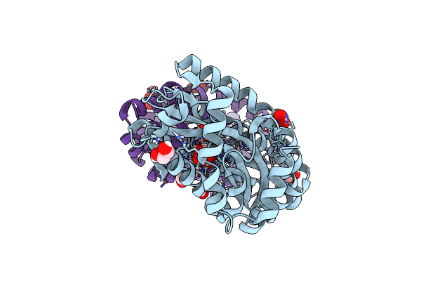 |
Crystal Structure Of An Isethionate Bound Substrate Binding Protein (Isep) From An Isethionate Trap Transporter
Organism: Oleidesulfovibrio alaskensis g20
Method: X-RAY DIFFRACTION Resolution:1.25 Å Release Date: 2024-07-10 Classification: TRANSPORT PROTEIN Ligands: 8X3, NO3, EDO |
Organism: Oleidesulfovibrio alaskensis g20
Method: X-RAY DIFFRACTION
Release Date: 2024-07-10
Ligands: 8X3, NO3, EDO
 |
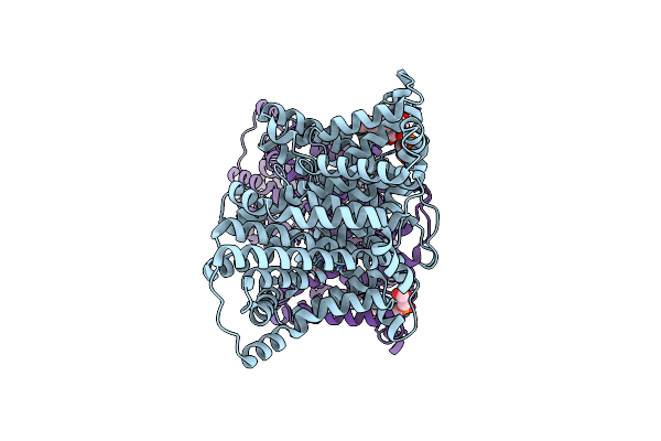
Apo Crystal Structure Of A Substrate Binding Protein (Isep) From An Isethionate Trap Transporter
Organism: Oleidesulfovibrio alaskensis g20
Method: X-RAY DIFFRACTION Resolution:1.48 Å Release Date: 2024-06-26 Classification: TRANSPORT PROTEIN Ligands: EDO, CL |
Organism: Oleidesulfovibrio alaskensis g20
Method: X-RAY DIFFRACTION
Release Date: 2024-06-26
Ligands: EDO, CL
 |
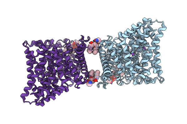
Cryo-Em Structure Of The Tripartite Atp-Independent Periplasmic (Trap) Transporter Siaqm From Haemophilus Influenzae (Parallel Dimer)
Organism: Haemophilus influenzae rd kw20
Method: ELECTRON MICROSCOPY Release Date: 2023-11-22 Classification: TRANSPORT PROTEIN Ligands: PGT, NA |
Organism: Haemophilus influenzae rd kw20
Method: ELECTRON MICROSCOPY
Release Date: 2023-11-22
Ligands: PGT, NA
 |
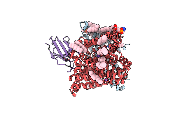
Cryo-Em Structure Of The Tripartite Atp-Independent Periplasmic (Trap) Transporter Siaqm From Haemophilus Influenzae (Antiparallel Dimer)
Organism: Haemophilus influenzae rd kw20
Method: ELECTRON MICROSCOPY Release Date: 2023-11-22 Classification: TRANSPORT PROTEIN |
Organism: Haemophilus influenzae rd kw20
Method: ELECTRON MICROSCOPY
Release Date: 2023-11-22
 |
Cryo-Em Structure Of The Tripartite Atp-Independent Periplasmic (Trap) Transporter Siaqm From Photobacterium Profundum In A Nanodisc
Organism: Photobacterium profundum ss9, Helicobacter pylori, Synthetic construct
Method: ELECTRON MICROSCOPY Release Date: 2023-03-15 Classification: TRANSPORT PROTEIN Ligands: OCT, PTY, TWT, D10, NA, TRD, HEX |
Organism: Photobacterium profundum ss9, Helicobacter pylori, Synthetic construct
Method: ELECTRON MICROSCOPY
Release Date: 2023-03-15
Ligands: OCT, PTY, TWT, D10, NA, TRD, HEX
 |
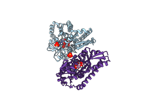
Cryo-Em Structure Of The Tripartite Atp-Independent Periplasmic (Trap) Transporter Siaqm From Photobacterium Profundum In Amphipol
Organism: Photobacterium profundum ss9, Helicobacter pylori, Synthetic construct
Method: ELECTRON MICROSCOPY Release Date: 2022-12-21 Classification: TRANSPORT PROTEIN |
Organism: Photobacterium profundum ss9, Helicobacter pylori, Synthetic construct
Method: ELECTRON MICROSCOPY
Release Date: 2022-12-21
 |
Structure Of The Sialic Acid Bound Tripartite Atp-Independent Periplasmic (Trap) Periplasmic Component Siap From Photobacterium Profundum
Organism: Photobacterium profundum
Method: X-RAY DIFFRACTION Resolution:1.04 Å Release Date: 2022-12-14 Classification: TRANSPORT PROTEIN Ligands: SLB, SO4 |
Organism: Photobacterium profundum
Method: X-RAY DIFFRACTION
Release Date: 2022-12-14
Ligands: SLB, SO4
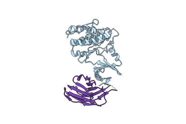 |
Tribbles (Trib2) Pseudokinase Bound To Nanobody Nb4.103
Organism: Homo sapiens, Synthetic construct
Method: X-RAY DIFFRACTION Resolution:2.70 Å Release Date: 2022-09-14 Classification: SIGNALING PROTEIN/IMMUNE SYSTEM |
Organism: Homo sapiens, Synthetic construct
Method: X-RAY DIFFRACTION
Release Date: 2022-09-14
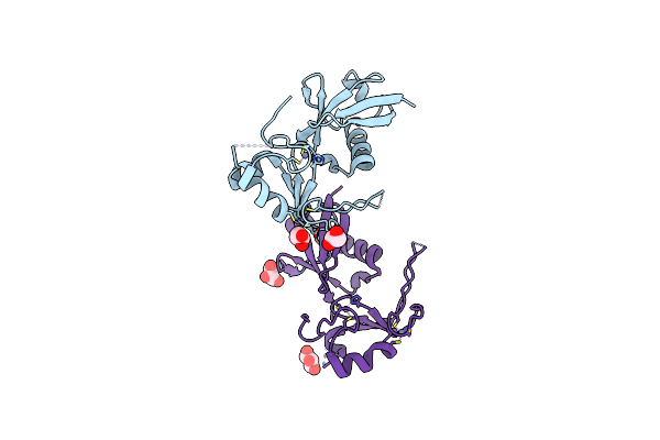 |
Crystal Structure Of The Ubark2C Fusion Protein
Organism: Homo sapiens
Method: X-RAY DIFFRACTION Resolution:2.50 Å Release Date: 2022-03-09 Classification: TRANSFERASE Ligands: GOL, ZN |
Organism: Homo sapiens
Method: X-RAY DIFFRACTION
Release Date: 2022-03-09
Ligands: GOL, ZN
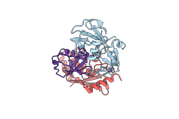 |
Crystal Structure Of The Ubark2C-Ubch5B~Ub Complex
Organism: Homo sapiens
Method: X-RAY DIFFRACTION Resolution:2.80 Å Release Date: 2022-03-09 Classification: TRANSFERASE Ligands: ZN |
Organism: Homo sapiens
Method: X-RAY DIFFRACTION
Release Date: 2022-03-09
Ligands: ZN
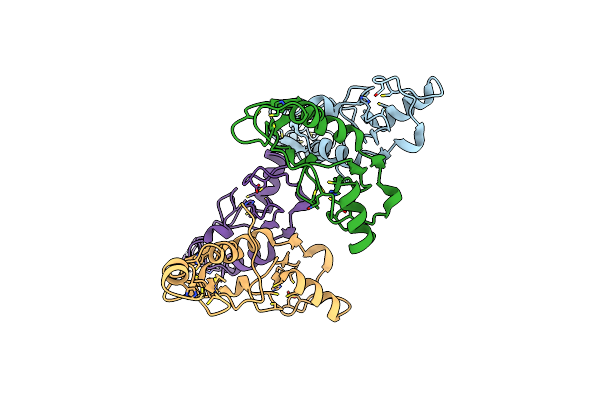 |
Structure Of Traf5 And Traf6 Ring Hetero Dimer
Organism: Homo sapiens
Method: X-RAY DIFFRACTION Resolution:2.80 Å Release Date: 2021-02-17 Classification: TRANSFERASE Ligands: ZN |
Organism: Homo sapiens
Method: X-RAY DIFFRACTION
Release Date: 2021-02-17
Ligands: ZN
 |
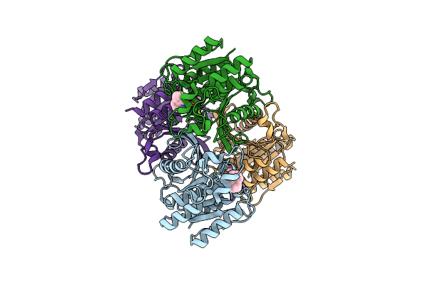
Fphf, Staphylococcus Aureus Fluorophosphonate-Binding Serine Hydrolases F, Apo Form
Organism: Staphylococcus aureus
Method: X-RAY DIFFRACTION Resolution:1.71 Å Release Date: 2020-09-16 Classification: HYDROLASE |
Organism: Staphylococcus aureus
Method: X-RAY DIFFRACTION
Release Date: 2020-09-16
 |
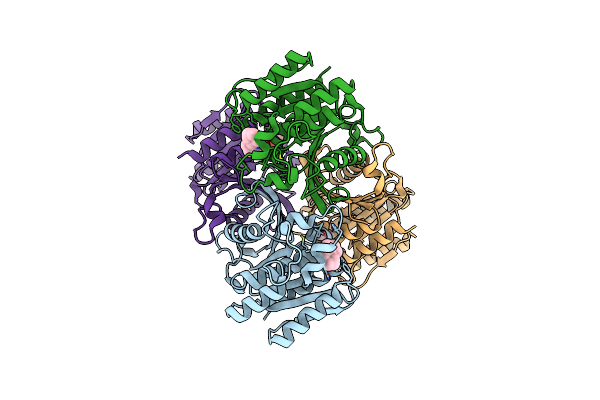
Fphf, Staphylococcus Aureus Fluorophosphonate-Binding Serine Hydrolases F, Kt129 Bound
Organism: Staphylococcus aureus
Method: X-RAY DIFFRACTION Resolution:1.98 Å Release Date: 2020-09-16 Classification: HYDROLASE Ligands: 6WG |
Organism: Staphylococcus aureus
Method: X-RAY DIFFRACTION
Release Date: 2020-09-16
Ligands: 6WG
 |
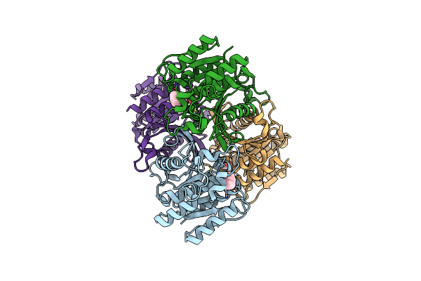
Fphf, Staphylococcus Aureus Fluorophosphonate-Binding Serine Hydrolases F, Kt130 Bound
Organism: Staphylococcus aureus
Method: X-RAY DIFFRACTION Resolution:1.94 Å Release Date: 2020-09-16 Classification: HYDROLASE Ligands: 6WG |
Organism: Staphylococcus aureus
Method: X-RAY DIFFRACTION
Release Date: 2020-09-16
Ligands: 6WG
 |
Fphf, Staphylococcus Aureus Fluorophosphonate-Binding Serine Hydrolases F, Substrate Bound
Organism: Staphylococcus aureus
Method: X-RAY DIFFRACTION Resolution:2.89 Å Release Date: 2020-09-16 Classification: HYDROLASE Ligands: HE4 |
Organism: Staphylococcus aureus
Method: X-RAY DIFFRACTION
Release Date: 2020-09-16
Ligands: HE4