
Search Count: 631
All
Selected
 |
Organism: Arabidopsis thaliana, Escherichia coli k12
Method: ELECTRON MICROSCOPY Release Date: 2026-04-08 Classification: SIGNALING PROTEIN Ligands: NAG, CU |
 |
Organism: Arabidopsis thaliana, Escherichia coli k12
Method: ELECTRON MICROSCOPY Release Date: 2026-04-08 Classification: SIGNALING PROTEIN Ligands: NAG |
 |
Organism: Pseudomonas aeruginosa
Method: ELECTRON MICROSCOPY Resolution:3.60 Å Release Date: 2026-03-11 Classification: MEMBRANE PROTEIN |
 |
Organism: Pseudomonas aeruginosa
Method: ELECTRON MICROSCOPY Resolution:3.55 Å Release Date: 2026-03-11 Classification: MEMBRANE PROTEIN |
 |
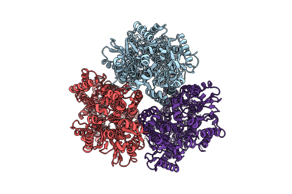
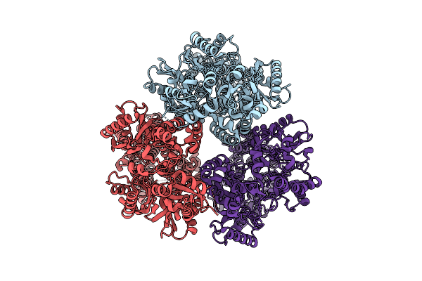
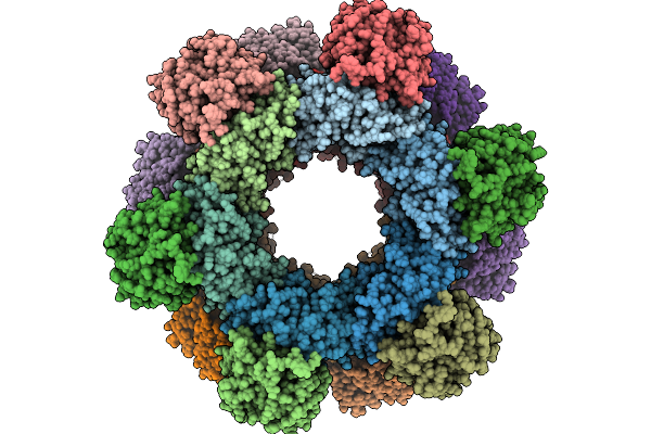
Cryo-Em Structure Of Self-Assembled Zymomonas Mobilis Levansucrase Nanotube
Organism: Zymomonas mobilis subsp. mobilis atcc 10988
Method: ELECTRON MICROSCOPY Release Date: 2026-03-04 Classification: TRANSFERASE |
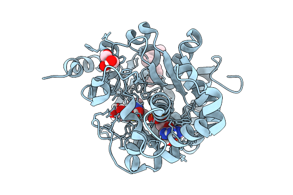 |
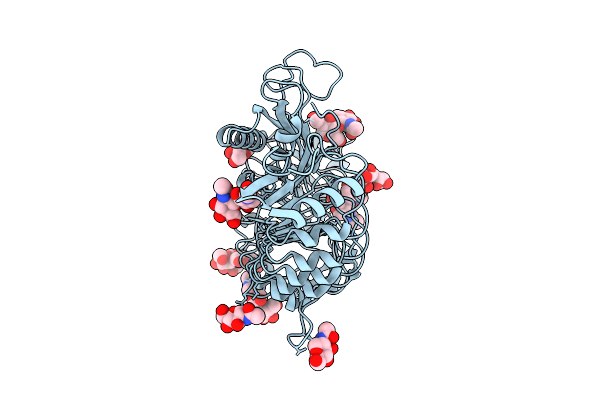
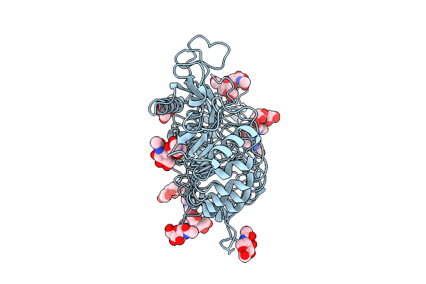
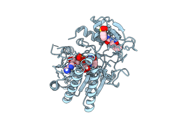
Crystal Structure Of Arabidopsis Thaliana Sulfotransferase Sot18 Complexed With Glucoraphanin Precursor Involved In Glucosinolate Biosynthesis
Organism: Arabidopsis thaliana
Method: X-RAY DIFFRACTION Resolution:1.39 Å Release Date: 2026-03-04 Classification: CYTOSOLIC PROTEIN Ligands: A3P, GOL, A1L7V, PLM |
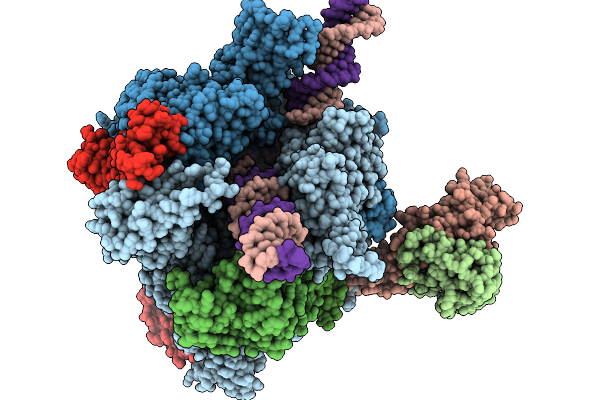 |
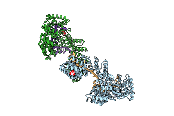
Structure Of A Native Drosophila Melanogaster Pol Ii Elongation Complex With A Well-Defined Rpb4/Rpb7 Stalk
Organism: Drosophila melanogaster
Method: ELECTRON MICROSCOPY Resolution:3.44 Å Release Date: 2026-01-07 Classification: TRANSCRIPTION Ligands: ZN |
 |
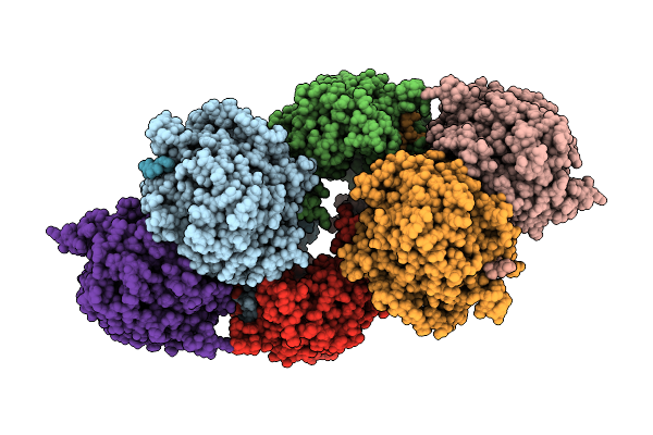
Cryo-Em Structure Of A Designed Pyridoxal Phosphate (Plp) Synthase Fused To A Designed Circumsporozoite Protein Antigen From Plasmodium Falciparum (Csp-P1-Csp And Csp-P2-Csp)
Organism: Plasmodium falciparum
Method: ELECTRON MICROSCOPY Release Date: 2025-12-31 Classification: BIOSYNTHETIC PROTEIN |
 |
Structure Of Udp-Galactose-4-Epimerase (Gale) Bound To Fragment From Diamond Xchem Experiment.
Organism: Homo sapiens
Method: X-RAY DIFFRACTION Resolution:1.65 Å Release Date: 2025-12-10 Classification: CARBOHYDRATE Ligands: NAD, JGA, MLI, PGE, EDO, CL |
 |
Organism: Avian orthoreovirus
Method: ELECTRON MICROSCOPY Release Date: 2025-12-03 Classification: VIRAL PROTEIN |
 |
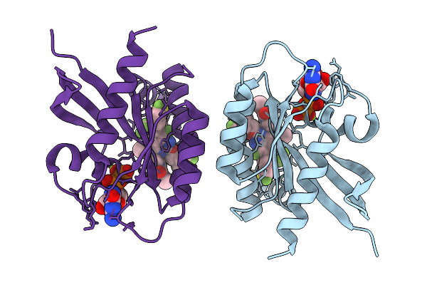
Structure Of Human Udp-Galactose 4-Epimerase In Complex With Compound Wbx04
Organism: Homo sapiens
Method: X-RAY DIFFRACTION Resolution:1.37 Å Release Date: 2025-12-03 Classification: ISOMERASE Ligands: EDO, NAD, A1IU2, MLT |
 |
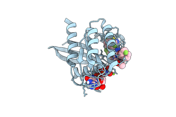
Structure Of Human Udp-Galactose 4-Epimerase In Complex With Compound Wbx09
Organism: Homo sapiens
Method: X-RAY DIFFRACTION Resolution:0.95 Å Release Date: 2025-12-03 Classification: ISOMERASE Ligands: EDO, NAD, A1IU5 |
 |
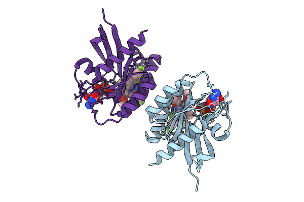
Structure Of Human Udp-Galactose 4-Epimerase In Complex With Covalent Compound Wbc10
Organism: Homo sapiens
Method: X-RAY DIFFRACTION Resolution:1.37 Å Release Date: 2025-12-03 Classification: ISOMERASE Ligands: NAI, A1IU6 |
 |
Organism: Homo sapiens
Method: X-RAY DIFFRACTION Resolution:1.70 Å Release Date: 2025-12-03 Classification: ONCOPROTEIN/INHIBITOR Ligands: GDP, MG, A1CG4 |
 |
Organism: Homo sapiens
Method: X-RAY DIFFRACTION Resolution:1.35 Å Release Date: 2025-12-03 Classification: ONCOPROTEIN/INHIBITOR Ligands: GNP, MG, A1CG4 |
 |
Crystal Structure Of Hras-G12D/Q95H (Gmppnp-Bound) In Complex With Bbo-11818
Organism: Homo sapiens
Method: X-RAY DIFFRACTION Resolution:2.02 Å Release Date: 2025-12-03 Classification: ONCOPROTEIN Ligands: GNP, MG, A1CG4 |
 |
Organism: Synthetic construct, Arabidopsis thaliana
Method: X-RAY DIFFRACTION Resolution:2.70 Å Release Date: 2025-11-12 Classification: HYDROLASE/RNA Ligands: ZN, GOL |
 |
Organism: Synthetic construct
Method: X-RAY DIFFRACTION Resolution:2.26 Å Release Date: 2025-11-12 Classification: HYDROLASE Ligands: GOL, ZN |
 |
Crystal Structure Of A Phgs Rhamnosyltransferase Ugt79G15 From Rehmannia Glutinosa
Organism: Rehmannia glutinosa
Method: X-RAY DIFFRACTION Resolution:1.88 Å Release Date: 2025-10-08 Classification: TRANSFERASE |
 |
Crystal Structure Of A Phgs Rhamnosyltransferase Ugt79G15 From Rehmannia Glutinosa In Complex With Udp
Organism: Rehmannia glutinosa
Method: X-RAY DIFFRACTION Resolution:2.40 Å Release Date: 2025-10-08 Classification: TRANSFERASE Ligands: UDP |