
Search Count: 834
All
Selected
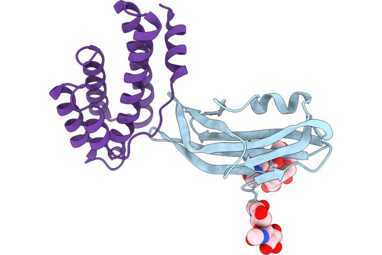 |
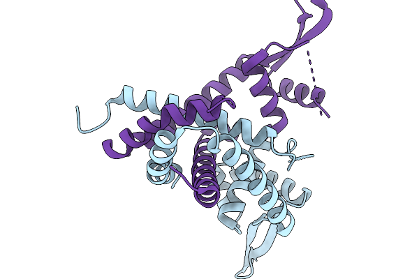
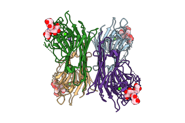
Structure Of The Porcine Deltacoronavirus (Pdcov) Receptor-Binding Domain Bound To The Rbd Minibinder 11, The Pd3 Fab, And The Kappa Light Chain Nanobody (Local Refinement)
Organism: Porcine deltacoronavirus, Synthetic construct
Method: ELECTRON MICROSCOPY Resolution:2.84 Å Release Date: 2026-05-13 Classification: VIRAL PROTEIN |
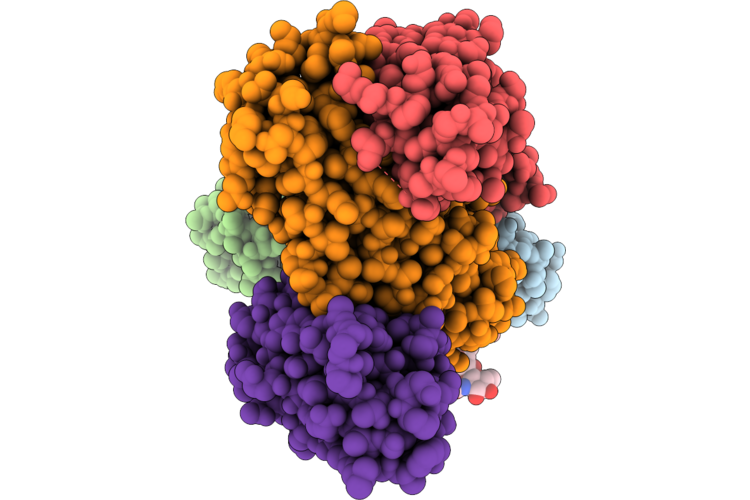 |
Structure Of The Porcine Deltacoronavirus (Pdcov) Receptor-Binding Domain Bound To The Rbd Minibinder 11, The Pd3 Fab, And The Kappa Light Chain Nanobody
Organism: Porcine deltacoronavirus, Synthetic construct, Mus musculus, Lama glama
Method: ELECTRON MICROSCOPY Resolution:2.80 Å Release Date: 2026-05-13 Classification: VIRAL PROTEIN |
 |
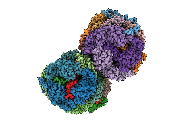
Organism: Influenza b virus (b/brisbane/60/2008), Homo sapiens
Method: ELECTRON MICROSCOPY Resolution:2.76 Å Release Date: 2026-04-29 Classification: VIRAL PROTEIN/IMMUNE SYSTEM Ligands: NAG |
 |
Organism: Influenza b virus (b/brisbane/60/2008), Homo sapiens
Method: ELECTRON MICROSCOPY Resolution:2.76 Å Release Date: 2026-04-29 Classification: VIRAL PROTEIN/IMMUNE SYSTEM Ligands: NAG |
 |
Organism: Influenza b virus (b/brisbane/60/2008), Homo sapiens
Method: ELECTRON MICROSCOPY Resolution:2.65 Å Release Date: 2026-04-29 Classification: VIRAL PROTEIN/IMMUNE SYSTEM Ligands: NAG |
 |
Organism: Influenza b virus, Homo sapiens
Method: ELECTRON MICROSCOPY Release Date: 2026-04-29 Classification: VIRAL PROTEIN/IMMUNE SYSTEM Ligands: NAG |
 |
Organism: Influenza b virus (b/hubei-wujiagang/158/2009), Homo sapiens
Method: ELECTRON MICROSCOPY Resolution:3.09 Å Release Date: 2026-04-29 Classification: VIRAL PROTEIN/IMMUNE SYSTEM Ligands: NAG |
 |
Organism: Influenza b virus (b/hubei-wujiagang/158/2009), Homo sapiens
Method: ELECTRON MICROSCOPY Resolution:2.99 Å Release Date: 2026-04-29 Classification: VIRAL PROTEIN/IMMUNE SYSTEM Ligands: NAG |
 |
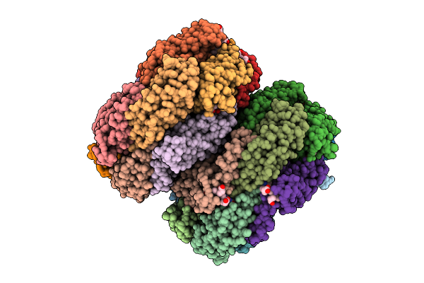
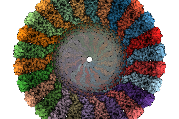
The Structure Of Phycobilisome With A Bicylindrical Core From The Cyanobacterium Synechococcus Elongatus Pcc 7942
Organism: Synechococcus elongatus pcc 7942 = fachb-805
Method: ELECTRON MICROSCOPY Resolution:3.20 Å Release Date: 2026-04-01 Classification: ELECTRON TRANSPORT Ligands: CYC |
 |
Organism: Streptomyces prunicolor
Method: X-RAY DIFFRACTION Resolution:1.96 Å Release Date: 2026-04-01 Classification: TRANSFERASE Ligands: GOL, PEG |
 |
Aminoacyl-Trna-Dependent Peptide Synthase, Sbb17, Complexed With Streptothrisamine
Organism: Streptomyces prunicolor
Method: X-RAY DIFFRACTION Resolution:2.50 Å Release Date: 2026-04-01 Classification: TRANSFERASE Ligands: A1L3W |
 |
Aminoacyl-Trna-Dependent Peptide Synthase, Sba18, Complexed With Streptothrisamine
Organism: Streptomyces sp.
Method: X-RAY DIFFRACTION Resolution:2.00 Å Release Date: 2026-04-01 Classification: TRANSFERASE Ligands: A1L3W, TRS |
 |
The Structure Of Phycobilisome With A Bicylindrical Core From The Cyanobacterium Synechococcus Elongatus Pcc 7942
Organism: Synechococcus elongatus pcc 7942 = fachb-805
Method: ELECTRON MICROSCOPY Release Date: 2026-04-01 Classification: ELECTRON TRANSPORT Ligands: CYC |
 |
The Cryo-Em Structure Of Phycobilisome Rod From Synechococcus Elongatus Pcc 7942
Organism: Synechococcus elongatus pcc 7942 = fachb-805
Method: ELECTRON MICROSCOPY Resolution:2.92 Å Release Date: 2026-04-01 Classification: ELECTRON TRANSPORT Ligands: CYC |
 |
Organism: Mycobacterium tuberculosis (strain atcc 25618 / h37rv)
Method: X-RAY DIFFRACTION Resolution:1.70 Å Release Date: 2026-03-25 Classification: GENE REGULATION |
 |
Organism: Homo sapiens
Method: ELECTRON MICROSCOPY Resolution:2.70 Å Release Date: 2026-03-25 Classification: MEMBRANE PROTEIN |
 |
Organism: Homo sapiens
Method: ELECTRON MICROSCOPY Resolution:2.60 Å Release Date: 2026-03-25 Classification: MEMBRANE PROTEIN |
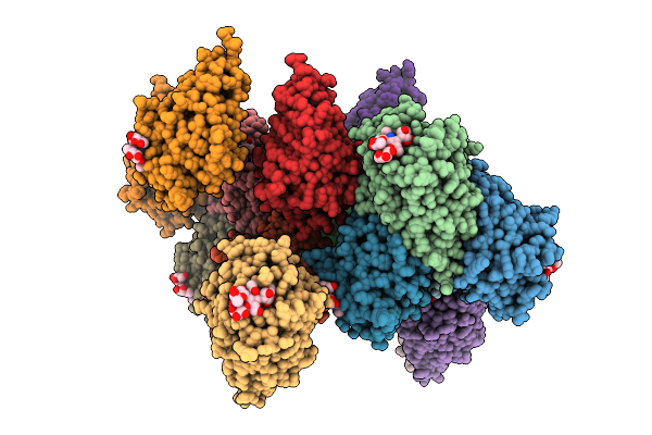 |
Organism: Lablab purpureus
Method: X-RAY DIFFRACTION Resolution:2.49 Å Release Date: 2026-03-18 Classification: PLANT PROTEIN Ligands: CA, MN |
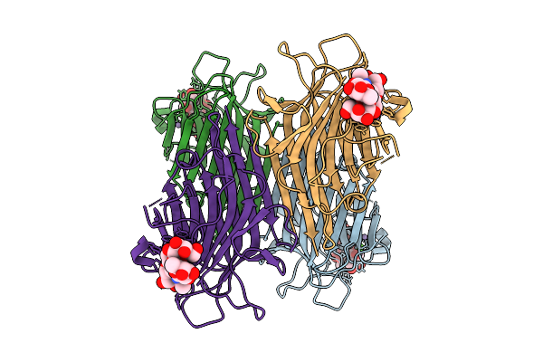 |
Organism: Lablab purpureus
Method: ELECTRON MICROSCOPY Release Date: 2026-03-18 Classification: PLANT PROTEIN Ligands: CA, MN |
 |
Organism: Lablab purpureus
Method: ELECTRON MICROSCOPY Release Date: 2026-03-18 Classification: PLANT PROTEIN Ligands: CA, MN |