
Search Count: 35
All
Selected
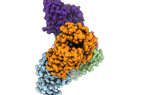 |
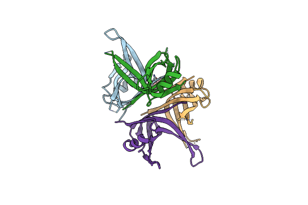
Organism: Homo sapiens, Mus musculus
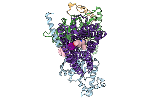
Method: ELECTRON MICROSCOPY Resolution:2.84 Å Release Date: 2026-01-14 Classification: MEMBRANE PROTEIN Ligands: CLR, A1EK5 |
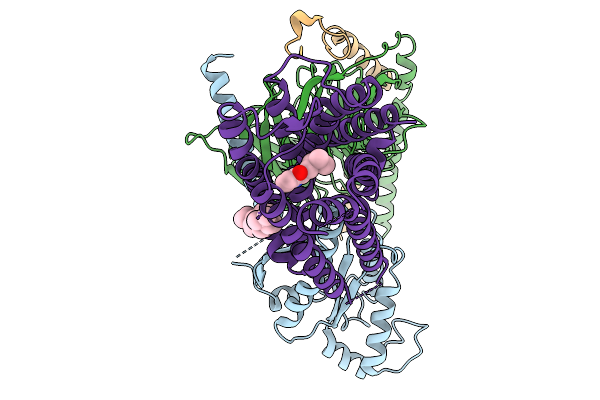 |
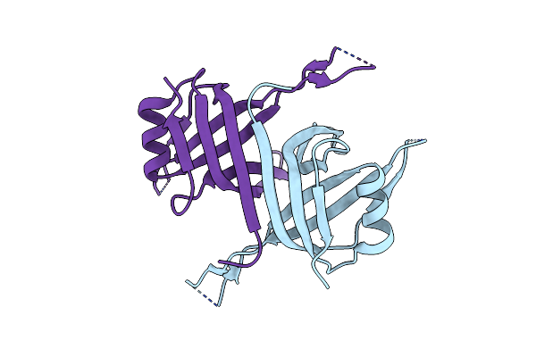
Organism: Homo sapiens
Method: ELECTRON MICROSCOPY Resolution:2.62 Å Release Date: 2026-01-14 Classification: MEMBRANE PROTEIN Ligands: 91Q, CLR |
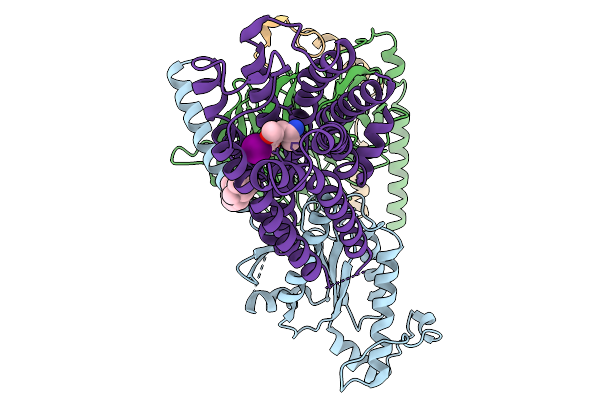 |
Organism: Homo sapiens
Method: ELECTRON MICROSCOPY Resolution:2.76 Å Release Date: 2026-01-14 Classification: MEMBRANE PROTEIN Ligands: A1EK5, CLR |
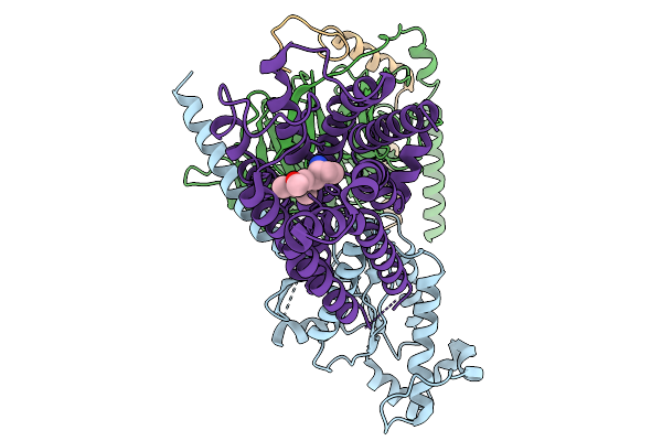 |
Organism: Homo sapiens
Method: ELECTRON MICROSCOPY Resolution:3.27 Å Release Date: 2026-01-14 Classification: MEMBRANE PROTEIN Ligands: A1EK8 |
 |
Organism: Homo sapiens
Method: ELECTRON MICROSCOPY Resolution:2.98 Å Release Date: 2026-01-14 Classification: MEMBRANE PROTEIN Ligands: CLR, A1ELA |
 |
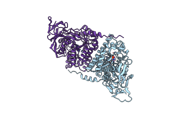
Organism: Severe acute respiratory syndrome coronavirus 2
Method: X-RAY DIFFRACTION Resolution:2.00 Å Release Date: 2023-08-16 Classification: VIRAL PROTEIN Ligands: OF9, CL |
 |
Structure Of Trpa1 In Drosophila Melanogaster In A State With 17 Ankyrin Repeats Determined
Organism: Drosophila melanogaster
Method: ELECTRON MICROSCOPY Release Date: 2023-07-26 Classification: MEMBRANE PROTEIN |
 |
Structure Of Trpa1 In Drosophila Melanogaster In A State With 5 Ankyrin Repeats Determined
Organism: Drosophila melanogaster
Method: ELECTRON MICROSCOPY Release Date: 2023-07-26 Classification: MEMBRANE PROTEIN |
 |
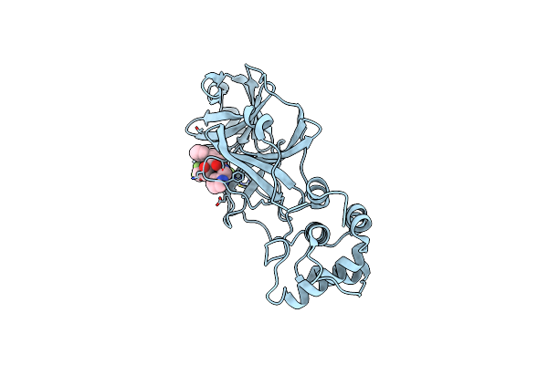
Crystal Structure Of The Recombinant Protein Hr121 From The S2 Protein Of Sars-Cov-2
Organism: Severe acute respiratory syndrome coronavirus 2
Method: X-RAY DIFFRACTION Resolution:2.41 Å Release Date: 2022-11-23 Classification: VIRAL PROTEIN |
 |
Organism: Salmonella typhimurium (strain lt2 / sgsc1412 / atcc 700720)
Method: X-RAY DIFFRACTION Resolution:2.87 Å Release Date: 2022-05-04 Classification: DNA BINDING PROTEIN Ligands: HOH |
 |
Organism: Pseudomonas aeruginosa pao1
Method: X-RAY DIFFRACTION Resolution:2.32 Å Release Date: 2022-03-09 Classification: DNA BINDING PROTEIN Ligands: QUE |
 |
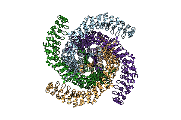
Crystal Structure Of The Pseudomonas Aeruginosa Dihydropyrimidinase Complexed With 5-Au
Organism: Pseudomonas aeruginosa (strain atcc 15692 / dsm 22644 / cip 104116 / jcm 14847 / lmg 12228 / 1c / prs 101 / pao1)
Method: X-RAY DIFFRACTION Resolution:2.16 Å Release Date: 2022-02-16 Classification: HYDROLASE Ligands: WBU, ZN |
 |
Organism: Severe acute respiratory syndrome coronavirus 2
Method: X-RAY DIFFRACTION Resolution:2.00 Å Release Date: 2020-10-07 Classification: VIRAL PROTEIN Ligands: GQU, TRS |
 |
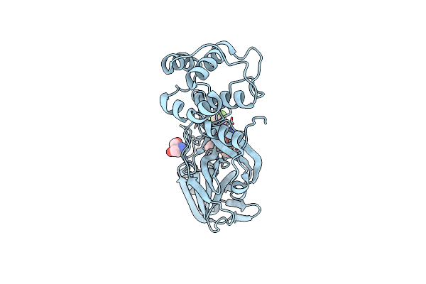
Crystal Structure Of The Sars-Cov-2 Main Protease In Complex With Boceprevir (Space Group P212121)
Organism: Severe acute respiratory syndrome coronavirus 2
Method: X-RAY DIFFRACTION Resolution:2.25 Å Release Date: 2020-08-19 Classification: VIRAL PROTEIN Ligands: U5G |
 |
Crystal Structure Of The Sars-Cov-2 Main Protease In Complex With Telaprevir
Organism: Severe acute respiratory syndrome coronavirus 2
Method: X-RAY DIFFRACTION Resolution:1.74 Å Release Date: 2020-07-29 Classification: VIRAL PROTEIN Ligands: SV6, CL |
 |
The Crystal Structure Of Human Chemokine Receptor Ccr5 In Complex With Compound 21
Organism: Homo sapiens, Clostridium pasteurianum
Method: X-RAY DIFFRACTION Resolution:2.80 Å Release Date: 2018-10-24 Classification: SIGNALING PROTEIN Ligands: ZN, NO3, A4R |
 |
The Crystal Structure Of Human Chemokine Receptor Ccr5 In Complex With Compound 34
Organism: Homo sapiens, Clostridium pasteurianum
Method: X-RAY DIFFRACTION Resolution:2.80 Å Release Date: 2018-10-24 Classification: SIGNALING PROTEIN Ligands: ZN, OLC, A4X |
 |
Organism: Streptomyces peucetius
Method: X-RAY DIFFRACTION Resolution:2.15 Å Release Date: 2018-04-18 Classification: TRANSFERASE Ligands: AFY |
 |
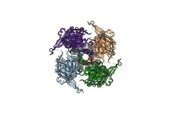
Crosslinked Crystal Structure Of Type Ii Fatty Acid Synthase Ketosynthase, Fabb, And Acyl Carrier Protein, Acpp
Organism: Escherichia coli
Method: X-RAY DIFFRACTION Resolution:2.40 Å Release Date: 2018-01-24 Classification: TRANSFERASE Ligands: 6W5 |
 |
Organism: Streptomyces flocculus
Method: X-RAY DIFFRACTION Resolution:2.71 Å Release Date: 2017-01-11 Classification: HYDROLASE |