

Search Count: 102
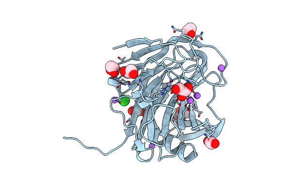 |
Crystal Structure Of The Gh12 Domain From Acidothermus Cellulolyticus Guxa
Organism: Acidothermus cellulolyticus
Method: X-RAY DIFFRACTION Resolution:1.50 Å Release Date: 2022-05-04 Classification: HYDROLASE |
Organism: Acidothermus cellulolyticus
Method: X-RAY DIFFRACTION
Release Date: 2022-05-04
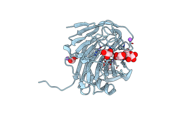 |
Crystal Structure Of The Gh12 Domain From Acidothermus Cellulolyticus Guxa Bound To Cellobiose
Organism: Acidothermus cellulolyticus
Method: X-RAY DIFFRACTION Resolution:1.85 Å Release Date: 2022-05-04 Classification: HYDROLASE Ligands: ZN, CL, NA, ACT, GOL |
Organism: Acidothermus cellulolyticus
Method: X-RAY DIFFRACTION
Release Date: 2022-05-04
Ligands: ZN, CL, NA, ACT, GOL
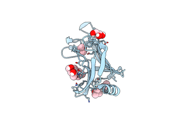 |
Structure Of Bdellovibrio Bacteriovorus Bd0675
Organism: Bdellovibrio bacteriovorus (strain atcc 15356 / dsm 50701 / ncib 9529 / hd100)
Method: X-RAY DIFFRACTION Resolution:1.50 Å Release Date: 2021-05-05 Classification: UNKNOWN FUNCTION Ligands: MPD, GOL, TBU |
Organism: Bdellovibrio bacteriovorus (strain atcc 15356 / dsm 50701 / ncib 9529 / hd100)
Method: X-RAY DIFFRACTION
Release Date: 2021-05-05
Ligands: MPD, GOL, TBU
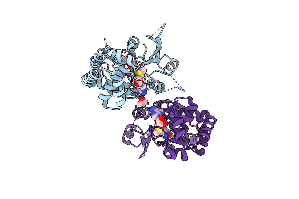 |
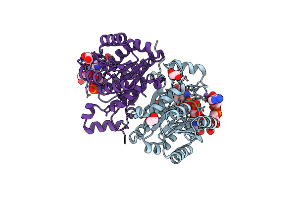
The Crystal Structure Of Mcbe1
Organism: Caldicellulosiruptor bescii (strain atcc baa-1888 / dsm 6725 / z-1320)
Method: X-RAY DIFFRACTION Resolution:1.90 Å Release Date: 2021-04-14 Classification: TRANSFERASE Ligands: SAM, SAH, EDO |
Organism: Caldicellulosiruptor bescii (strain atcc baa-1888 / dsm 6725 / z-1320)
Method: X-RAY DIFFRACTION
Release Date: 2021-04-14
Ligands: SAM, SAH, EDO
 |
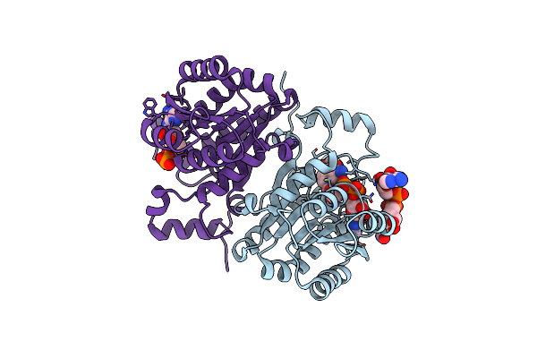
Structure Of Serratia Marcescens 2,3-Butanediol Dehydrogenase Mutant Q247A
Organism: Serratia marcescens
Method: X-RAY DIFFRACTION Resolution:1.90 Å Release Date: 2020-12-23 Classification: OXIDOREDUCTASE Ligands: NAD, GOL, EDO, NA, ADP |
Organism: Serratia marcescens
Method: X-RAY DIFFRACTION
Release Date: 2020-12-23
Ligands: NAD, GOL, EDO, NA, ADP
 |
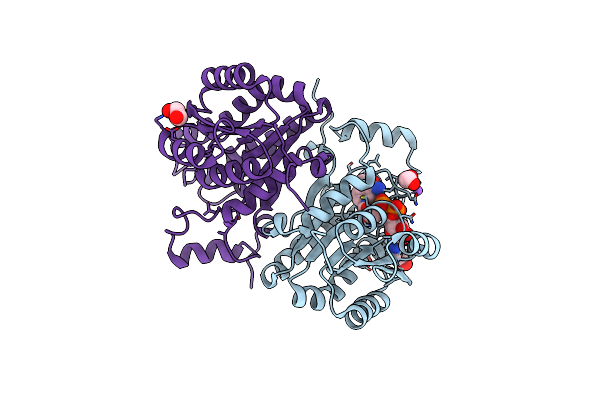
Structure Of Serratia Marcescens 2,3-Butanediol Dehydrogenase
Organism: Serratia marcescens
Method: X-RAY DIFFRACTION Resolution:2.00 Å Release Date: 2020-12-23 Classification: OXIDOREDUCTASE Ligands: NAD, HBR, HBS, ADP |
Organism: Serratia marcescens
Method: X-RAY DIFFRACTION
Release Date: 2020-12-23
Ligands: NAD, HBR, HBS, ADP
 |
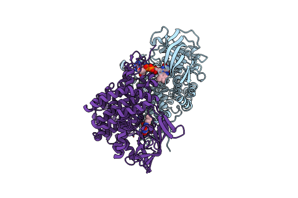
Structure Of Serratia Marcescens 2,3-Butanediol Dehydrogenase Mutant Q247A/V139Q
Organism: Serratia marcescens
Method: X-RAY DIFFRACTION Resolution:1.80 Å Release Date: 2020-12-23 Classification: OXIDOREDUCTASE Ligands: NAD, NA, EDO |
Organism: Serratia marcescens
Method: X-RAY DIFFRACTION
Release Date: 2020-12-23
Ligands: NAD, NA, EDO
 |
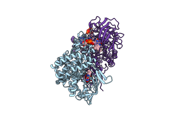
Bd2924 Apo-Form
Organism: Bdellovibrio bacteriovorus (strain atcc 15356 / dsm 50701 / ncib 9529 / hd100)
Method: X-RAY DIFFRACTION Resolution:1.51 Å Release Date: 2019-09-11 Classification: OXIDOREDUCTASE Ligands: FAD |
Organism: Bdellovibrio bacteriovorus (strain atcc 15356 / dsm 50701 / ncib 9529 / hd100)
Method: X-RAY DIFFRACTION
Release Date: 2019-09-11
Ligands: FAD
 |
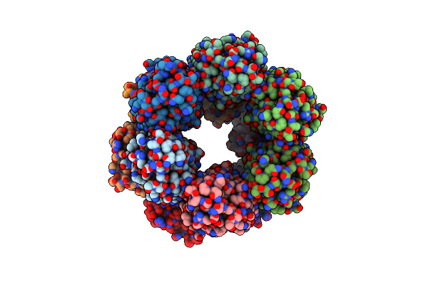
Bd2924 C10 Acyl-Coenzymea Bound Form
Organism: Bdellovibrio bacteriovorus hd100
Method: X-RAY DIFFRACTION Resolution:1.87 Å Release Date: 2019-09-11 Classification: OXIDOREDUCTASE Ligands: FAD, MFK |
Organism: Bdellovibrio bacteriovorus hd100
Method: X-RAY DIFFRACTION
Release Date: 2019-09-11
Ligands: FAD, MFK
 |
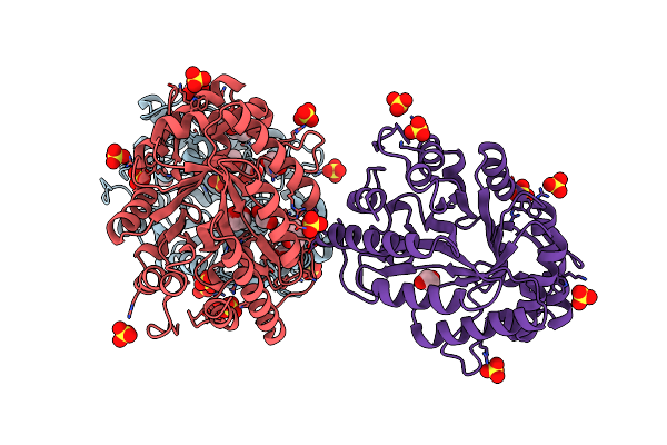
Structure Of Caldicellulosiruptor Danielii Cbm3 Module Of Glycoside Hydrolase Wp_045175321
Organism: Caldicellulosiruptor sp. wai35.b1
Method: X-RAY DIFFRACTION Resolution:2.00 Å Release Date: 2019-04-24 Classification: SUGAR BINDING PROTEIN Ligands: CA |
Organism: Caldicellulosiruptor sp. wai35.b1
Method: X-RAY DIFFRACTION
Release Date: 2019-04-24
Ligands: CA
 |
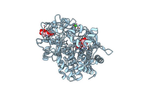
Structure Of Caldicellulosiruptor Danielii Gh10 Module Of Glycoside Hydrolase Wp_045175321
Organism: Caldicellulosiruptor sp. wai35.b1
Method: X-RAY DIFFRACTION Resolution:1.90 Å Release Date: 2019-04-24 Classification: HYDROLASE Ligands: SO4, EDO, GOL, FMT |
Organism: Caldicellulosiruptor sp. wai35.b1
Method: X-RAY DIFFRACTION
Release Date: 2019-04-24
Ligands: SO4, EDO, GOL, FMT
 |
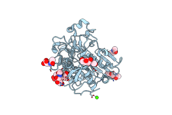
Structure Of Caldicellulosiruptor Danielii Gh48 Module Of Glycoside Hydrolase Wp_045175321
Organism: Caldicellulosiruptor sp. wai35.b1
Method: X-RAY DIFFRACTION Resolution:1.90 Å Release Date: 2019-04-24 Classification: HYDROLASE Ligands: CA |
Organism: Caldicellulosiruptor sp. wai35.b1
Method: X-RAY DIFFRACTION
Release Date: 2019-04-24
Ligands: CA
 |
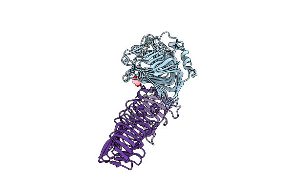
The Crystal Structure Of Xoat1
Organism: Arabidopsis thaliana
Method: X-RAY DIFFRACTION Resolution:1.85 Å Release Date: 2019-02-20 Classification: TRANSFERASE Ligands: CO, NAG, CA |
Organism: Arabidopsis thaliana
Method: X-RAY DIFFRACTION
Release Date: 2019-02-20
Ligands: CO, NAG, CA
 |
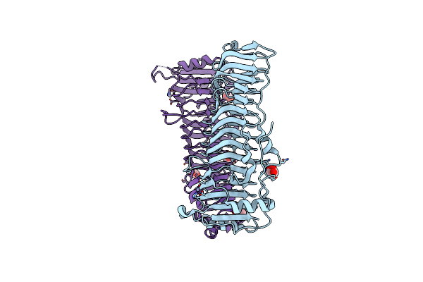
The Crystal Structure Of Caldicellulosiruptor Kristjanssonii Tapirin C-Terminal Domain
Organism: Caldicellulosiruptor kristjanssonii (strain atcc 700853 / dsm 12137 / i77r1b)
Method: X-RAY DIFFRACTION Resolution:2.65 Å Release Date: 2018-12-19 Classification: CELL ADHESION Ligands: CA, GOL |
Organism: Caldicellulosiruptor kristjanssonii (strain atcc 700853 / dsm 12137 / i77r1b)
Method: X-RAY DIFFRACTION
Release Date: 2018-12-19
Ligands: CA, GOL
 |
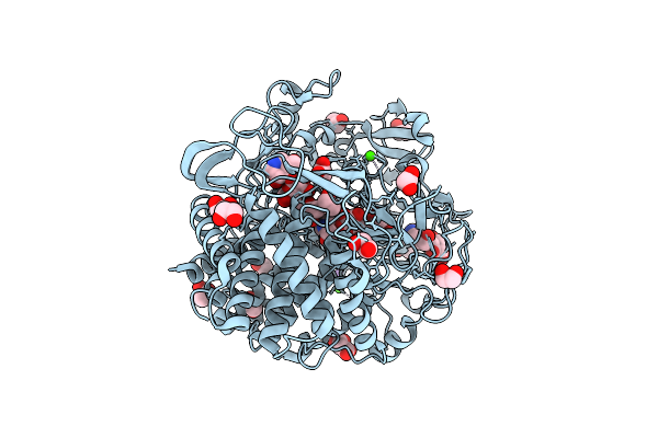
The Crystal Structure Of Caldicellulosiruptor Hydrothermalis Tapirin C-Terminal Domain
Organism: Caldicellulosiruptor hydrothermalis (strain dsm 18901 / vkm b-2411 / 108)
Method: X-RAY DIFFRACTION Resolution:1.75 Å Release Date: 2018-12-19 Classification: CELL ADHESION Ligands: ZN, EDO, GOL, CL, GOA, OXL |
Organism: Caldicellulosiruptor hydrothermalis (strain dsm 18901 / vkm b-2411 / 108)
Method: X-RAY DIFFRACTION
Release Date: 2018-12-19
Ligands: ZN, EDO, GOL, CL, GOA, OXL
 |
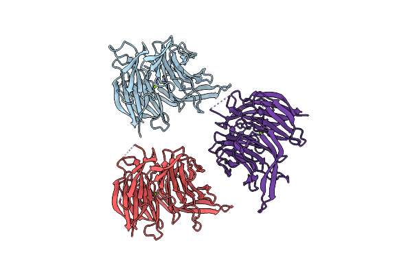
Structure Of B. Pumilus Gh48 In Complex With A Cellobio-Derived Isofagomine
Organism: Bacillus pumilus
Method: X-RAY DIFFRACTION Resolution:1.95 Å Release Date: 2018-10-31 Classification: HYDROLASE Ligands: G2I, 9MR, CA, NA, EDO, MLI, ACT |
Organism: Bacillus pumilus
Method: X-RAY DIFFRACTION
Release Date: 2018-10-31
Ligands: G2I, 9MR, CA, NA, EDO, MLI, ACT
 |
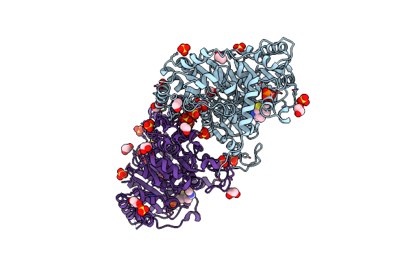
Crystal Structure Of The Wd40-Repeat Of Rfwd3
Organism: Homo sapiens
Method: X-RAY DIFFRACTION Resolution:1.80 Å Release Date: 2018-06-27 Classification: TRANSFERASE Ligands: MG |
Organism: Homo sapiens
Method: X-RAY DIFFRACTION
Release Date: 2018-06-27
Ligands: MG
 |
Zymomonas Mobilis Pyruvate Decarboxylase Mutant Pdc-2.3
Organism: Zymomonas mobilis
Method: X-RAY DIFFRACTION Resolution:1.67 Å Release Date: 2017-10-18 Classification: CELL ADHESION Ligands: TPP, MG, SO4, EDO |
Organism: Zymomonas mobilis
Method: X-RAY DIFFRACTION
Release Date: 2017-10-18
Ligands: TPP, MG, SO4, EDO
 |
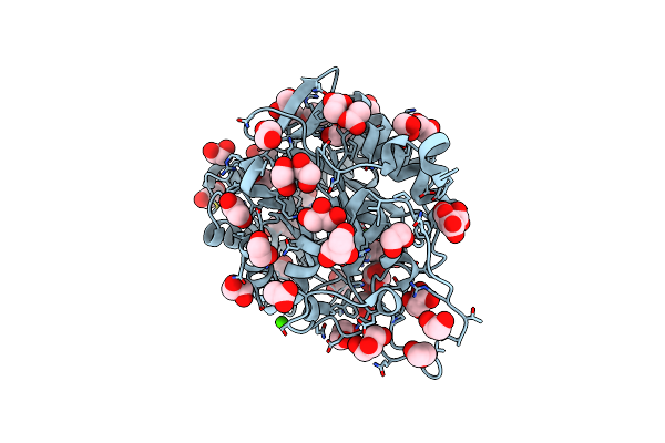
Structure Of T.Fusca Aa10A
Organism: Thermobifida fusca
Method: X-RAY DIFFRACTION Resolution:2.00 Å Release Date: 2017-02-01 Classification: LYASE Ligands: CU, GOL, IOD |
Organism: Thermobifida fusca
Method: X-RAY DIFFRACTION
Release Date: 2017-02-01
Ligands: CU, GOL, IOD
 |
The Unliganded Structure Of Caldicellulosiruptor Saccharolyticus Gh5
Organism: Caldicellulosiruptor saccharolyticus
Method: X-RAY DIFFRACTION Resolution:1.50 Å Release Date: 2016-11-02 Classification: HYDROLASE Ligands: EDO, GOL, MLI, ACT, CA |
Organism: Caldicellulosiruptor saccharolyticus
Method: X-RAY DIFFRACTION
Release Date: 2016-11-02
Ligands: EDO, GOL, MLI, ACT, CA