

Search Count: 14
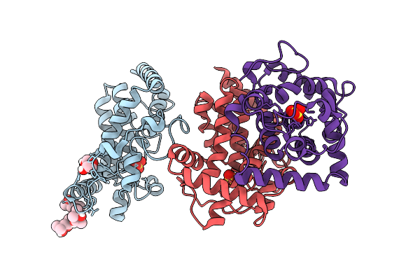 |
Crystal Structure Of Phoc Mutant Acid Phosphatase
Organism: Morganella morganii
Method: X-RAY DIFFRACTION Resolution:2.00 Å Release Date: 2026-01-14 Classification: BIOSYNTHETIC PROTEIN Ligands: MPD, PEG, SO4 |
Organism: Morganella morganii
Method: X-RAY DIFFRACTION
Release Date: 2026-01-14
Ligands: MPD, PEG, SO4
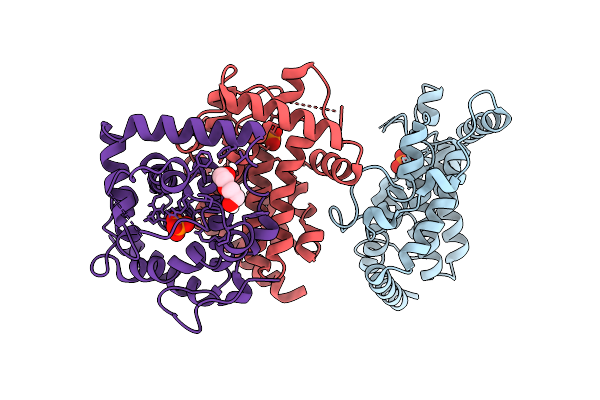 |
Crystal Structure Of Phoc Wild Type Acid Phosphatase
Organism: Morganella morganii
Method: X-RAY DIFFRACTION Resolution:2.24 Å Release Date: 2026-01-14 Classification: BIOSYNTHETIC PROTEIN Ligands: PO4, PEG |
Organism: Morganella morganii
Method: X-RAY DIFFRACTION
Release Date: 2026-01-14
Ligands: PO4, PEG
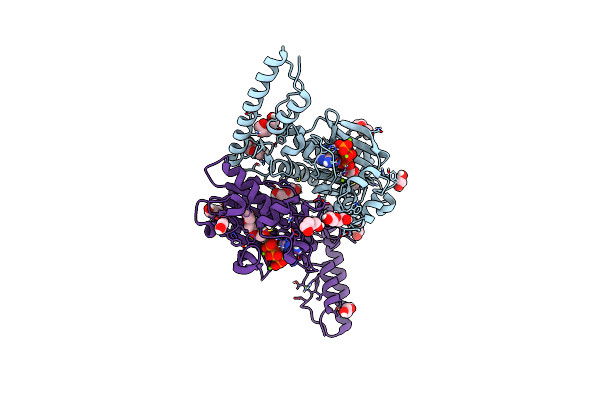 |
Crystal Structure Of Pyrrolysyl-Trna Synthetase From Methanomethylophilus Alvus Engineered For 3-Methyl-L-Histidine, Bound To Amppnp
Organism: Candidatus methanomethylophilus alvus
Method: X-RAY DIFFRACTION Resolution:1.82 Å Release Date: 2023-07-19 Classification: LIGASE Ligands: ANP, PEG, PGE, EDO, MG |
Organism: Candidatus methanomethylophilus alvus
Method: X-RAY DIFFRACTION
Release Date: 2023-07-19
Ligands: ANP, PEG, PGE, EDO, MG
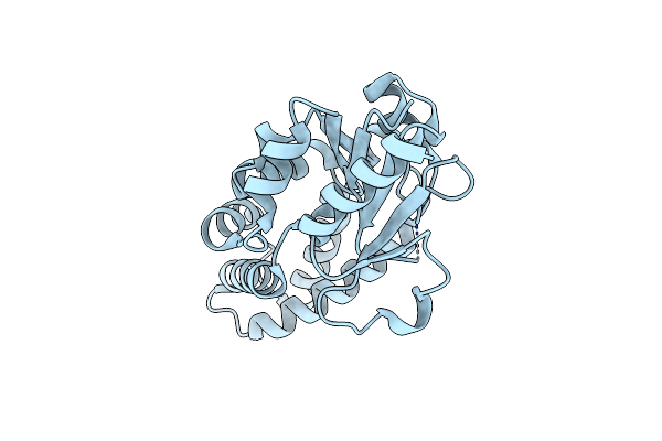 |
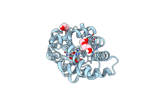
A De Novo Enzyme For The Morita-Baylis-Hillman Reaction Bh32.7
Organism: Synthetic construct
Method: X-RAY DIFFRACTION Resolution:1.80 Å Release Date: 2021-11-03 Classification: BIOSYNTHETIC PROTEIN |
Organism: Synthetic construct
Method: X-RAY DIFFRACTION
Release Date: 2021-11-03
 |
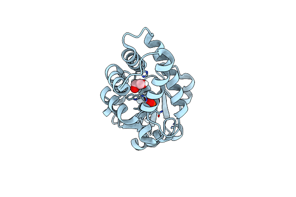
A De Novo Enzyme For The Morita-Baylis-Hillman Reaction Bh32.6
Organism: Synthetic construct
Method: X-RAY DIFFRACTION Resolution:1.48 Å Release Date: 2021-08-25 Classification: BIOSYNTHETIC PROTEIN Ligands: PEG, EDO, CA, FMT |
Organism: Synthetic construct
Method: X-RAY DIFFRACTION
Release Date: 2021-08-25
Ligands: PEG, EDO, CA, FMT
 |
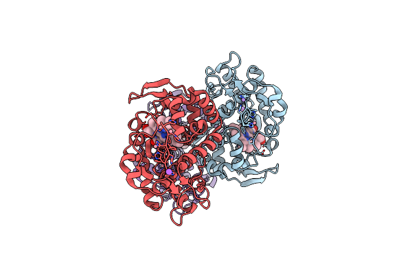
A De Novo Enzyme For The Morita-Baylis-Hillman Reaction Bh32.12
Organism: Synthetic construct
Method: X-RAY DIFFRACTION Resolution:2.29 Å Release Date: 2021-08-25 Classification: BIOSYNTHETIC PROTEIN Ligands: PO4, EDO |
Organism: Synthetic construct
Method: X-RAY DIFFRACTION
Release Date: 2021-08-25
Ligands: PO4, EDO
 |
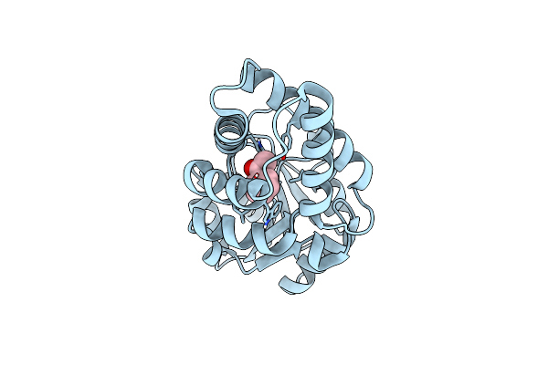
The Crystal Structure Of Engineered Cytochrome C Peroxidase From Saccharomyces Cerevisiae With A His175Me-His Proximal Ligand Substitution
Organism: Saccharomyces cerevisiae
Method: X-RAY DIFFRACTION Resolution:1.90 Å Release Date: 2020-02-12 Classification: OXIDOREDUCTASE Ligands: HEM, CO, MN, NA |
Organism: Saccharomyces cerevisiae
Method: X-RAY DIFFRACTION
Release Date: 2020-02-12
Ligands: HEM, CO, MN, NA
 |
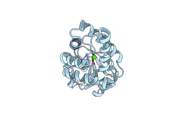
Crystal Structure Of Bh32 Alkylated With The Mechanistic Inhibitor 2-Bromoacetophenone
Organism: Pyrococcus horikoshii
Method: X-RAY DIFFRACTION Resolution:2.02 Å Release Date: 2019-06-05 Classification: HYDROLASE Ligands: AC0 |
Organism: Pyrococcus horikoshii
Method: X-RAY DIFFRACTION
Release Date: 2019-06-05
Ligands: AC0
 |
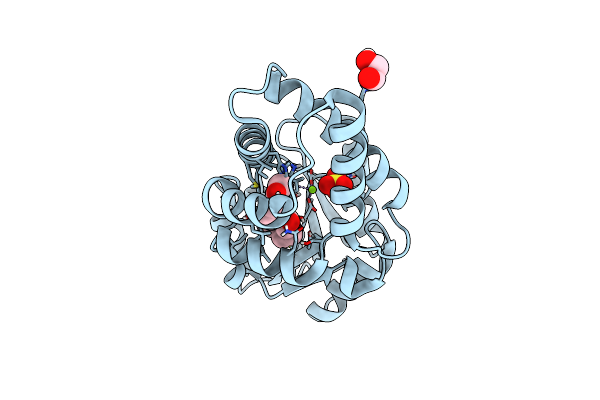
Crystal Structure Of Oe1
Organism: Pyrococcus horikoshii
Method: X-RAY DIFFRACTION Resolution:2.00 Å Release Date: 2019-06-05 Classification: HYDROLASE Ligands: CA |
Organism: Pyrococcus horikoshii
Method: X-RAY DIFFRACTION
Release Date: 2019-06-05
Ligands: CA
 |
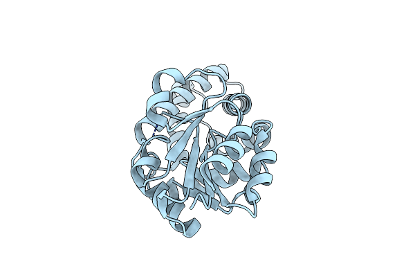
Crystal Structure Of Oe1.2
Organism: Pyrococcus horikoshii
Method: X-RAY DIFFRACTION Resolution:1.96 Å Release Date: 2019-06-05 Classification: HYDROLASE Ligands: PGE, AC0, SO4, EDO, MG |
Organism: Pyrococcus horikoshii
Method: X-RAY DIFFRACTION
Release Date: 2019-06-05
Ligands: PGE, AC0, SO4, EDO, MG
 |
Crystal Structure Of Oe1.3
Organism: Pyrococcus horikoshii
Method: X-RAY DIFFRACTION Resolution:1.90 Å Release Date: 2019-06-05 Classification: HYDROLASE |
Organism: Pyrococcus horikoshii
Method: X-RAY DIFFRACTION
Release Date: 2019-06-05
 |
Crystal Structure Of Oe1.3 Alkylated With The Mechanistic Inhibitor 2-Bromoacetophenone
Organism: Pyrococcus horikoshii
Method: X-RAY DIFFRACTION Resolution:1.50 Å Release Date: 2019-06-05 Classification: HYDROLASE Ligands: AC0, ACT, PEG |
Organism: Pyrococcus horikoshii
Method: X-RAY DIFFRACTION
Release Date: 2019-06-05
Ligands: AC0, ACT, PEG
 |
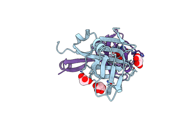
Structure Of Amide Bond Synthetase Mcba K483A Mutant From Marinactinospora Thermotolerans
Organism: Marinactinospora thermotolerans
Method: X-RAY DIFFRACTION Resolution:2.80 Å Release Date: 2018-09-05 Classification: LIGASE Ligands: AMP, EQ2 |
Organism: Marinactinospora thermotolerans
Method: X-RAY DIFFRACTION
Release Date: 2018-09-05
Ligands: AMP, EQ2
 |
T57V Mutant Of Aspartate Decarboxylase
Organism: Escherichia coli
Method: X-RAY DIFFRACTION Resolution:1.62 Å Release Date: 2012-07-25 Classification: LYASE Ligands: MLI |
Organism: Escherichia coli
Method: X-RAY DIFFRACTION
Release Date: 2012-07-25
Ligands: MLI