

Search Count: 21
 |
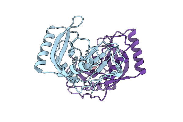
Phosphodiesterase From Burkholderia Phage Bcsr5 In The Closed Lid Conformation
Organism: Burkholderia phage bcsr5
Method: X-RAY DIFFRACTION Resolution:2.00 Å Release Date: 2025-11-05 Classification: VIRAL PROTEIN Ligands: FLC |
Organism: Burkholderia phage bcsr5
Method: X-RAY DIFFRACTION
Release Date: 2025-11-05
Ligands: FLC
 |
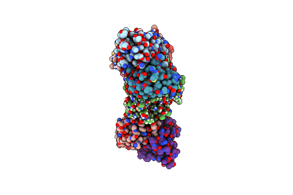
Phosphodiesterase From Burkholderia Phage Bcsr5 In The Open Lid Conformation
Organism: Burkholderia phage bcsr5
Method: X-RAY DIFFRACTION Resolution:2.03 Å Release Date: 2025-11-05 Classification: VIRAL PROTEIN Ligands: CAC |
Organism: Burkholderia phage bcsr5
Method: X-RAY DIFFRACTION
Release Date: 2025-11-05
Ligands: CAC
 |
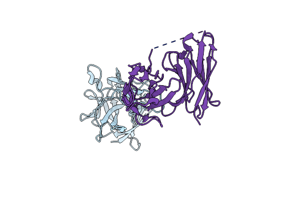
Hastv1 Spike In Complex With Neutralizing Fabs 3H4 And 3B4
Organism: Mus musculus, Human astrovirus 1
Method: ELECTRON MICROSCOPY Release Date: 2024-12-25 Classification: VIRAL PROTEIN/IMMUNE SYSTEM Ligands: NAG |
Organism: Mus musculus, Human astrovirus 1
Method: ELECTRON MICROSCOPY
Release Date: 2024-12-25
Ligands: NAG
 |
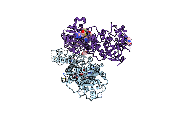
Human Astrovirus 2 Spike Bound To Neutralizing Scfv 4B6
Organism: Human astrovirus 2, Mus musculus
Method: X-RAY DIFFRACTION Resolution:2.67 Å Release Date: 2024-12-25 Classification: VIRAL PROTEIN |
Organism: Human astrovirus 2, Mus musculus
Method: X-RAY DIFFRACTION
Release Date: 2024-12-25
 |
Sars-Cov-2 Nsp14 In Complex With Sah And Tdi-015051
Organism: Sars-cov-2 pseudovirus
Method: X-RAY DIFFRACTION Resolution:2.18 Å Release Date: 2024-10-23 Classification: VIRAL PROTEIN Ligands: ZN, SAH, EDO, IMD, YDT, CL |
Organism: Sars-cov-2 pseudovirus
Method: X-RAY DIFFRACTION
Release Date: 2024-10-23
Ligands: ZN, SAH, EDO, IMD, YDT, CL
 |
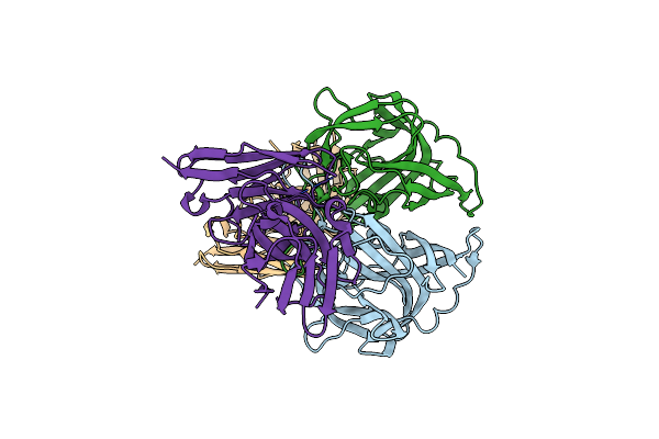
Crystal Structure Of The Human Astrovirus Serotype 8 Capsid Spike In Complex With Scfv 3E8, An Astrovirus-Neutralizing Antibody, At 2.05-A Resolution
Organism: Human astrovirus-8, Mus musculus
Method: X-RAY DIFFRACTION Resolution:2.05 Å Release Date: 2021-10-13 Classification: VIRAL PROTEIN |
Organism: Human astrovirus-8, Mus musculus
Method: X-RAY DIFFRACTION
Release Date: 2021-10-13
 |
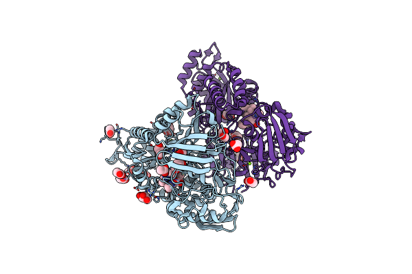
Crystal Structure Of The Human Astrovirus Serotype 8 Capsid Spike In Complex With Scfv 2D9, An Astrovirus-Neutralizing Antibody, At 2.65-A Resolution
Organism: Human astrovirus-8, Mus musculus
Method: X-RAY DIFFRACTION Resolution:2.65 Å Release Date: 2021-10-13 Classification: VIRAL PROTEIN |
Organism: Human astrovirus-8, Mus musculus
Method: X-RAY DIFFRACTION
Release Date: 2021-10-13
 |
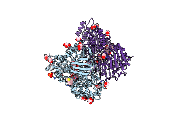
X-Ray Structure Of Nika From Escherichia Coli In Complex With Fe-6-Me2-Bpmcn
Organism: Escherichia coli (strain k12)
Method: X-RAY DIFFRACTION Resolution:1.90 Å Release Date: 2020-11-04 Classification: METAL BINDING PROTEIN Ligands: FE, QTT, ACT, GOL, MG, CL |
Organism: Escherichia coli (strain k12)
Method: X-RAY DIFFRACTION
Release Date: 2020-11-04
Ligands: FE, QTT, ACT, GOL, MG, CL
 |
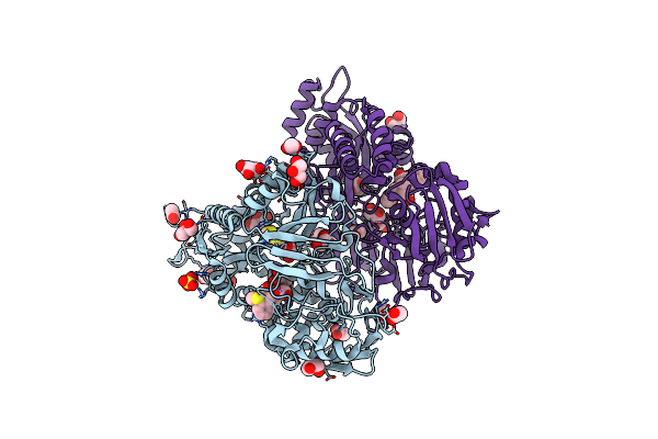
Crystal Structure Of Nika In Complex With Fe-L2 (N-(2-Hydroxy-3Methoxybenzyl)-N-N'-Ethylenediaminediacetic Acid)
Organism: Escherichia coli (strain k12)
Method: X-RAY DIFFRACTION Resolution:1.90 Å Release Date: 2017-12-13 Classification: METAL BINDING PROTEIN Ligands: FE, 9YH, ACT, SO4, GOL, CL |
Organism: Escherichia coli (strain k12)
Method: X-RAY DIFFRACTION
Release Date: 2017-12-13
Ligands: FE, 9YH, ACT, SO4, GOL, CL
 |
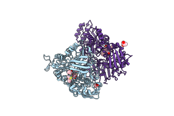
Crystal Structure Of Nika In Complex With Hydroxylated Fe-L1 (N-(2-Hydroxybenzyl)-N'-(2-Thiomethylbenzyl)-N,N'-Ethylenediamine Diacetic Acid)
Organism: Escherichia coli (strain k12)
Method: X-RAY DIFFRACTION Resolution:1.70 Å Release Date: 2017-12-13 Classification: METAL BINDING PROTEIN Ligands: FE, 9YK, ACT, SO4, GOL, CL, DTD |
Organism: Escherichia coli (strain k12)
Method: X-RAY DIFFRACTION
Release Date: 2017-12-13
Ligands: FE, 9YK, ACT, SO4, GOL, CL, DTD
 |
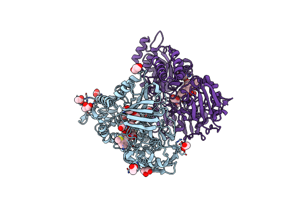
Crystal Structure Of Nika In Complex With Fe-L1 (N-(2-Hydroxybenzyl)-N'-(2-Thiomethylbenzyl)-N,N'-Ethylenediamine Diacetic Acid)
Organism: Escherichia coli (strain k12)
Method: X-RAY DIFFRACTION Resolution:2.30 Å Release Date: 2017-12-13 Classification: METAL BINDING PROTEIN Ligands: FE, 9YK, GOL |
Organism: Escherichia coli (strain k12)
Method: X-RAY DIFFRACTION
Release Date: 2017-12-13
Ligands: FE, 9YK, GOL
 |
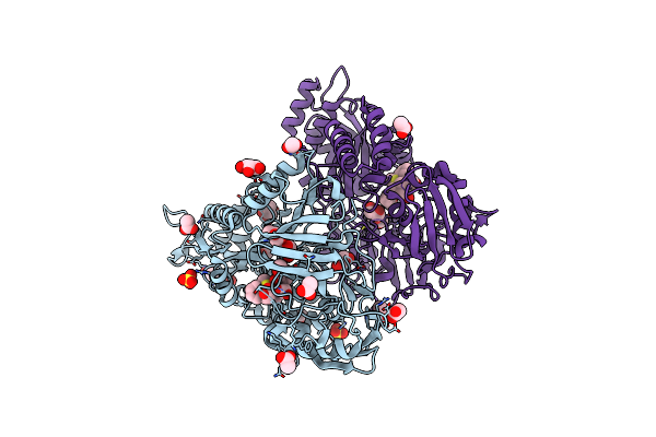
Crystal Structure Of Nika In Complex With Fe-L2 (Fe-L2 (N-(2-Hydroxy-3-Methoxybenzyl)-N'-(2-Thiomethylbenzyl)- N,N'-Ethylenediamine Diacetic Acid) After Dioxygen Oxidation
Organism: Escherichia coli (strain k12)
Method: X-RAY DIFFRACTION Resolution:1.70 Å Release Date: 2017-12-13 Classification: METAL BINDING PROTEIN Ligands: FE, 9YH, GOL, ACT, CL |
Organism: Escherichia coli (strain k12)
Method: X-RAY DIFFRACTION
Release Date: 2017-12-13
Ligands: FE, 9YH, GOL, ACT, CL
 |
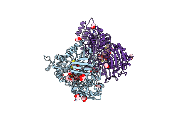
Crystal Structure Of Nika In Complex With Reduced Fe-L2 (N-(2-Hydroxy-3-Methoxybenzyl)-N'-(2-Thiomethylbenzyl)- N,N'-Ethylenediamine Diacetic Acid)
Organism: Escherichia coli (strain k12)
Method: X-RAY DIFFRACTION Resolution:1.60 Å Release Date: 2017-12-13 Classification: METAL BINDING PROTEIN Ligands: FE, 9YH, ACT, GOL, SO4, CL |
Organism: Escherichia coli (strain k12)
Method: X-RAY DIFFRACTION
Release Date: 2017-12-13
Ligands: FE, 9YH, ACT, GOL, SO4, CL
 |
Crystal Structure Of Nika In Complex With Reduced Fe-L1 (N-(2-Hydroxybenzyl)-N'-(2-Thiomethylbenzyl)-N,N'-Ethylenediamine Diacetic Acid)
Organism: Escherichia coli (strain k12)
Method: X-RAY DIFFRACTION Resolution:1.70 Å Release Date: 2017-12-13 Classification: METAL BINDING PROTEIN Ligands: FE, 9YH, DTT, GOL, CL, ACT |
Organism: Escherichia coli (strain k12)
Method: X-RAY DIFFRACTION
Release Date: 2017-12-13
Ligands: FE, 9YH, DTT, GOL, CL, ACT
 |
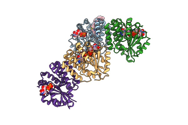
Crystal Structure Of Dck Mutant C3S
Organism: Homo sapiens
Method: X-RAY DIFFRACTION Resolution:3.70 Å Release Date: 2017-11-22 Classification: TRANSFERASE Ligands: UDP, UMP, MG |
Organism: Homo sapiens
Method: X-RAY DIFFRACTION
Release Date: 2017-11-22
Ligands: UDP, UMP, MG
 |
Crystal Structure Of Dck Mutant C3S In Complex With Masitinib And Udp
Organism: Homo sapiens
Method: X-RAY DIFFRACTION Resolution:3.25 Å Release Date: 2017-11-22 Classification: TRANSFERASE Ligands: UDP, G65, DCM, MG |
Organism: Homo sapiens
Method: X-RAY DIFFRACTION
Release Date: 2017-11-22
Ligands: UDP, G65, DCM, MG
 |
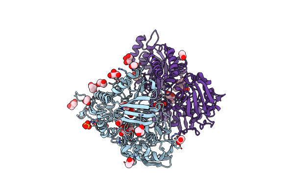
Crystal Structure Of Dck Mutant C3S In Complex With Imatinib And Udp
Organism: Homo sapiens
Method: X-RAY DIFFRACTION Resolution:3.20 Å Release Date: 2017-11-22 Classification: TRANSFERASE Ligands: UDP, STI, MG, DCM |
Organism: Homo sapiens
Method: X-RAY DIFFRACTION
Release Date: 2017-11-22
Ligands: UDP, STI, MG, DCM
 |
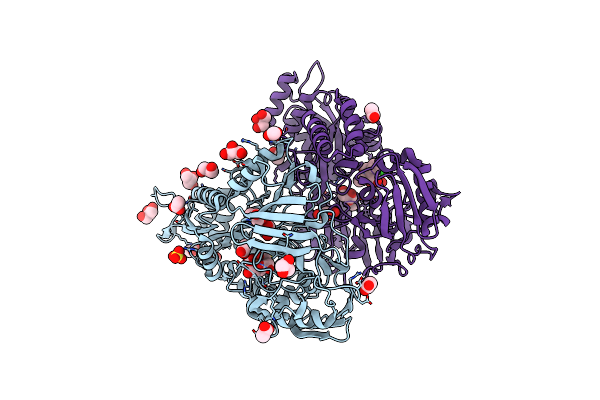
X-Ray Structure Of Nika From Escherichia Coli In Complex With Ru(Bis(Pyrzol-1-Yl)Acetate Scorpionate)(Co)2Cl
Organism: Escherichia coli (strain k12)
Method: X-RAY DIFFRACTION Resolution:1.80 Å Release Date: 2017-03-29 Classification: METAL BINDING PROTEIN Ligands: ACT, SO4, GOL, FE, EDT, RU, CL, 6RP, CMO |
Organism: Escherichia coli (strain k12)
Method: X-RAY DIFFRACTION
Release Date: 2017-03-29
Ligands: ACT, SO4, GOL, FE, EDT, RU, CL, 6RP, CMO
 |
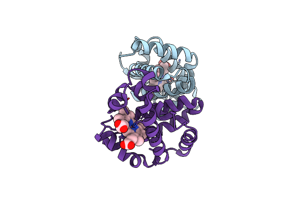
Crystal Structure Of The Periplasmic Nickel-Binding Protein Nika From Escherichia Coli In Complex With Ru(Bpza)(Co)2Cl
Organism: Escherichia coli k12
Method: X-RAY DIFFRACTION Resolution:1.80 Å Release Date: 2017-02-22 Classification: METAL BINDING PROTEIN Ligands: ACT, SO4, GOL, FE, EDT, RU, CL, 6RP, CMO |
Organism: Escherichia coli k12
Method: X-RAY DIFFRACTION
Release Date: 2017-02-22
Ligands: ACT, SO4, GOL, FE, EDT, RU, CL, 6RP, CMO
 |
Structure Of Greyhound Hemoglobin: Origin Of High Oxygen Affinity
Organism: Canis lupus familiaris
Method: X-RAY DIFFRACTION Resolution:1.90 Å Release Date: 2010-11-03 Classification: OXYGEN TRANSPORT Ligands: HEM |
Organism: Canis lupus familiaris
Method: X-RAY DIFFRACTION
Release Date: 2010-11-03
Ligands: HEM