

Search Count: 22
 |
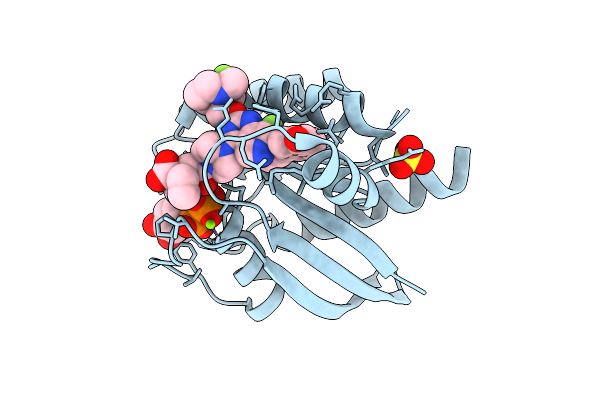
Kras-Wt(1-169) - Gdp In Complex With Compound (R)-1
Organism: Homo sapiens
Method: X-RAY DIFFRACTION Resolution:1.31 Å Release Date: 2025-07-23 Classification: HYDROLASE Ligands: MG, SO4, A1I89, GDP |
Organism: Homo sapiens
Method: X-RAY DIFFRACTION
Release Date: 2025-07-23
Ligands: MG, SO4, A1I89, GDP
 |
Kras-G12D(1-169) - Gdp In Covalent Complex With Compound (3R,4R)-3
Organism: Homo sapiens
Method: X-RAY DIFFRACTION Resolution:1.55 Å Release Date: 2025-07-23 Classification: HYDROLASE Ligands: MG, SO4, A1I9E, GDP |
Organism: Homo sapiens
Method: X-RAY DIFFRACTION
Release Date: 2025-07-23
Ligands: MG, SO4, A1I9E, GDP
 |
Kras-G12D(1-169) - Gdp In Covalent Complex With Compound (3S,4R)-8
Organism: Homo sapiens
Method: X-RAY DIFFRACTION Resolution:1.30 Å Release Date: 2025-07-23 Classification: HYDROLASE Ligands: MG, A1I9F, GDP |
Organism: Homo sapiens
Method: X-RAY DIFFRACTION
Release Date: 2025-07-23
Ligands: MG, A1I9F, GDP
 |
Structure Of Human Shoc2 In Complex With A Small Molecule Inhibitor (R)-5
Organism: Homo sapiens
Method: X-RAY DIFFRACTION Resolution:2.68 Å Release Date: 2025-06-25 Classification: STRUCTURAL PROTEIN Ligands: A1CF1, K |
Organism: Homo sapiens
Method: X-RAY DIFFRACTION
Release Date: 2025-06-25
Ligands: A1CF1, K
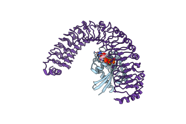 |
Nras 1-169 Q61R In Complex With Shoc2 80-582
Organism: Homo sapiens
Method: X-RAY DIFFRACTION Resolution:2.73 Å Release Date: 2025-04-02 Classification: SIGNALING PROTEIN/HYDROLASE Ligands: GTP, MG |
Organism: Homo sapiens
Method: X-RAY DIFFRACTION
Release Date: 2025-04-02
Ligands: GTP, MG
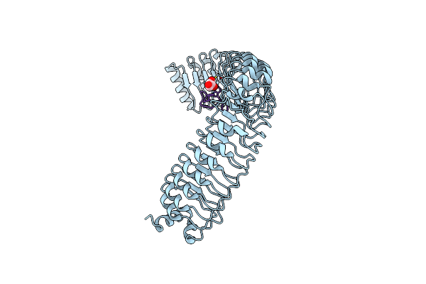 |
Structure Of Human Shoc2 In Complex With A Cyclic Peptide
Organism: Homo sapiens, Synthetic construct
Method: X-RAY DIFFRACTION Resolution:2.04 Å Release Date: 2025-04-02 Classification: STRUCTURAL PROTEIN Ligands: GOL |
Organism: Homo sapiens, Synthetic construct
Method: X-RAY DIFFRACTION
Release Date: 2025-04-02
Ligands: GOL
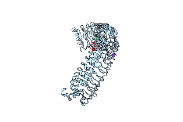 |
Structure Of Human Shoc2 In Complex With A Small Molecule Inhibitor (S)-5
Organism: Homo sapiens
Method: X-RAY DIFFRACTION Resolution:2.37 Å Release Date: 2025-04-02 Classification: STRUCTURAL PROTEIN Ligands: A1ATZ, K |
Organism: Homo sapiens
Method: X-RAY DIFFRACTION
Release Date: 2025-04-02
Ligands: A1ATZ, K
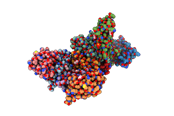 |
Crystal Structure Of Cnab2 Domain From Lactobacillus Plantarum
Organism: Lactiplantibacillus plantarum
Method: X-RAY DIFFRACTION Resolution:1.86 Å Release Date: 2023-02-22 Classification: PEPTIDE BINDING PROTEIN Ligands: GOL, SO4, TRS |
Organism: Lactiplantibacillus plantarum
Method: X-RAY DIFFRACTION
Release Date: 2023-02-22
Ligands: GOL, SO4, TRS
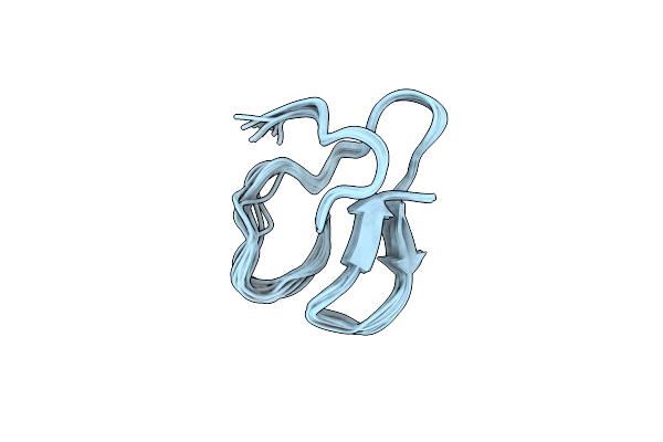 |
Solution Structure Of The Cellulose-Binding Domain Of Endoglucanase I From Trichoderma Reesei And Its Interaction With Cello- Oligosaccharides
|
Organism: Hypocrea jecorina
Method: SOLUTION NMR
Release Date: 2013-05-22
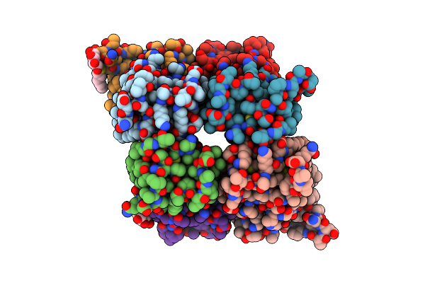 |
Monoclinic Crystal Structure Of Hydrophobin Hfbii In Presence Of A Detergent
Organism: Hypocrea jecorina
Method: X-RAY DIFFRACTION Resolution:2.20 Å Release Date: 2007-07-17 Classification: STRUCTURAL PROTEIN Ligands: HTG |
Organism: Hypocrea jecorina
Method: X-RAY DIFFRACTION
Release Date: 2007-07-17
Ligands: HTG
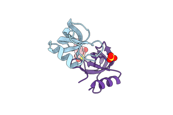 |
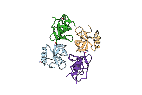
Orhorhombic Crystal Structure Of Hydrophobin Hfbii In The Presence Of A Detergent
Organism: Hypocrea jecorina
Method: X-RAY DIFFRACTION Resolution:1.00 Å Release Date: 2007-07-17 Classification: SURFACE ACTIVE PROTEIN Ligands: SO4, HTG |
Organism: Hypocrea jecorina
Method: X-RAY DIFFRACTION
Release Date: 2007-07-17
Ligands: SO4, HTG
 |
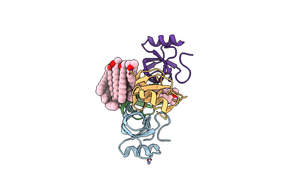
Crystal Structure Of Hydrophobin Hfbi
Organism: Hypocrea jecorina
Method: X-RAY DIFFRACTION Resolution:2.10 Å Release Date: 2006-08-15 Classification: SURFACE ACTIVE PROTEIN Ligands: ZN |
Organism: Hypocrea jecorina
Method: X-RAY DIFFRACTION
Release Date: 2006-08-15
Ligands: ZN
 |
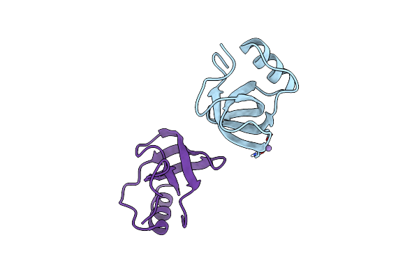
Crystal Structure Of Hydrophobin Hfbi With Detergent
Organism: Hypocrea jecorina
Method: X-RAY DIFFRACTION Resolution:2.30 Å Release Date: 2006-08-15 Classification: SURFACE ACTIVE PROTEIN Ligands: ZN, LDA |
Organism: Hypocrea jecorina
Method: X-RAY DIFFRACTION
Release Date: 2006-08-15
Ligands: ZN, LDA
 |
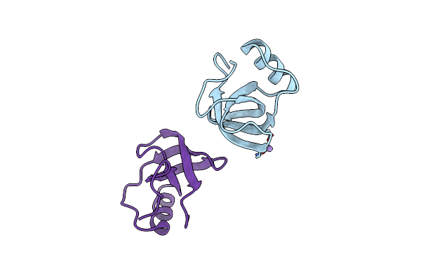
Ultra-High Resolution Structure Of Hydrophobin Hfbii
Organism: Hypocrea jecorina
Method: X-RAY DIFFRACTION Resolution:0.75 Å Release Date: 2006-03-28 Classification: SURFACE ACTIVE PROTEIN Ligands: MN |
Organism: Hypocrea jecorina
Method: X-RAY DIFFRACTION
Release Date: 2006-03-28
Ligands: MN
 |
Atomic Resolution Structure Of The Hfbii Hydrophobin: A Self-Assembling Amphiphile
Organism: Hypocrea jecorina
Method: X-RAY DIFFRACTION Resolution:1.00 Å Release Date: 2004-01-13 Classification: surface active protein Ligands: MN |
Organism: Hypocrea jecorina
Method: X-RAY DIFFRACTION
Release Date: 2004-01-13
Ligands: MN
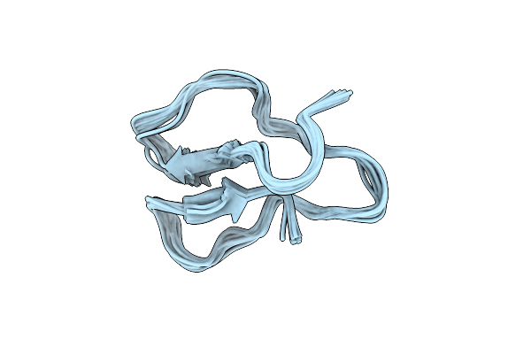 |
Three-Dimensional Structures Of Three Engineered Cellulose-Binding Domains Of Cellobiohydrolase I From Trichoderma Reesei, Nmr, 18 Structures
|
Organism: Hypocrea jecorina
Method: SOLUTION NMR
Release Date: 1998-04-29
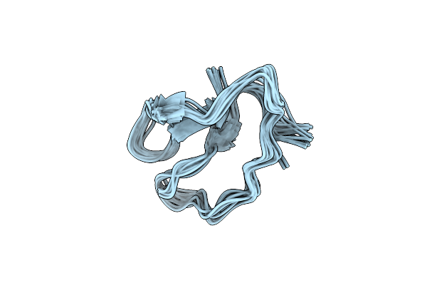 |
Three-Dimensional Structures Of Three Engineered Cellulose-Binding Domains Of Cellobiohydrolase I From Trichoderma Reesei, Nmr, 19 Structures
|
Organism: Hypocrea jecorina
Method: SOLUTION NMR
Release Date: 1998-04-29
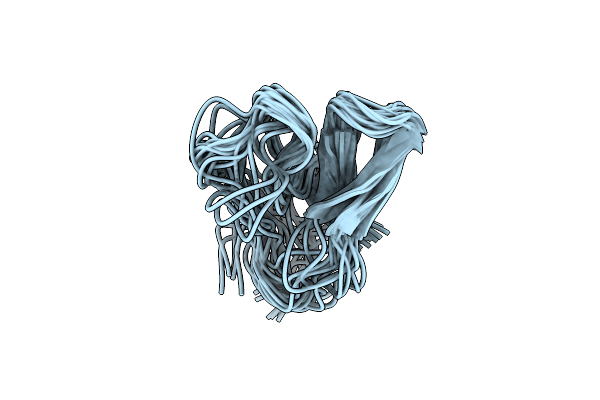 |
Three-Dimensional Structures Of Three Engineered Cellulose-Binding Domains Of Cellobiohydrolase I From Trichoderma Reesei, Nmr, 23 Structures
|
Organism: Hypocrea jecorina
Method: SOLUTION NMR
Release Date: 1998-04-08
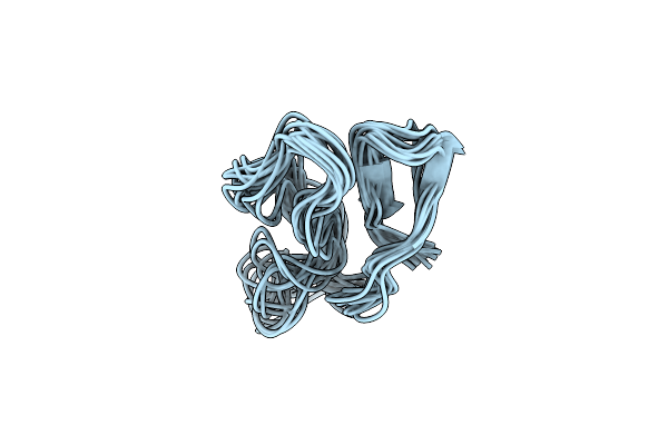 |
Three-Dimensional Structures Of Three Engineered Cellulose-Binding Domains Of Cellobiohydrolase I From Trichoderma Reesei, Nmr, 14 Structures
|
Organism: Hypocrea jecorina
Method: SOLUTION NMR
Release Date: 1998-04-08
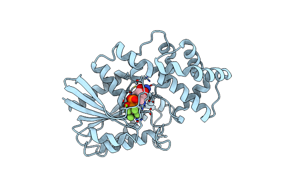 |
Structures Of Active Conformations Of Gi Alpha 1 And The Mechanism Of Gtp Hydrolysis
Organism: Rattus norvegicus
Method: X-RAY DIFFRACTION Resolution:2.20 Å Release Date: 1995-03-31 Classification: SIGNAL TRANSDUCTION PROTEIN Ligands: MG, ALF, GDP |
Organism: Rattus norvegicus
Method: X-RAY DIFFRACTION
Release Date: 1995-03-31
Ligands: MG, ALF, GDP