

Search Count: 23
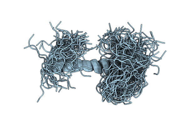 |
Serf1_Human Short Isoform Of Small Edrk-Rich Factor 1, Serf1A At Ph 6.0.
Organism: Homo sapiens
Method: SOLUTION NMR Release Date: 2026-01-14 Classification: NUCLEAR PROTEIN |
Organism: Homo sapiens
Method: SOLUTION NMR
Release Date: 2026-01-14
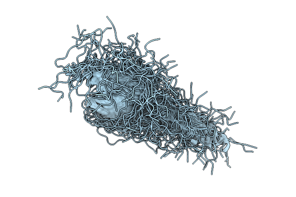 |
Serf1_Human Short Isoform Of Small Edrk-Rich Factor 1, Serf1A At Ph 6.8.
Organism: Homo sapiens
Method: SOLUTION NMR Release Date: 2026-01-14 Classification: NUCLEAR PROTEIN |
Organism: Homo sapiens
Method: SOLUTION NMR
Release Date: 2026-01-14
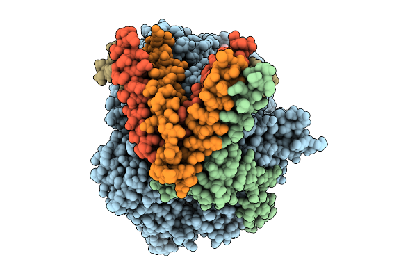 |
Cryoem Structure Of Respiratory Syncytial Virus Polymerase In Complex With Novel Non-Nucleoside Inhibitor Compound 16
Organism: Human respiratory syncytial virus, Synthetic construct
Method: ELECTRON MICROSCOPY Release Date: 2025-09-24 Classification: VIRAL PROTEIN,TRANSFERASE/INHIBITOR |
Organism: Human respiratory syncytial virus, Synthetic construct
Method: ELECTRON MICROSCOPY
Release Date: 2025-09-24
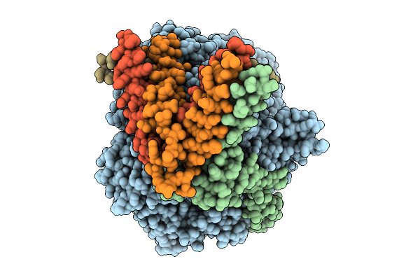 |
Cryoem Map Of Respiratory Syncytial Virus Polymerase With Non-Nucleoside Inhibitor Compound 21
Organism: Human respiratory syncytial virus, Synthetic construct
Method: ELECTRON MICROSCOPY Release Date: 2025-09-24 Classification: VIRAL PROTEIN/INHIBITOR |
Organism: Human respiratory syncytial virus, Synthetic construct
Method: ELECTRON MICROSCOPY
Release Date: 2025-09-24
 |
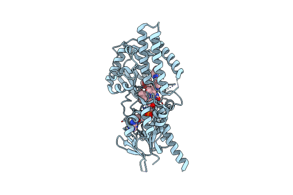
Cryoem Structure Of Respiratory Syncytial Virus Polymerase With Novel Non-Nucleoside Inhibitor Compound 22
Organism: Human respiratory syncytial virus
Method: ELECTRON MICROSCOPY Release Date: 2025-05-28 Classification: TRANSFERASE/INHIBITOR Ligands: A1BVR |
Organism: Human respiratory syncytial virus
Method: ELECTRON MICROSCOPY
Release Date: 2025-05-28
Ligands: A1BVR
 |
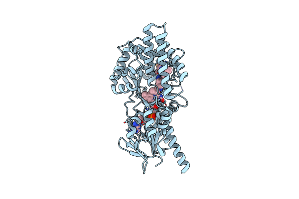
Crystal Structure Of Human Spermine Oxidase
Organism: Homo sapiens
Method: X-RAY DIFFRACTION Resolution:2.40 Å Release Date: 2022-07-13 Classification: UNKNOWN FUNCTION Ligands: 6YU, CL, MPD |
Organism: Homo sapiens
Method: X-RAY DIFFRACTION
Release Date: 2022-07-13
Ligands: 6YU, CL, MPD
 |
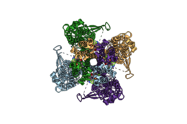
Structure Of Human Spermine Oxidase In Complex With A Highly Selective Allosteric Inhibitor
Organism: Homo sapiens
Method: X-RAY DIFFRACTION Resolution:2.09 Å Release Date: 2022-07-13 Classification: UNKNOWN FUNCTION Ligands: 6YU, 3EI, CL |
Organism: Homo sapiens
Method: X-RAY DIFFRACTION
Release Date: 2022-07-13
Ligands: 6YU, 3EI, CL
 |
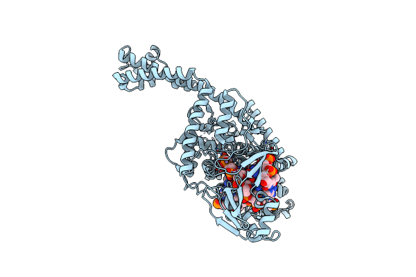
Structure Of Human Trpa1 In Complex With Inhibitor Gdc-0334
Organism: Homo sapiens
Method: ELECTRON MICROSCOPY Release Date: 2021-02-17 Classification: MEMBRANE PROTEIN/Inhibitor Ligands: LXY |
Organism: Homo sapiens
Method: ELECTRON MICROSCOPY
Release Date: 2021-02-17
Ligands: LXY
 |
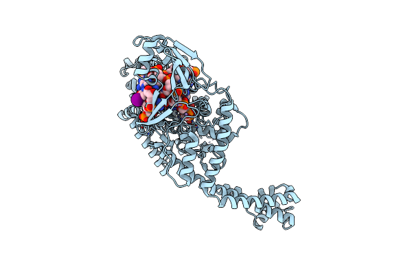
Crystal Structure Of Human Topoisomerase Ii Beta In Complex With Dna: A New Quaternary Conformation Showing Opening Of The Protein-Linked Dna-Gate
Organism: Homo sapiens, Synthetic construct
Method: X-RAY DIFFRACTION Resolution:2.75 Å Release Date: 2018-08-08 Classification: ISOMERASE/DNA Ligands: MN |
Organism: Homo sapiens, Synthetic construct
Method: X-RAY DIFFRACTION
Release Date: 2018-08-08
Ligands: MN
 |
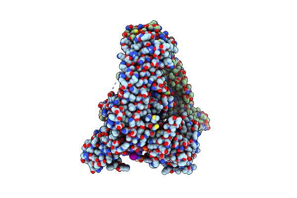
Crystal Structure Of Human Topoisomerase Ii Beta In Complex With 5-Iodouridine-Containing-Dna In Space Group P3221
Organism: Homo sapiens, Synthetic construct
Method: X-RAY DIFFRACTION Resolution:3.87 Å Release Date: 2018-08-08 Classification: ISOMERASE/DNA Ligands: MN |
Organism: Homo sapiens, Synthetic construct
Method: X-RAY DIFFRACTION
Release Date: 2018-08-08
Ligands: MN
 |
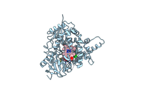
Crystal Structure Of Human Topoisomerase Ii Beta In Complex With 5-Iodouridine-Containing-Dna And Etoposide In Space Group P21
Organism: Homo sapiens, Synthetic construct
Method: X-RAY DIFFRACTION Resolution:2.30 Å Release Date: 2018-08-08 Classification: ISOMERASE/DNA Ligands: MG, EVP |
Organism: Homo sapiens, Synthetic construct
Method: X-RAY DIFFRACTION
Release Date: 2018-08-08
Ligands: MG, EVP
 |
Sterol 14 Alpha-Demethylase (Cyp51) From Trypanosoma Cruzi In Complex With The Pyridine Inhibitor (S)-2-(4-Chlorophenyl)-2-(Pyridin-3-Yl)-1- (4-(4-(Trifluoromethyl)Phenyl)Piperazin-1-Yl)Ethanone (Epl-Bs1246,Udo)
Organism: Trypanosoma cruzi
Method: X-RAY DIFFRACTION Resolution:2.80 Å Release Date: 2013-09-25 Classification: OXIDOREDUCTASE Ligands: HEM, UDO |
Organism: Trypanosoma cruzi
Method: X-RAY DIFFRACTION
Release Date: 2013-09-25
Ligands: HEM, UDO
 |
Sterol 14-Alpha Demethylase (Cyp51)From Trypanosoma Cruzi In Complex With The Pyridine Inhibitor N-(1-(5-(Trifluoromethyl)(Pyridin-2-Yl)) Piperidin-4Yl)-N-(4-(Trifluoromethyl)Phenyl)Pyridin-3-Amine (Epl- Bs967, Udd)
Organism: Trypanosoma cruzi
Method: X-RAY DIFFRACTION Resolution:2.90 Å Release Date: 2013-09-25 Classification: OXIDOREDUCTASE Ligands: HEM, UDD |
Organism: Trypanosoma cruzi
Method: X-RAY DIFFRACTION
Release Date: 2013-09-25
Ligands: HEM, UDD
 |
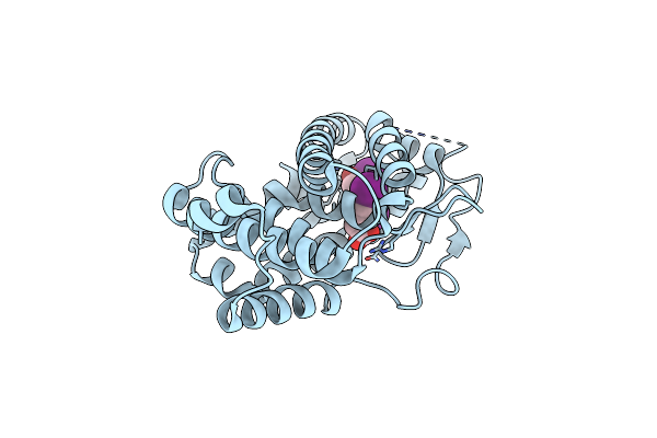
Crystal Structure Of Tr-Alfa Bound To The Selective Thyromimetic Triac
Organism: Homo sapiens
Method: X-RAY DIFFRACTION Resolution:2.01 Å Release Date: 2009-12-08 Classification: TRANSCRIPTION Ligands: 4HY |
Organism: Homo sapiens
Method: X-RAY DIFFRACTION
Release Date: 2009-12-08
Ligands: 4HY
 |
Crystal Structure Of Tr-Beta Bound To The Selective Thyromimetic Triac
Organism: Homo sapiens
Method: X-RAY DIFFRACTION Resolution:2.50 Å Release Date: 2009-12-08 Classification: TRANSCRIPTION Ligands: 4HY |
Organism: Homo sapiens
Method: X-RAY DIFFRACTION
Release Date: 2009-12-08
Ligands: 4HY
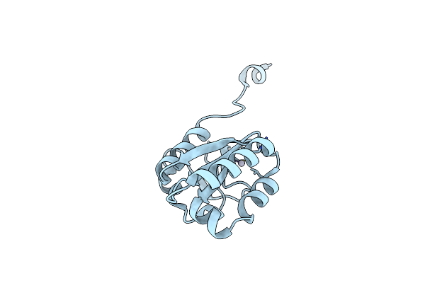 |
Crystal Structure Of The Central Domain Of Human Ercc1
Organism: Homo sapiens
Method: X-RAY DIFFRACTION Resolution:1.90 Å Release Date: 2005-08-02 Classification: DNA BINDING PROTEIN Ligands: HG |
Organism: Homo sapiens
Method: X-RAY DIFFRACTION
Release Date: 2005-08-02
Ligands: HG
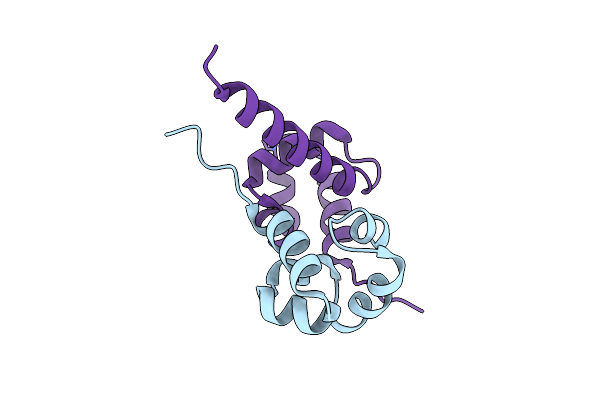 |
Crystal Structure Of The Complex Between The C-Terminal Domains Of Human Xpf And Ercc1
Organism: Homo sapiens
Method: X-RAY DIFFRACTION Resolution:2.70 Å Release Date: 2005-08-02 Classification: DNA BINDING PROTEIN Ligands: HG |
Organism: Homo sapiens
Method: X-RAY DIFFRACTION
Release Date: 2005-08-02
Ligands: HG
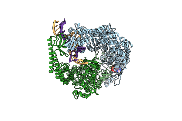 |
The Crystal Structure Of E. Coli Muts Binding To Dna With A C:A Mismatch
Organism: Escherichia coli, Synthetic construct
Method: X-RAY DIFFRACTION Resolution:2.90 Å Release Date: 2003-08-08 Classification: DNA BINDING Ligands: ADP, MG |
Organism: Escherichia coli, Synthetic construct
Method: X-RAY DIFFRACTION
Release Date: 2003-08-08
Ligands: ADP, MG
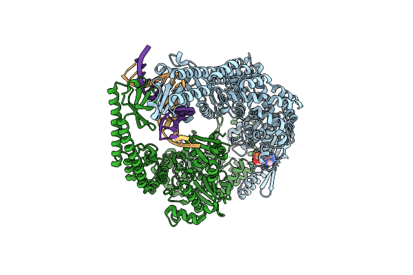 |
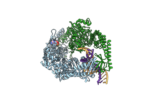
The Crystal Structure Of E. Coli Muts Binding To Dna With An A:A Mismatch
Organism: Escherichia coli, Synthetic construct
Method: X-RAY DIFFRACTION Resolution:2.40 Å Release Date: 2003-08-08 Classification: DNA BINDING Ligands: ADP, MG |
Organism: Escherichia coli, Synthetic construct
Method: X-RAY DIFFRACTION
Release Date: 2003-08-08
Ligands: ADP, MG
 |
The Crystal Structure Of E. Coli Muts Binding To Dna With A G:G Mismatch
Organism: Escherichia coli, Synthetic construct
Method: X-RAY DIFFRACTION Resolution:2.50 Å Release Date: 2003-08-08 Classification: DNA BINDING Ligands: ADP, MG |
Organism: Escherichia coli, Synthetic construct
Method: X-RAY DIFFRACTION
Release Date: 2003-08-08
Ligands: ADP, MG