

Search Count: 11
 |
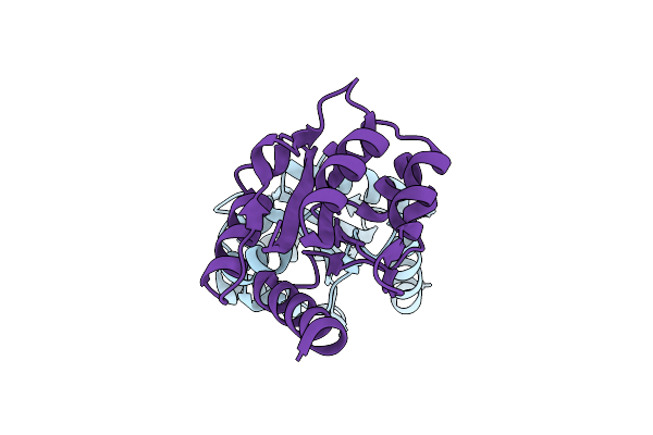
The Crystal Structure Of The Tir Domain-Containing Protein From Acinetobacter Baumannii (Abtir)
Organism: Acinetobacter baumannii
Method: X-RAY DIFFRACTION Resolution:2.16 Å Release Date: 2022-09-07 Classification: HYDROLASE Ligands: SO4, P6G |
Organism: Acinetobacter baumannii
Method: X-RAY DIFFRACTION
Release Date: 2022-09-07
Ligands: SO4, P6G
 |
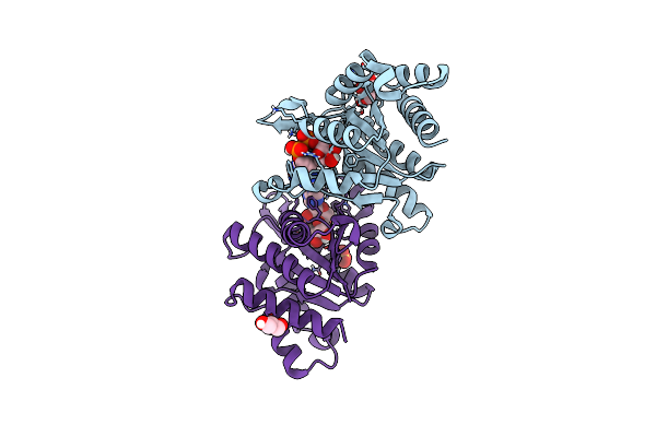
Crystal Structure Of The Bttir Tir Domain
Organism: Bacteroides thetaiotaomicron
Method: X-RAY DIFFRACTION Resolution:1.42 Å Release Date: 2022-09-07 Classification: HYDROLASE |
Organism: Bacteroides thetaiotaomicron
Method: X-RAY DIFFRACTION
Release Date: 2022-09-07
 |
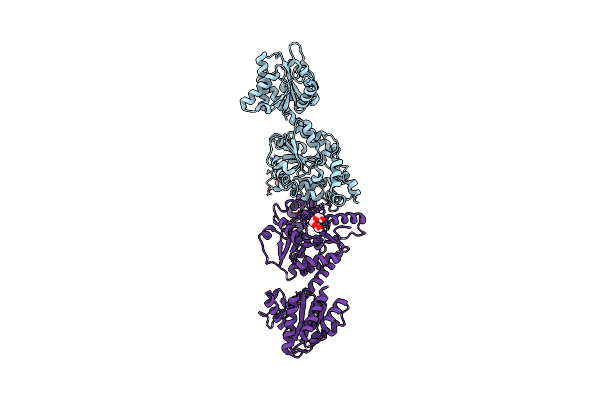
Crystal Structure Of The Bcthsa Slog Domain In Complex With 3'Cadpr
Organism: Bacillus subtilis
Method: X-RAY DIFFRACTION Resolution:1.57 Å Release Date: 2022-09-07 Classification: HYDROLASE Ligands: OJC, GOL, SO4 |
Organism: Bacillus subtilis
Method: X-RAY DIFFRACTION
Release Date: 2022-09-07
Ligands: OJC, GOL, SO4
 |
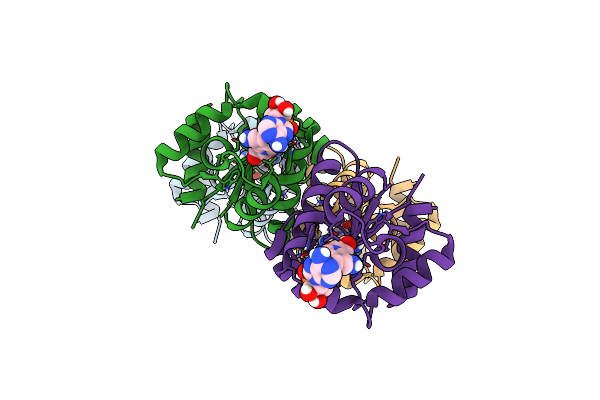
Crystal Structure Of Ligand-Free Sethsa
Organism: Streptococcus equi
Method: X-RAY DIFFRACTION Resolution:3.40 Å Release Date: 2022-09-07 Classification: HYDROLASE Ligands: PGE, GOL |
Organism: Streptococcus equi
Method: X-RAY DIFFRACTION
Release Date: 2022-09-07
Ligands: PGE, GOL
 |
Cryoem Structure Of The Tir Domain From Abtir In Complex With 3Ad
Organism: Acinetobacter baumannii
Method: ELECTRON MICROSCOPY Release Date: 2022-09-07 Classification: HYDROLASE Ligands: 1O4 |
Organism: Acinetobacter baumannii
Method: ELECTRON MICROSCOPY
Release Date: 2022-09-07
Ligands: 1O4
 |
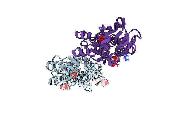
Crystal Structure Of The Molybdate-Binding Periplasmic Protein Moda From The Bacteria Pseudomonsa Aeruginosa In Ligand-Free Form
Organism: Pseudomonas aeruginosa pa1
Method: X-RAY DIFFRACTION Resolution:1.78 Å Release Date: 2022-05-04 Classification: METAL BINDING PROTEIN Ligands: GOL, NH4, SO4 |
Organism: Pseudomonas aeruginosa pa1
Method: X-RAY DIFFRACTION
Release Date: 2022-05-04
Ligands: GOL, NH4, SO4
 |
Crystal Structure Of The Molybdate-Binding Periplasmic Protein Moda From The Bacteria Pseudomonsa Aeruginosa In Chromate-Bound Form
Organism: Pseudomonas aeruginosa pa1
Method: X-RAY DIFFRACTION Resolution:1.90 Å Release Date: 2022-05-04 Classification: METAL BINDING PROTEIN Ligands: CQ4, NH4, GOL |
Organism: Pseudomonas aeruginosa pa1
Method: X-RAY DIFFRACTION
Release Date: 2022-05-04
Ligands: CQ4, NH4, GOL
 |
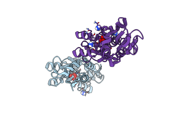
Crystal Structure Of The Molybdate-Binding Periplasmic Protein Moda From The Bacteria Pseudomonsa Aeruginosa In Molybdate-Bound Form
Organism: Pseudomonas aeruginosa pa1
Method: X-RAY DIFFRACTION Resolution:2.50 Å Release Date: 2022-05-04 Classification: METAL BINDING PROTEIN Ligands: NH4, MOO, GOL |
Organism: Pseudomonas aeruginosa pa1
Method: X-RAY DIFFRACTION
Release Date: 2022-05-04
Ligands: NH4, MOO, GOL
 |
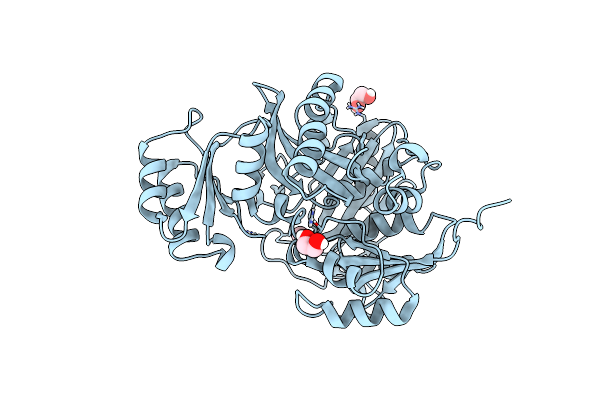
Crystal Structure Of The Molybdate-Binding Periplasmic Protein Moda From The Bacteria Pseudomonsa Aeruginosa In Tungstate-Bound Form
Organism: Pseudomonas aeruginosa pa1
Method: X-RAY DIFFRACTION Resolution:2.16 Å Release Date: 2022-05-04 Classification: METAL BINDING PROTEIN Ligands: WO4, NH4 |
Organism: Pseudomonas aeruginosa pa1
Method: X-RAY DIFFRACTION
Release Date: 2022-05-04
Ligands: WO4, NH4
 |
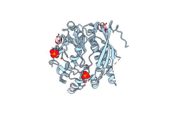
Cryptococcus Neoformans Gar Synthetase
Organism: Cryptococcus neoformans var. grubii serotype a
Method: X-RAY DIFFRACTION Resolution:2.00 Å Release Date: 2021-08-25 Classification: BIOSYNTHETIC PROTEIN, LIGASE Ligands: EDO |
Organism: Cryptococcus neoformans var. grubii serotype a
Method: X-RAY DIFFRACTION
Release Date: 2021-08-25
Ligands: EDO
 |
Cryptococcus Neoformans Air Synthetase
Organism: Cryptococcus neoformans var. grubii serotype a
Method: X-RAY DIFFRACTION Resolution:2.24 Å Release Date: 2021-08-25 Classification: BIOSYNTHETIC PROTEIN, LIGASE Ligands: EDO, SIN, PO4, GLY |
Organism: Cryptococcus neoformans var. grubii serotype a
Method: X-RAY DIFFRACTION
Release Date: 2021-08-25
Ligands: EDO, SIN, PO4, GLY