

Search Count: 21
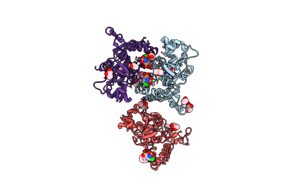 |
Crystal Structure Of The Iglur2 Ligand-Binding Core (S1S2J-N754S) In Complex With Glutamate And Cyclothiazide At 2.25 A Resolution
Organism: Rattus norvegicus
Method: X-RAY DIFFRACTION Resolution:2.25 Å Release Date: 2009-07-28 Classification: MEMBRANE PROTEIN Ligands: GLU, CYZ, ZN, GOL, ACT, DMS, CAC |
Organism: Rattus norvegicus
Method: X-RAY DIFFRACTION
Release Date: 2009-07-28
Ligands: GLU, CYZ, ZN, GOL, ACT, DMS, CAC
 |
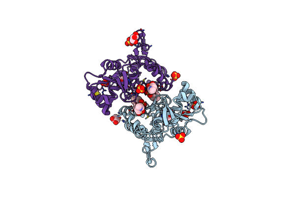
Crystal Structure Of The Iglur2 Ligand-Binding Core (S1S2J-N754S) In Complex With Glutamate And Ns1493 At 1.85 A Resolution
Organism: Rattus norvegicus
Method: X-RAY DIFFRACTION Resolution:1.85 Å Release Date: 2009-07-28 Classification: MEMBRANE PROTEIN Ligands: GLU, NS3, SO4, GOL, FLC |
Organism: Rattus norvegicus
Method: X-RAY DIFFRACTION
Release Date: 2009-07-28
Ligands: GLU, NS3, SO4, GOL, FLC
 |
Crystal Structure Of The Iglur2 Ligand-Binding Core (S1S2J-N754S) In Complex With Glutamate And Ns5206 At 2.10 A Resolution
Organism: Rattus norvegicus
Method: X-RAY DIFFRACTION Resolution:2.10 Å Release Date: 2009-07-28 Classification: MEMBRANE PROTEIN Ligands: GLU, NS6, GOL, SO4, DMS |
Organism: Rattus norvegicus
Method: X-RAY DIFFRACTION
Release Date: 2009-07-28
Ligands: GLU, NS6, GOL, SO4, DMS
 |
Crystal Structure Of The Iglur2 Ligand-Binding Core (S1S2J-N754S) In Complex With Glutamate And Ns5217 At 1.50 A Resolution
Organism: Rattus norvegicus
Method: X-RAY DIFFRACTION Resolution:1.49 Å Release Date: 2009-07-28 Classification: MEMBRANE PROTEIN Ligands: GLU, NS7, SO4, GOL, DMS |
Organism: Rattus norvegicus
Method: X-RAY DIFFRACTION
Release Date: 2009-07-28
Ligands: GLU, NS7, SO4, GOL, DMS
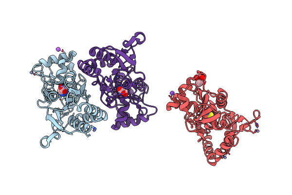 |
Structure Of The Ligand-Binding Core Of Glur2 In Complex With The Agonist (S)-Tdpa At 2.25 A Resolution
Organism: Rattus norvegicus
Method: X-RAY DIFFRACTION Resolution:2.27 Å Release Date: 2008-10-28 Classification: MEMBRANE PROTEIN Ligands: S2P, ZN, NA, CL, CAC |
Organism: Rattus norvegicus
Method: X-RAY DIFFRACTION
Release Date: 2008-10-28
Ligands: S2P, ZN, NA, CL, CAC
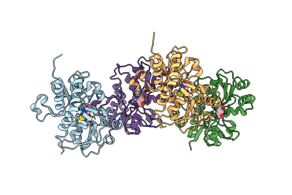 |
Structure Of The Ligand-Binding Core Of Glur2 In Complex With The Agonist (R)-Tdpa At 1.95 A Resolution
Organism: Rattus norvegicus
Method: X-RAY DIFFRACTION Resolution:1.95 Å Release Date: 2008-10-14 Classification: MEMBRANE PROTEIN Ligands: R2P |
Organism: Rattus norvegicus
Method: X-RAY DIFFRACTION
Release Date: 2008-10-14
Ligands: R2P
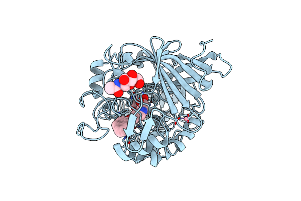 |
Hypocrea Jecorina Cel7A In Complex With (R)-Dihydroxy-Phenanthrenolol
Organism: Hypocrea jecorina
Method: X-RAY DIFFRACTION Resolution:1.05 Å Release Date: 2008-07-01 Classification: HYDROLASE Ligands: NAG, CO, GOL, XX6 |
Organism: Hypocrea jecorina
Method: X-RAY DIFFRACTION
Release Date: 2008-07-01
Ligands: NAG, CO, GOL, XX6
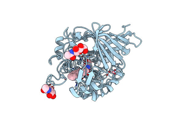 |
Hypocrea Jecorina Cel7A In Complex With (S)-Dihydroxy-Phenanthrenolol
Organism: Hypocrea jecorina
Method: X-RAY DIFFRACTION Resolution:1.60 Å Release Date: 2008-07-01 Classification: HYDROLASE Ligands: NAG, CO, XX7 |
Organism: Hypocrea jecorina
Method: X-RAY DIFFRACTION
Release Date: 2008-07-01
Ligands: NAG, CO, XX7
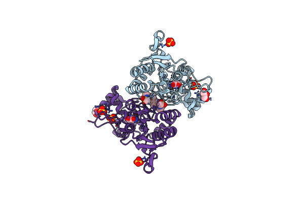 |
Crystal Structure Of The Iglur2 Ligand Binding Core (S1S2J-N775S) In Complex With A Dimeric Positive Modulator As Well As Glutamate At 2.25 A Resolution
Organism: Rattus norvegicus
Method: X-RAY DIFFRACTION Resolution:2.25 Å Release Date: 2007-12-04 Classification: MEMBRANE PROTEIN Ligands: SO4, CL, GLU, BHY, GOL |
Organism: Rattus norvegicus
Method: X-RAY DIFFRACTION
Release Date: 2007-12-04
Ligands: SO4, CL, GLU, BHY, GOL
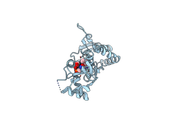 |
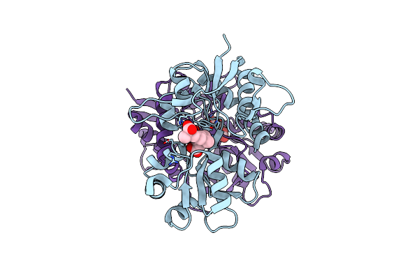
Crystal Structure Of The Ligand-Binding Core Of Iglur5 In Complex With The Antagonist (S)-Atpo At 1.85 A Resolution
Organism: Rattus norvegicus
Method: X-RAY DIFFRACTION Resolution:1.85 Å Release Date: 2007-07-03 Classification: MEMBRANE PROTEIN Ligands: AT1, GOL |
Organism: Rattus norvegicus
Method: X-RAY DIFFRACTION
Release Date: 2007-07-03
Ligands: AT1, GOL
 |
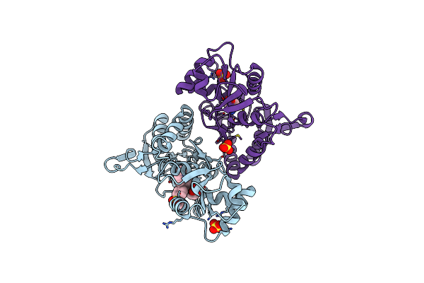
Crystal Structure Of The Ligand-Binding Core Of Iglur5 In Complex With The Partial Agonist Domoic Acid At 2.5 A Resolution
Organism: Rattus norvegicus
Method: X-RAY DIFFRACTION Resolution:2.50 Å Release Date: 2007-07-03 Classification: MEMBRANE PROTEIN Ligands: DOQ |
Organism: Rattus norvegicus
Method: X-RAY DIFFRACTION
Release Date: 2007-07-03
Ligands: DOQ
 |
The Structure Of A Mixed Glur2 Ligand-Binding Core Dimer In Complex With (S)-Glutamate And The Antagonist (S)-Ns1209
Organism: Rattus norvegicus
Method: X-RAY DIFFRACTION Resolution:2.65 Å Release Date: 2006-06-06 Classification: ION CHANNEL Ligands: SO4, M1L, GLU |
Organism: Rattus norvegicus
Method: X-RAY DIFFRACTION
Release Date: 2006-06-06
Ligands: SO4, M1L, GLU
 |
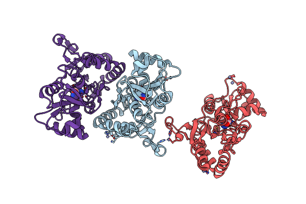
The Influence Of A Chiral Amino Acid On The Helical Handedness Of Pna In Solution And In Crystals
Method: X-RAY DIFFRACTION
Resolution:2.35 Å Release Date: 2004-01-13 Classification: PEPTIDE NUCLEIC ACID |
Method: X-RAY DIFFRACTION
Release Date: 2004-01-13
 |
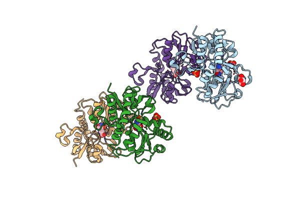
X-Ray Structure Of The Glur2 Ligand-Binding Core (S1S2J) In Complex With (S)-Des-Me-Ampa At 1.97 A Resolution, Crystallization In The Presence Of Zinc Acetate
Organism: Rattus norvegicus
Method: X-RAY DIFFRACTION Resolution:1.97 Å Release Date: 2003-07-08 Classification: MEMBRANE PROTEIN Ligands: ZN, SHI |
Organism: Rattus norvegicus
Method: X-RAY DIFFRACTION
Release Date: 2003-07-08
Ligands: ZN, SHI
 |
X-Ray Structure Of The Glur2 Ligand-Binding Core (S1S2J) In Complex With (S)-Des-Me-Ampa At 1.46 A Resolution. Crystallization In The Presence Of Lithium Sulfate.
Organism: Rattus norvegicus
Method: X-RAY DIFFRACTION Resolution:1.46 Å Release Date: 2003-07-01 Classification: MEMBRANE PROTEIN Ligands: SO4, SHI, GOL |
Organism: Rattus norvegicus
Method: X-RAY DIFFRACTION
Release Date: 2003-07-01
Ligands: SO4, SHI, GOL
 |
X-Ray Structure Of The Glur2 Ligand-Binding Core (S1S2J) In Complex With The Antagonist (S)-Atpo At 2.1 A Resolution.
Organism: Rattus norvegicus
Method: X-RAY DIFFRACTION Resolution:2.10 Å Release Date: 2003-03-04 Classification: MEMBRANE PROTEIN Ligands: AT1, SO4, ACT |
Organism: Rattus norvegicus
Method: X-RAY DIFFRACTION
Release Date: 2003-03-04
Ligands: AT1, SO4, ACT
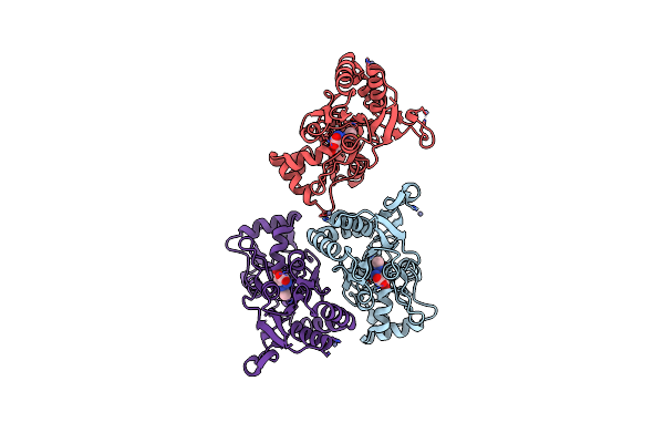 |
X-Ray Structure Of The Glur2 Ligand Binding Core (S1S2J) In Complex With 2-Me-Tet-Ampa At 1.85 A Resolution.
Organism: Rattus norvegicus
Method: X-RAY DIFFRACTION Resolution:1.85 Å Release Date: 2002-09-18 Classification: MEMBRANE PROTEIN Ligands: ZN, BN1 |
Organism: Rattus norvegicus
Method: X-RAY DIFFRACTION
Release Date: 2002-09-18
Ligands: ZN, BN1
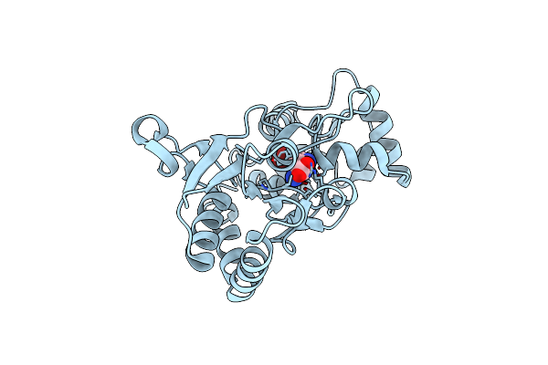 |
X-Ray Structure Of The Glur2 Ligand Binding Core (S1S2J) In Complex With Br-Hibo At 1.65 A Resolution
Organism: Rattus norvegicus
Method: X-RAY DIFFRACTION Resolution:1.65 Å Release Date: 2002-09-18 Classification: MEMBRANE PROTEIN Ligands: BRH |
Organism: Rattus norvegicus
Method: X-RAY DIFFRACTION
Release Date: 2002-09-18
Ligands: BRH
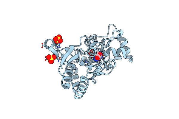 |
X-Ray Structure Of The Glur2 Ligand Binding Core (S1S2J-Y702F) In Complex With Br-Hibo At 1.73 A Resolution
Organism: Rattus norvegicus
Method: X-RAY DIFFRACTION Resolution:1.73 Å Release Date: 2002-09-18 Classification: MEMBRANE PROTEIN Ligands: SO4, BRH |
Organism: Rattus norvegicus
Method: X-RAY DIFFRACTION
Release Date: 2002-09-18
Ligands: SO4, BRH
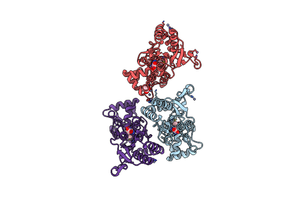 |
X-Ray Structure Of The Glur2 Ligand Binding Core (S1S2J) In Complex With Acpa At 1.46 A Resolution
Organism: Rattus norvegicus
Method: X-RAY DIFFRACTION Resolution:1.46 Å Release Date: 2002-09-18 Classification: MEMBRANE PROTEIN Ligands: ZN, AM1, ACT |
Organism: Rattus norvegicus
Method: X-RAY DIFFRACTION
Release Date: 2002-09-18
Ligands: ZN, AM1, ACT