

Search Count: 20
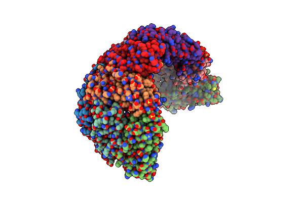 |
Crystal Structure Of E. Coli W35F Bfr Mutant
Organism: Escherichia coli k-12
Method: X-RAY DIFFRACTION Resolution:2.70 Å Release Date: 2009-10-06 Classification: METAL BINDING PROTEIN Ligands: SO4, FE, HEM |
Organism: Escherichia coli k-12
Method: X-RAY DIFFRACTION
Release Date: 2009-10-06
Ligands: SO4, FE, HEM
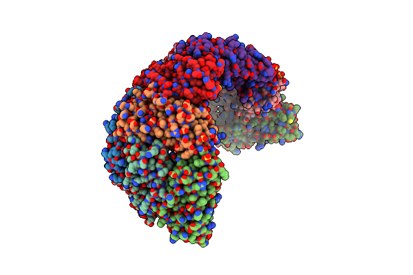 |
Crystal Structure Of W133F Variant E. Coli Bacterioferritn With Iron.
Organism: Escherichia coli
Method: X-RAY DIFFRACTION Resolution:2.60 Å Release Date: 2009-08-18 Classification: METAL BINDING PROTEIN Ligands: SO4, FE2, HEM |
Organism: Escherichia coli
Method: X-RAY DIFFRACTION
Release Date: 2009-08-18
Ligands: SO4, FE2, HEM
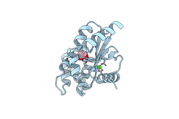 |
Crystal Structure Of Bdbd From Bacillus Subtilis (Reduced)
Organism: Bacillus subtilis
Method: X-RAY DIFFRACTION Resolution:1.50 Å Release Date: 2009-06-16 Classification: OXIDOREDUCTASE Ligands: CA, EDO |
Organism: Bacillus subtilis
Method: X-RAY DIFFRACTION
Release Date: 2009-06-16
Ligands: CA, EDO
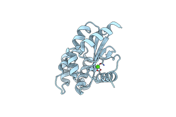 |
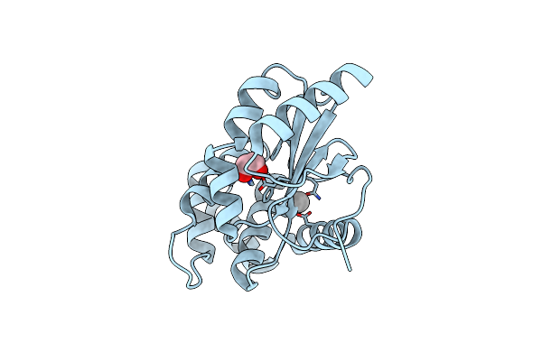
Crystal Structure Of Bdbd From Bacillus Subtilis (Oxidised)
Organism: Bacillus subtilis
Method: X-RAY DIFFRACTION Resolution:2.30 Å Release Date: 2009-06-16 Classification: OXIDOREDUCTASE Ligands: CA |
Organism: Bacillus subtilis
Method: X-RAY DIFFRACTION
Release Date: 2009-06-16
Ligands: CA
 |
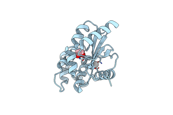
Crystal Structure Of Edta-Treated Bdbd (Oxidised)
Organism: Bacillus subtilis
Method: X-RAY DIFFRACTION Resolution:1.69 Å Release Date: 2009-06-16 Classification: OXIDOREDUCTASE Ligands: EDO, UNX |
Organism: Bacillus subtilis
Method: X-RAY DIFFRACTION
Release Date: 2009-06-16
Ligands: EDO, UNX
 |
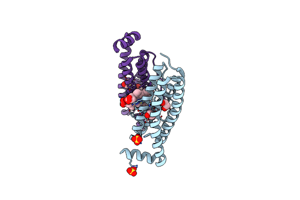
Crystal Structure Of Etda-Treated Bdbd (Reduced)
Organism: Bacillus subtilis
Method: X-RAY DIFFRACTION Resolution:1.40 Å Release Date: 2009-06-16 Classification: OXIDOREDUCTASE Ligands: EDO, UNX |
Organism: Bacillus subtilis
Method: X-RAY DIFFRACTION
Release Date: 2009-06-16
Ligands: EDO, UNX
 |
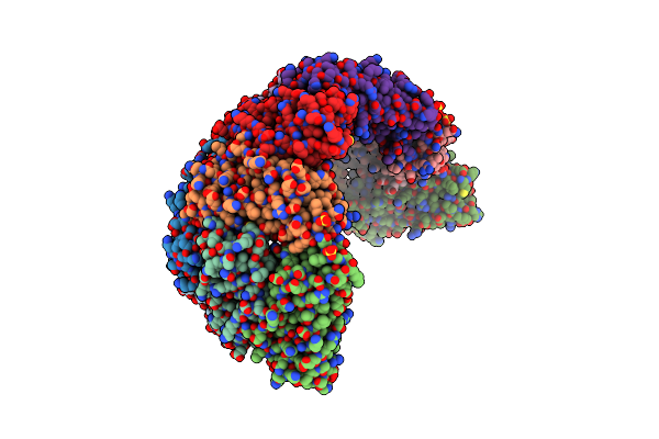
Escherichia Coli Bacterioferritin Mutant E128R/E135R
Organism: Escherichia coli
Method: X-RAY DIFFRACTION Resolution:1.80 Å Release Date: 2009-05-12 Classification: OXIDOREDUCTASE Ligands: ZN, SO4, HEM, EDO |
Organism: Escherichia coli
Method: X-RAY DIFFRACTION
Release Date: 2009-05-12
Ligands: ZN, SO4, HEM, EDO
 |
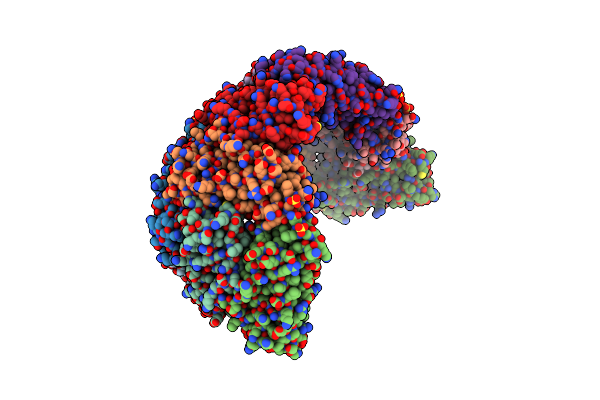
Crystal Structure Of E. Coli Bacterioferritin (Bfr) With An Unoccupied Ferroxidase Centre (Apo-Bfr).
Organism: Escherichia coli k12
Method: X-RAY DIFFRACTION Resolution:2.70 Å Release Date: 2009-05-05 Classification: METAL BINDING PROTEIN Ligands: SO4, HEM |
Organism: Escherichia coli k12
Method: X-RAY DIFFRACTION
Release Date: 2009-05-05
Ligands: SO4, HEM
 |
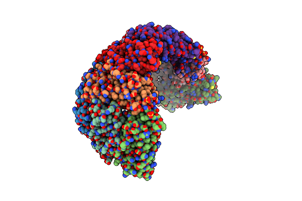
Crystal Structure Of E. Coli Bacterioferritin (Bfr) Soaked In Phosphate With An Alternative Conformation Of The Unoccupied Ferroxidase Centre (Apo-Bfr Ii).
Organism: Escherichia coli
Method: X-RAY DIFFRACTION Resolution:2.50 Å Release Date: 2009-05-05 Classification: METAL BINDING PROTEIN Ligands: SO4, HEM |
Organism: Escherichia coli
Method: X-RAY DIFFRACTION
Release Date: 2009-05-05
Ligands: SO4, HEM
 |
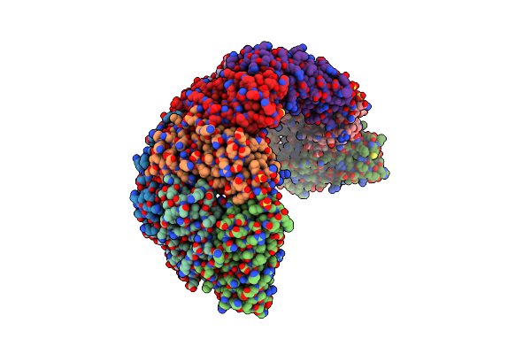
Crystal Structure Of E. Coli Bacterioferritin (Bfr) Obtained After Soaking Apo-Bfr Crystals For 2.5 Minutes In Fe2+ (2.5M Fe(Ii)-Bfr)
Organism: Escherichia coli
Method: X-RAY DIFFRACTION Resolution:2.70 Å Release Date: 2009-05-05 Classification: METAL BINDING PROTEIN Ligands: SO4, FE2, HEM |
Organism: Escherichia coli
Method: X-RAY DIFFRACTION
Release Date: 2009-05-05
Ligands: SO4, FE2, HEM
 |
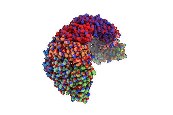
Crystal Structure Of E. Coli Bacterioferritin (Bfr) After A 65 Minute (Aerobic) Exposure To Fe(Ii) Revealing A Possible Mu-Oxo Bridge/Mu-Hydroxy Bridged Diiron Intermediate At The Ferroxidase Centre. (Fe(Iii)-O-Fe(Iii)-Bfr).
Organism: Escherichia coli
Method: X-RAY DIFFRACTION Resolution:2.80 Å Release Date: 2009-05-05 Classification: METAL BINDING PROTEIN Ligands: SO4, FE2, UNL, HEM |
Organism: Escherichia coli
Method: X-RAY DIFFRACTION
Release Date: 2009-05-05
Ligands: SO4, FE2, UNL, HEM
 |
Crystal Structure Of E. Coli Bacterioferritin (Bfr) With Two Zn(Ii) Ion Sites At The Ferroxidase Centre (Zn-Bfr).
Organism: Escherichia coli
Method: X-RAY DIFFRACTION Resolution:2.95 Å Release Date: 2009-05-05 Classification: METAL BINDING PROTEIN Ligands: SO4, ZN, HEM |
Organism: Escherichia coli
Method: X-RAY DIFFRACTION
Release Date: 2009-05-05
Ligands: SO4, ZN, HEM
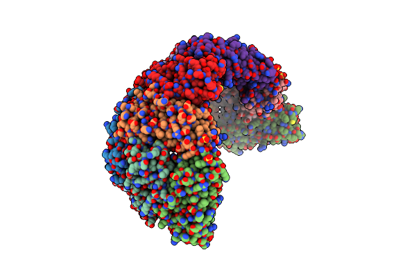 |
Crystal Structure Of E. Coli Bacterioferritin (Bfr) In Which The Ferroxidase Centre Is Inhibited With Zn(Ii) And High Occupancy Iron Is Bound Within The Cavity.
Organism: Escherichia coli
Method: X-RAY DIFFRACTION Resolution:2.40 Å Release Date: 2009-05-05 Classification: METAL BINDING PROTEIN Ligands: SO4, ZN, FE2, HEM |
Organism: Escherichia coli
Method: X-RAY DIFFRACTION
Release Date: 2009-05-05
Ligands: SO4, ZN, FE2, HEM
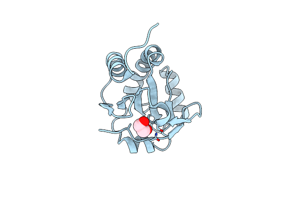 |
Structure Of A Resa Variant With A Dsba-Like Active Site Motif (Cphc)
Organism: Bacillus subtilis
Method: X-RAY DIFFRACTION Resolution:1.90 Å Release Date: 2008-08-12 Classification: OXIDOREDUCTASE Ligands: EDO |
Organism: Bacillus subtilis
Method: X-RAY DIFFRACTION
Release Date: 2008-08-12
Ligands: EDO
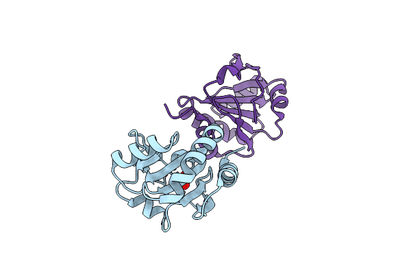 |
Structure Of Cehc Variant Resa
Organism: Bacillus subtilis
Method: X-RAY DIFFRACTION Resolution:2.50 Å Release Date: 2008-08-12 Classification: OXIDOREDUCTASE Ligands: SO4 |
Organism: Bacillus subtilis
Method: X-RAY DIFFRACTION
Release Date: 2008-08-12
Ligands: SO4
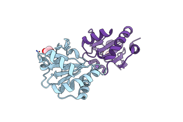 |
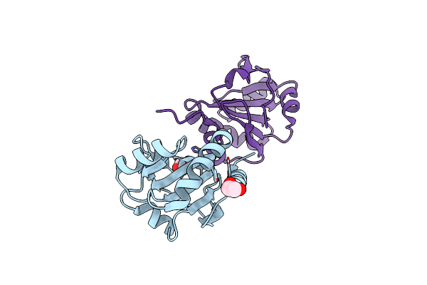
Crystal Structure Of Resa Cys77Ala Variant
Organism: Bacillus subtilis
Method: X-RAY DIFFRACTION Resolution:2.00 Å Release Date: 2006-09-19 Classification: OXIDOREDUCTASE Ligands: EDO |
Organism: Bacillus subtilis
Method: X-RAY DIFFRACTION
Release Date: 2006-09-19
Ligands: EDO
 |
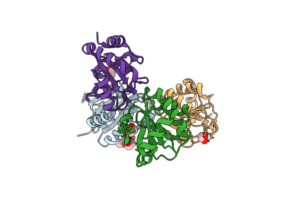
Resa C74A Variant
Organism: Bacillus subtilis
Method: X-RAY DIFFRACTION Resolution:2.40 Å Release Date: 2006-09-19 Classification: OXIDOREDUCTASE Ligands: EDO |
Organism: Bacillus subtilis
Method: X-RAY DIFFRACTION
Release Date: 2006-09-19
Ligands: EDO
 |
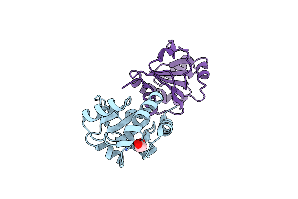
Resa E80Q
Organism: Bacillus subtilis
Method: X-RAY DIFFRACTION Resolution:1.95 Å Release Date: 2006-09-19 Classification: OXIDOREDUCTASE Ligands: ACT, EDO |
Organism: Bacillus subtilis
Method: X-RAY DIFFRACTION
Release Date: 2006-09-19
Ligands: ACT, EDO
 |
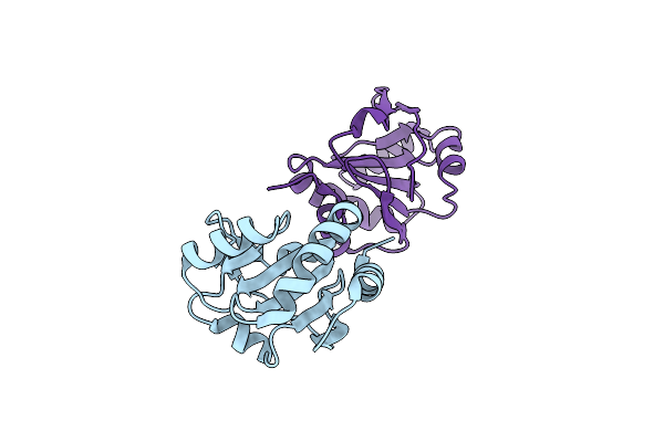
Resa Ph 9.25
Organism: Bacillus subtilis
Method: X-RAY DIFFRACTION Resolution:2.60 Å Release Date: 2006-09-19 Classification: OXIDOREDUCTASE Ligands: EDO |
Organism: Bacillus subtilis
Method: X-RAY DIFFRACTION
Release Date: 2006-09-19
Ligands: EDO
 |
Resa C74A/C77A
Organism: Bacillus subtilis
Method: X-RAY DIFFRACTION Resolution:3.10 Å Release Date: 2006-09-19 Classification: OXIDOREDUCTASE |
Organism: Bacillus subtilis
Method: X-RAY DIFFRACTION
Release Date: 2006-09-19