

Search Count: 39
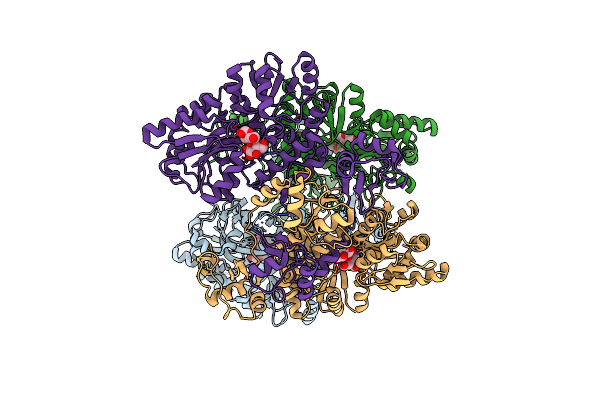 |
Crystal Structure Of Apo Hpv16 E6
Organism: Escherichia coli (strain k12), Human papillomavirus type 16
Method: X-RAY DIFFRACTION Resolution:3.25 Å Release Date: 2023-03-15 Classification: VIRAL PROTEIN Ligands: ZN |
Organism: Escherichia coli (strain k12), Human papillomavirus type 16
Method: X-RAY DIFFRACTION
Release Date: 2023-03-15
Ligands: ZN
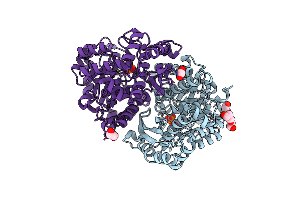 |
Structure Of Human Enolase 2 In Complex With Phosphoserine
Organism: Homo sapiens
Method: X-RAY DIFFRACTION Resolution:2.10 Å Release Date: 2022-11-09 Classification: LYASE Ligands: PO4, MG, ACT, EDO, PGE, SEP |
Organism: Homo sapiens
Method: X-RAY DIFFRACTION
Release Date: 2022-11-09
Ligands: PO4, MG, ACT, EDO, PGE, SEP
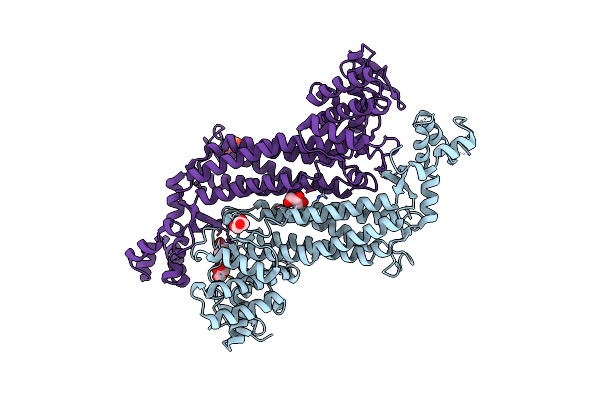 |
Crystal Structure Of Recombinant Human Fumarase In Complex With D-2-Amino-3-Phosphono-Propionic Acid
Organism: Homo sapiens
Method: X-RAY DIFFRACTION Resolution:2.15 Å Release Date: 2022-02-23 Classification: LYASE Ligands: APO, GOL |
Organism: Homo sapiens
Method: X-RAY DIFFRACTION
Release Date: 2022-02-23
Ligands: APO, GOL
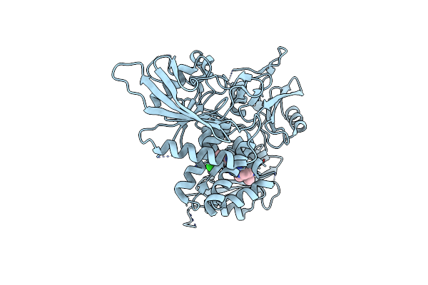 |
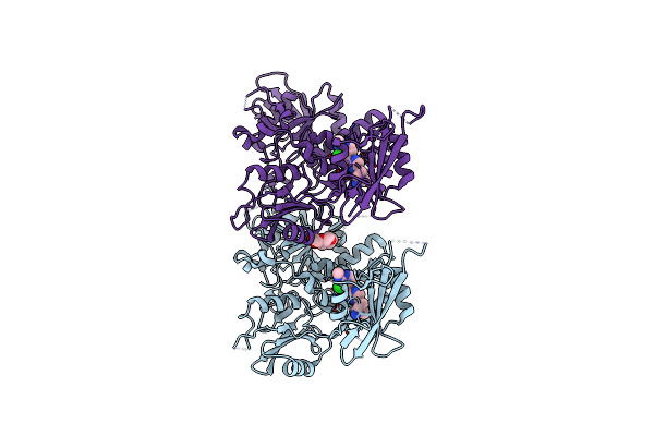
Structure Of Human Shp2 In Complex With Compound 16
Organism: Homo sapiens
Method: X-RAY DIFFRACTION Resolution:2.83 Å Release Date: 2021-10-27 Classification: HYDROLASE/INHIBITOR Ligands: 33I |
Organism: Homo sapiens
Method: X-RAY DIFFRACTION
Release Date: 2021-10-27
Ligands: 33I
 |
Structure Of Human Shp2 In Complex With Compound 22
Organism: Homo sapiens
Method: X-RAY DIFFRACTION Resolution:2.60 Å Release Date: 2021-10-27 Classification: HYDROLASE/INHIBITOR Ligands: 37I, PG4 |
Organism: Homo sapiens
Method: X-RAY DIFFRACTION
Release Date: 2021-10-27
Ligands: 37I, PG4
 |
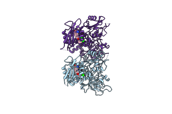
Structure Of Human Shp2 In Complex With Compound 27
Organism: Homo sapiens
Method: X-RAY DIFFRACTION Resolution:2.85 Å Release Date: 2021-10-27 Classification: HYDROLASE/INHIBITOR Ligands: 3CW |
Organism: Homo sapiens
Method: X-RAY DIFFRACTION
Release Date: 2021-10-27
Ligands: 3CW
 |
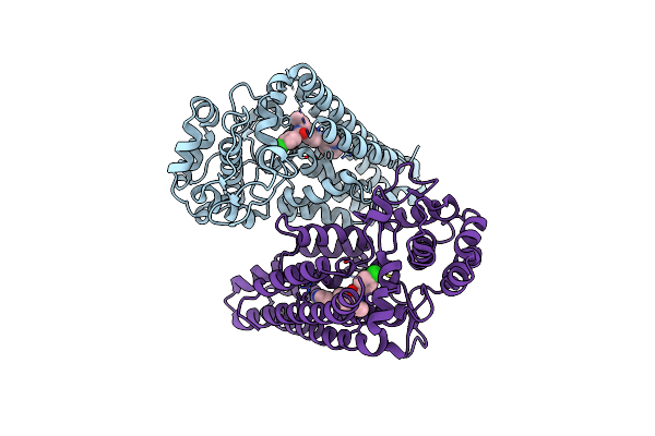
Structure Of Human Shp2 In Complex With Compound 30
Organism: Homo sapiens
Method: X-RAY DIFFRACTION Resolution:3.00 Å Release Date: 2021-10-27 Classification: HYDROLASE/INHIBITOR Ligands: 3ED |
Organism: Homo sapiens
Method: X-RAY DIFFRACTION
Release Date: 2021-10-27
Ligands: 3ED
 |
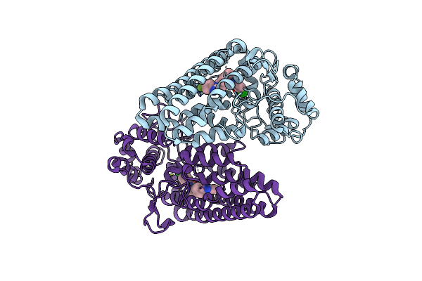
Crystal Structure Of The Indoleamine 2,3-Dioxygenase 1 (Ido1) In Complex With Compound 22
Organism: Homo sapiens
Method: X-RAY DIFFRACTION Resolution:2.58 Å Release Date: 2021-09-29 Classification: IMMUNOSUPPRESSANT Ligands: SLW |
Organism: Homo sapiens
Method: X-RAY DIFFRACTION
Release Date: 2021-09-29
Ligands: SLW
 |
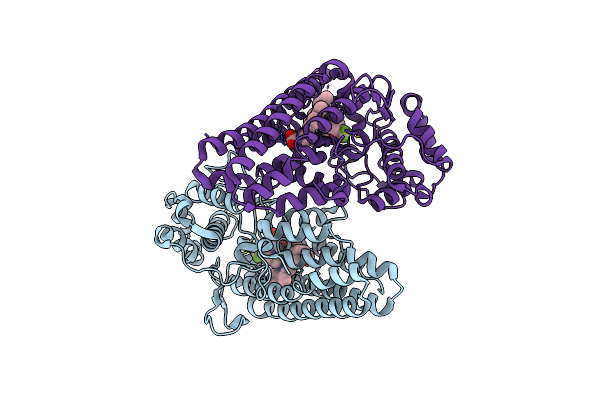
Crystal Structure Of The Indoleamine 2,3-Dioxygenagse 1 (Ido1) Complexed With Iacs-70099
Organism: Homo sapiens
Method: X-RAY DIFFRACTION Resolution:3.10 Å Release Date: 2021-09-01 Classification: HYDROLASE/HYDROLASE INHIBITOR Ligands: YRP |
Organism: Homo sapiens
Method: X-RAY DIFFRACTION
Release Date: 2021-09-01
Ligands: YRP
 |
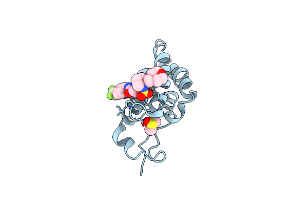
Crystal Structure Of The Indoleamine 2,3-Dioxygenagse 1 (Ido1) Complexed With Iacs-8968
Organism: Homo sapiens
Method: X-RAY DIFFRACTION Resolution:2.60 Å Release Date: 2021-09-01 Classification: HYDROLASE/HYDROLASE INHIBITOR Ligands: YRM, HEM |
Organism: Homo sapiens
Method: X-RAY DIFFRACTION
Release Date: 2021-09-01
Ligands: YRM, HEM
 |
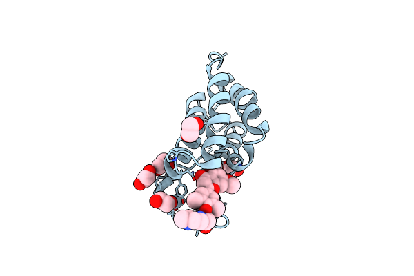
Crystal Structure Of The First Bromodomain Of Human Brd4 In Complex With The Inhibitor Zl0590
Organism: Homo sapiens
Method: X-RAY DIFFRACTION Resolution:1.43 Å Release Date: 2020-08-19 Classification: GENE REGULATION Ligands: DMS, POJ |
Organism: Homo sapiens
Method: X-RAY DIFFRACTION
Release Date: 2020-08-19
Ligands: DMS, POJ
 |
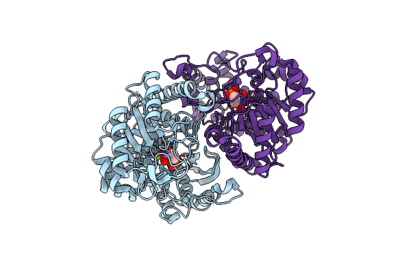
Crystal Structure Of The First Bromodomain Of Human Brd4 In Complex With The Inhibitor Zl0516
Organism: Homo sapiens
Method: X-RAY DIFFRACTION Resolution:2.00 Å Release Date: 2020-04-15 Classification: TRANSCRIPTION Ligands: EDO, QKP |
Organism: Homo sapiens
Method: X-RAY DIFFRACTION
Release Date: 2020-04-15
Ligands: EDO, QKP
 |
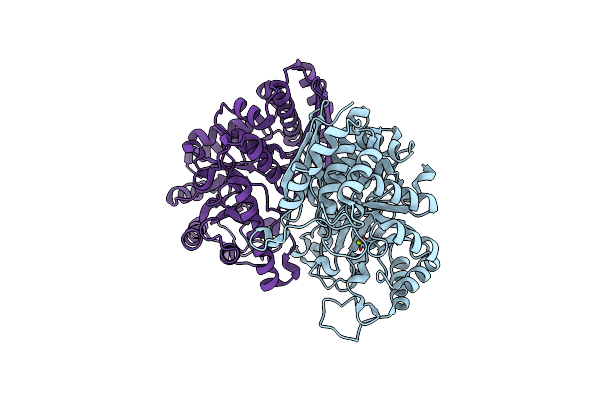
Structure Of Human Enolase 2 With ((3S,5S)-1,5-Dihydroxy-3-Methyl-2-Oxopyrrolidin-3-Yl)Phosphonate (Purified Enantiomer)
Organism: Homo sapiens
Method: X-RAY DIFFRACTION Resolution:2.63 Å Release Date: 2017-10-18 Classification: LYASE/LYASE INHIBITOR Ligands: 5TX, MG |
Organism: Homo sapiens
Method: X-RAY DIFFRACTION
Release Date: 2017-10-18
Ligands: 5TX, MG
 |
Structure Of Human Enolase 2
Organism: Homo sapiens
Method: X-RAY DIFFRACTION Resolution:2.32 Å Release Date: 2017-09-27 Classification: LYASE Ligands: MG, CL |
Organism: Homo sapiens
Method: X-RAY DIFFRACTION
Release Date: 2017-09-27
Ligands: MG, CL
 |
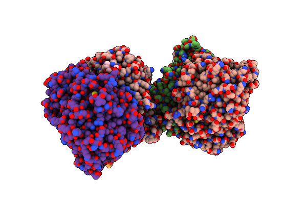
Structure Of Human Enolase 2 In Complex With (S)-(1-Hydroxy-2-Oxopiperidin-3-Yl)Phosphonate
Organism: Homo sapiens
Method: X-RAY DIFFRACTION Resolution:2.63 Å Release Date: 2017-02-22 Classification: LYASE/LYASE INHIBITOR Ligands: MG, 6BM, PGE |
Organism: Homo sapiens
Method: X-RAY DIFFRACTION
Release Date: 2017-02-22
Ligands: MG, 6BM, PGE
 |
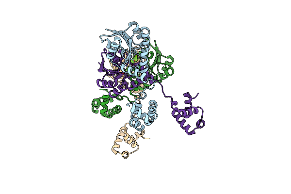
Structure Of Human Enolase 2 In Complex With ((3S,5S)-1,5-Dihydroxy-3-Methyl-2-Oxopyrrolidin-3-Yl)Phosphonic Acid
Organism: Homo sapiens
Method: X-RAY DIFFRACTION Resolution:2.05 Å Release Date: 2016-11-23 Classification: LYASE/LYASE INHIBITOR Ligands: MG, 5TX, PGE |
Organism: Homo sapiens
Method: X-RAY DIFFRACTION
Release Date: 2016-11-23
Ligands: MG, 5TX, PGE
 |
Crystal Structure Of The Beryllofluoride-Activated Liar From Enterococcus Faecium
Organism: Enterococcus faecium sd3b-2
Method: X-RAY DIFFRACTION Resolution:3.19 Å Release Date: 2016-11-16 Classification: TRANSCRIPTION Ligands: MG, BEF |
Organism: Enterococcus faecium sd3b-2
Method: X-RAY DIFFRACTION
Release Date: 2016-11-16
Ligands: MG, BEF
 |
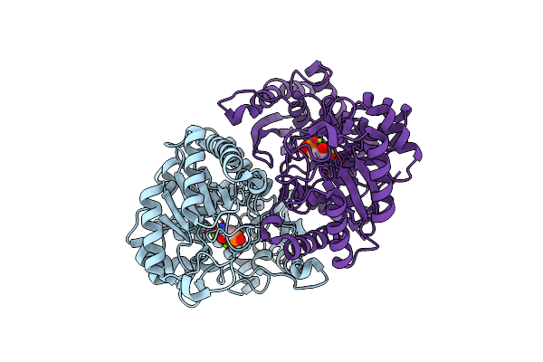
Structure Of Human Enolase 2 In Complex With Sf2312
Organism: Homo sapiens
Method: X-RAY DIFFRACTION Resolution:1.99 Å Release Date: 2016-07-20 Classification: LYASE/LYASE INHIBITOR Ligands: 4NG, MG |
Organism: Homo sapiens
Method: X-RAY DIFFRACTION
Release Date: 2016-07-20
Ligands: 4NG, MG
 |
Structure Of Human Enolase 2 In Complex With Phosphonoacetohydroxamate
Organism: Homo sapiens
Method: X-RAY DIFFRACTION Resolution:2.31 Å Release Date: 2016-04-13 Classification: LYASE/LYASE INHIBITOR Ligands: MG, PAH |
Organism: Homo sapiens
Method: X-RAY DIFFRACTION
Release Date: 2016-04-13
Ligands: MG, PAH
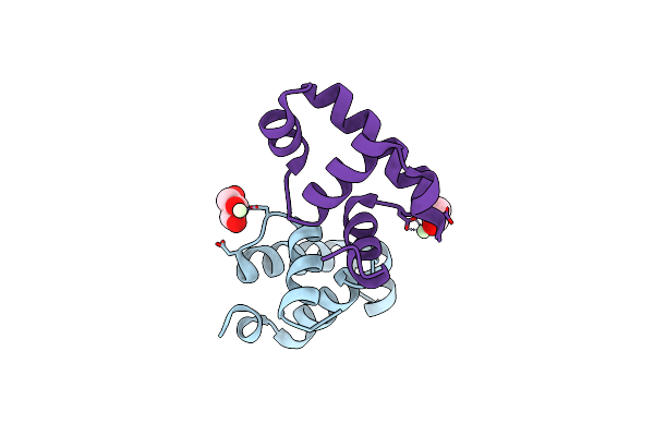 |
Crystal Structure Of The Dna Binding Domains Of Wild Type Liar From E. Faecalis
Method: X-RAY DIFFRACTION
Resolution:1.77 Å Release Date: 2015-05-06 Classification: DNA BINDING PROTEIN Ligands: PR, GOL, ACT |
Method: X-RAY DIFFRACTION
Release Date: 2015-05-06
Ligands: PR, GOL, ACT