

Search Count: 50
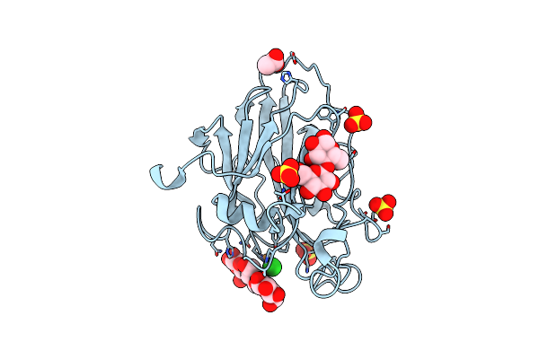 |
Structure Of An Lpmo (Expressed In E.Coli) At 1.49X10^4 Gy
Organism: Lentinus similis
Method: X-RAY DIFFRACTION Resolution:1.40 Å Release Date: 2022-08-31 Classification: OXIDOREDUCTASE Ligands: CU, CL, SO4, ACT |
Organism: Lentinus similis
Method: X-RAY DIFFRACTION
Release Date: 2022-08-31
Ligands: CU, CL, SO4, ACT
 |
Lsaa9A Expressed In E. Coli
Organism: Lentinus similis
Method: X-RAY DIFFRACTION Resolution:1.30 Å Release Date: 2022-08-24 Classification: OXIDOREDUCTASE Ligands: ACT, CU, CL, SO4 |
Organism: Lentinus similis
Method: X-RAY DIFFRACTION
Release Date: 2022-08-24
Ligands: ACT, CU, CL, SO4
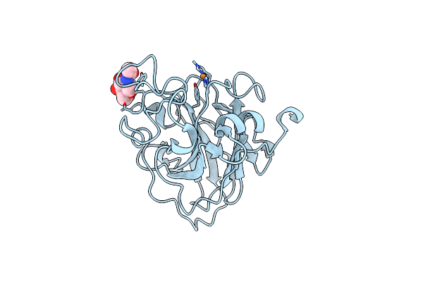 |
X-Ray Structure Of Lpmo At 7.88X10^3 Gy
Organism: Lentinus similis
Method: X-RAY DIFFRACTION Resolution:1.63 Å Release Date: 2022-08-24 Classification: OXIDOREDUCTASE Ligands: CU, NAG |
Organism: Lentinus similis
Method: X-RAY DIFFRACTION
Release Date: 2022-08-24
Ligands: CU, NAG
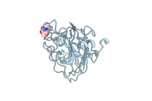 |
X-Ray Structure Of Lpmo At 5.99X10^4 Gy
Organism: Lentinus similis
Method: X-RAY DIFFRACTION Resolution:1.75 Å Release Date: 2022-08-24 Classification: OXIDOREDUCTASE Ligands: CU, NAG |
Organism: Lentinus similis
Method: X-RAY DIFFRACTION
Release Date: 2022-08-24
Ligands: CU, NAG
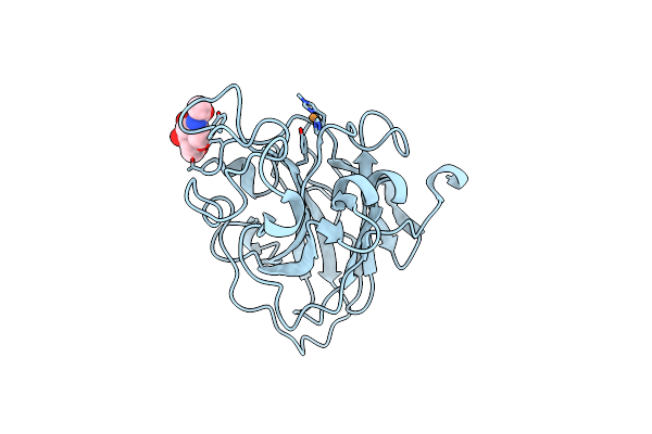 |
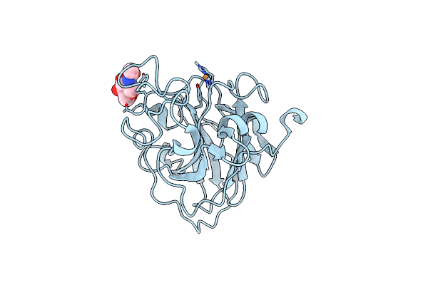
X-Ray Structure Of Lpmo At 1.39X10^5 Gy
Organism: Lentinus similis
Method: X-RAY DIFFRACTION Resolution:1.40 Å Release Date: 2022-08-24 Classification: OXIDOREDUCTASE Ligands: CU, NAG |
Organism: Lentinus similis
Method: X-RAY DIFFRACTION
Release Date: 2022-08-24
Ligands: CU, NAG
 |
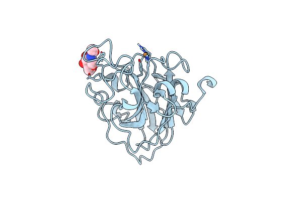
X-Ray Structure Of Lpmo At 3.6X10^5 Gy
Organism: Lentinus similis
Method: X-RAY DIFFRACTION Resolution:1.35 Å Release Date: 2022-08-24 Classification: OXIDOREDUCTASE Ligands: CU, NAG, CL |
Organism: Lentinus similis
Method: X-RAY DIFFRACTION
Release Date: 2022-08-24
Ligands: CU, NAG, CL
 |
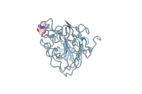
X-Ray Structure Of Lpmo At 1.45X10^6 Gy
Organism: Lentinus similis
Method: X-RAY DIFFRACTION Resolution:1.30 Å Release Date: 2022-08-24 Classification: OXIDOREDUCTASE Ligands: CU, NAG |
Organism: Lentinus similis
Method: X-RAY DIFFRACTION
Release Date: 2022-08-24
Ligands: CU, NAG
 |
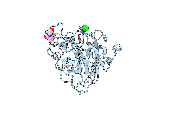
X-Ray Structure Of Lpmo At 6.65X10^6 Gy
Organism: Lentinus similis
Method: X-RAY DIFFRACTION Resolution:1.65 Å Release Date: 2022-08-24 Classification: OXIDOREDUCTASE Ligands: CU, NAG |
Organism: Lentinus similis
Method: X-RAY DIFFRACTION
Release Date: 2022-08-24
Ligands: CU, NAG
 |
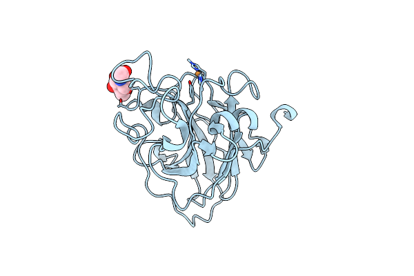
Room Temperature Structure Of An Lpmo.
Organism: Lentinus similis
Method: X-RAY DIFFRACTION Resolution:1.80 Å Release Date: 2022-08-24 Classification: OXIDOREDUCTASE Ligands: CU, NAG, CL |
Organism: Lentinus similis
Method: X-RAY DIFFRACTION
Release Date: 2022-08-24
Ligands: CU, NAG, CL
 |
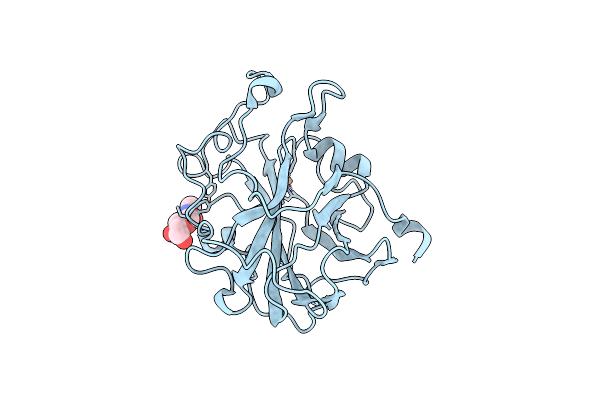
Room Temperature X-Ray Structure Of Lpmo At 1.91X10^3 Gy
Organism: Lentinus similis
Method: X-RAY DIFFRACTION Resolution:1.90 Å Release Date: 2022-08-24 Classification: OXIDOREDUCTASE Ligands: CU, NAG |
Organism: Lentinus similis
Method: X-RAY DIFFRACTION
Release Date: 2022-08-24
Ligands: CU, NAG
 |
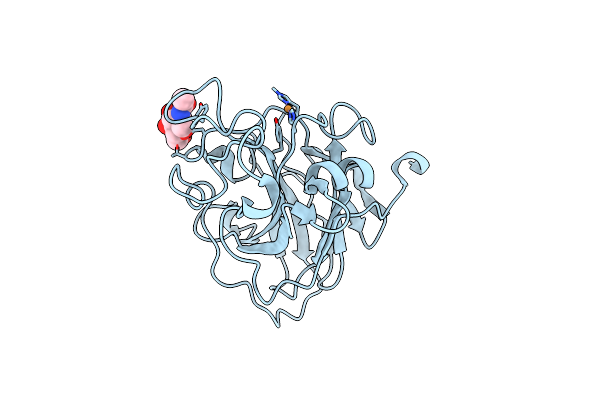
Structure Of An Lpmo, Collected From Serial Synchrotron Crystallography Data.
Organism: Lentinus similis
Method: X-RAY DIFFRACTION Resolution:2.40 Å Release Date: 2022-08-24 Classification: OXIDOREDUCTASE Ligands: CU, NAG |
Organism: Lentinus similis
Method: X-RAY DIFFRACTION
Release Date: 2022-08-24
Ligands: CU, NAG
 |
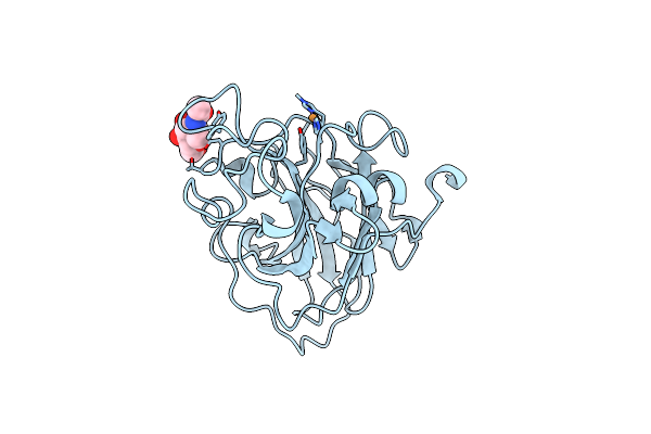
Lsaa9_A Chemically Reduced With Ascorbic Acid (Low X-Ray Dose)
Organism: Lentinus similis
Method: X-RAY DIFFRACTION Resolution:1.80 Å Release Date: 2022-08-24 Classification: OXIDOREDUCTASE Ligands: CU, NAG, CL |
Organism: Lentinus similis
Method: X-RAY DIFFRACTION
Release Date: 2022-08-24
Ligands: CU, NAG, CL
 |
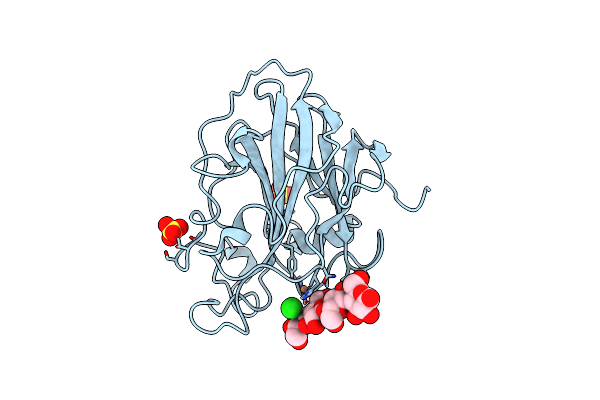
Lsaa9_A Chemically Reduced With Ascorbic Acid (High X-Ray Dose)
Organism: Lentinus similis
Method: X-RAY DIFFRACTION Resolution:1.50 Å Release Date: 2022-08-24 Classification: OXIDOREDUCTASE Ligands: CU, NAG, CL |
Organism: Lentinus similis
Method: X-RAY DIFFRACTION
Release Date: 2022-08-24
Ligands: CU, NAG, CL
 |
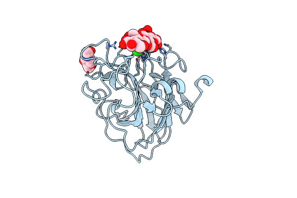
Lpmo, Expressed In E.Coli, In Complex With Cellotetraose
Organism: Lentinus similis
Method: X-RAY DIFFRACTION Resolution:1.40 Å Release Date: 2022-08-24 Classification: OXIDOREDUCTASE Ligands: SO4, CU, CL |
Organism: Lentinus similis
Method: X-RAY DIFFRACTION
Release Date: 2022-08-24
Ligands: SO4, CU, CL
 |
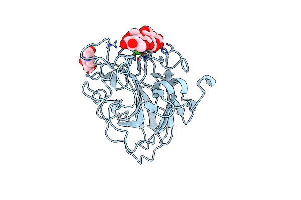
Structure Of Lpmo In Complex With Cellotetraose At 7.88X10^3 Gy
Organism: Lentinus similis
Method: X-RAY DIFFRACTION Resolution:2.21 Å Release Date: 2022-08-24 Classification: OXIDOREDUCTASE Ligands: CU, NAG, CL |
Organism: Lentinus similis
Method: X-RAY DIFFRACTION
Release Date: 2022-08-24
Ligands: CU, NAG, CL
 |
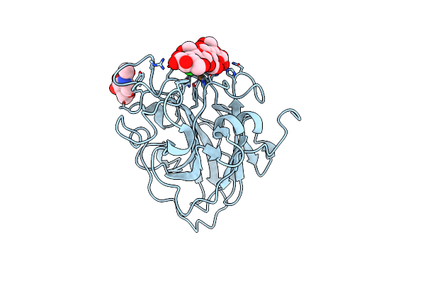
Structure Of Lpmo In Complex With Cellotetraose At 5.99X10^4 Gy
Organism: Lentinus similis
Method: X-RAY DIFFRACTION Resolution:2.10 Å Release Date: 2022-08-24 Classification: OXIDOREDUCTASE Ligands: CU, NAG, CL |
Organism: Lentinus similis
Method: X-RAY DIFFRACTION
Release Date: 2022-08-24
Ligands: CU, NAG, CL
 |
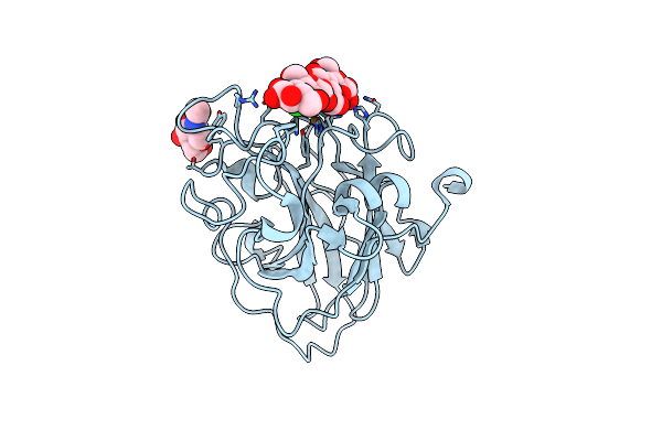
Structure Of Lpmo In Complex With Cellotetraose At 1.39X10^5 Gy
Organism: Lentinus similis
Method: X-RAY DIFFRACTION Resolution:1.90 Å Release Date: 2022-08-24 Classification: OXIDOREDUCTASE Ligands: CU, NAG, CL |
Organism: Lentinus similis
Method: X-RAY DIFFRACTION
Release Date: 2022-08-24
Ligands: CU, NAG, CL
 |
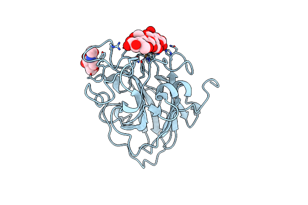
Structure Of Lpmo In Complex With Cellotetraose At 3.6X10^5 Gy
Organism: Lentinus similis
Method: X-RAY DIFFRACTION Resolution:1.90 Å Release Date: 2022-08-24 Classification: OXIDOREDUCTASE Ligands: CU, NAG, CL |
Organism: Lentinus similis
Method: X-RAY DIFFRACTION
Release Date: 2022-08-24
Ligands: CU, NAG, CL
 |
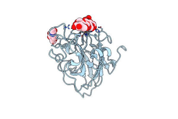
Structure Of Lpmo In Complex With Cellotetraose At 1.45X10^6 Gy
Organism: Lentinus similis
Method: X-RAY DIFFRACTION Resolution:1.90 Å Release Date: 2022-08-24 Classification: OXIDOREDUCTASE Ligands: CU, NAG, CL |
Organism: Lentinus similis
Method: X-RAY DIFFRACTION
Release Date: 2022-08-24
Ligands: CU, NAG, CL
 |
Structure Of Lpmo In Complex With Cellotetraose At 6.65X10^6 Gy
Organism: Lentinus similis
Method: X-RAY DIFFRACTION Resolution:2.05 Å Release Date: 2022-08-24 Classification: OXIDOREDUCTASE Ligands: CU, NAG, CL |
Organism: Lentinus similis
Method: X-RAY DIFFRACTION
Release Date: 2022-08-24
Ligands: CU, NAG, CL