

Search Count: 59
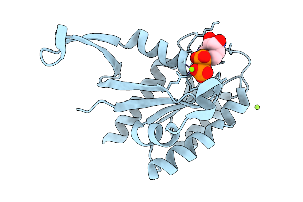 |
Crystal Structure Of Gdp-Bound Kras G12D/I55E: Suppressing G12D Oncogenicity Via Second-Site I55E Mutation
Organism: Homo sapiens
Method: X-RAY DIFFRACTION Resolution:1.23 Å Release Date: 2026-03-25 Classification: ONCOPROTEIN Ligands: GDP, MG, CL |
Organism: Homo sapiens
Method: X-RAY DIFFRACTION
Release Date: 2026-03-25
Ligands: GDP, MG, CL
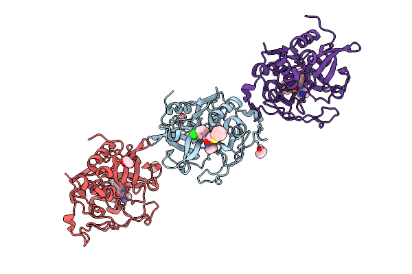 |
Structure Of Cathepsin B1 From Schistosoma Mansoni (Smcb1) In Complex With A Carborane Inhibitor
Organism: Schistosoma mansoni
Method: X-RAY DIFFRACTION Resolution:2.20 Å Release Date: 2025-10-29 Classification: HYDROLASE Ligands: A1IHQ, EDO |
Organism: Schistosoma mansoni
Method: X-RAY DIFFRACTION
Release Date: 2025-10-29
Ligands: A1IHQ, EDO
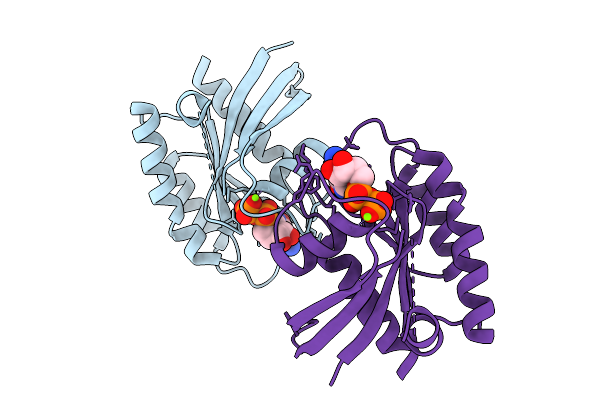 |
Crystal Structure Of Gdp-Bound Kras G12D/M67R: Suppressing G12D Oncogenicity Via Second-Site M67R Mutation
Organism: Homo sapiens
Method: X-RAY DIFFRACTION Resolution:1.70 Å Release Date: 2025-10-08 Classification: ONCOPROTEIN Ligands: GDP, MG |
Organism: Homo sapiens
Method: X-RAY DIFFRACTION
Release Date: 2025-10-08
Ligands: GDP, MG
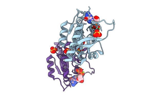 |
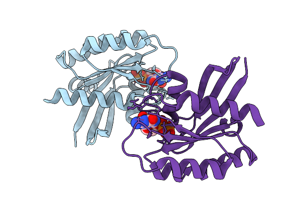
Crystal Structure Of Gdp-Bound Kras G12D/F28K: Suppressing G12D Oncogenicity Via Second-Site F28K Mutation
Organism: Homo sapiens
Method: X-RAY DIFFRACTION Resolution:1.74 Å Release Date: 2025-10-08 Classification: ONCOPROTEIN Ligands: GDP, MG, SO4 |
Organism: Homo sapiens
Method: X-RAY DIFFRACTION
Release Date: 2025-10-08
Ligands: GDP, MG, SO4
 |
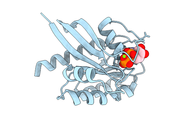
Crystal Structure Of Gdp-Bound Kras G12D/P34R: Suppressing G12D Oncogenicity Via Second-Site P34R Mutation
Organism: Homo sapiens
Method: X-RAY DIFFRACTION Resolution:1.50 Å Release Date: 2025-10-08 Classification: ONCOPROTEIN Ligands: MG, GDP |
Organism: Homo sapiens
Method: X-RAY DIFFRACTION
Release Date: 2025-10-08
Ligands: MG, GDP
 |
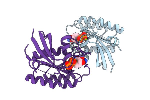
Crystal Structure Of Gdp-Bound Kras G12D/R41Q: Suppressing G12D Oncogenicity Via Second-Site R41Q Mutation
Organism: Homo sapiens
Method: X-RAY DIFFRACTION Resolution:1.22 Å Release Date: 2025-10-08 Classification: ONCOPROTEIN Ligands: GDP, MG |
Organism: Homo sapiens
Method: X-RAY DIFFRACTION
Release Date: 2025-10-08
Ligands: GDP, MG
 |
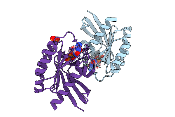
Crystal Structure Of Gdp-Bound Kras G12D/V45E: Suppressing G12D Oncogenicity Via Second-Site V45E Mutation
Organism: Homo sapiens
Method: X-RAY DIFFRACTION Resolution:2.20 Å Release Date: 2025-10-08 Classification: ONCOPROTEIN Ligands: GDP, MG |
Organism: Homo sapiens
Method: X-RAY DIFFRACTION
Release Date: 2025-10-08
Ligands: GDP, MG
 |
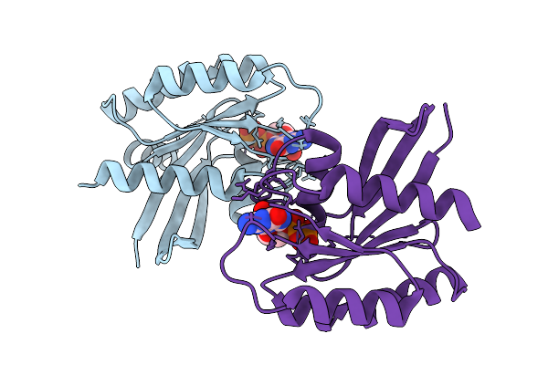
Crystal Structure Of Gdp-Bound Kras G12D/D54R: Suppressing G12D Oncogenicity Via Second-Site D54R Mutation
Organism: Homo sapiens
Method: X-RAY DIFFRACTION Resolution:2.51 Å Release Date: 2025-10-08 Classification: ONCOPROTEIN Ligands: GDP, MG, SO4 |
Organism: Homo sapiens
Method: X-RAY DIFFRACTION
Release Date: 2025-10-08
Ligands: GDP, MG, SO4
 |
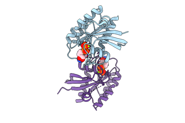
Crystal Structure Of Gdp-Bound Kras G12D/G60R: Suppressing G12D Oncogenicity Via Second-Site G60R Mutation
Organism: Homo sapiens
Method: X-RAY DIFFRACTION Resolution:1.80 Å Release Date: 2025-10-08 Classification: ONCOPROTEIN Ligands: MG, GDP |
Organism: Homo sapiens
Method: X-RAY DIFFRACTION
Release Date: 2025-10-08
Ligands: MG, GDP
 |
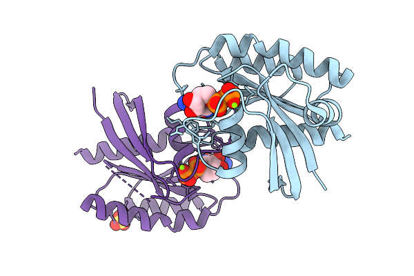
Crystal Structure Of Gdp-Bound Kras G12D/V103Y: Suppressing G12D Oncogenicity Via Second-Site V103Y Mutation
Organism: Homo sapiens
Method: X-RAY DIFFRACTION Resolution:1.80 Å Release Date: 2025-10-08 Classification: ONCOPROTEIN Ligands: MG, GDP |
Organism: Homo sapiens
Method: X-RAY DIFFRACTION
Release Date: 2025-10-08
Ligands: MG, GDP
 |
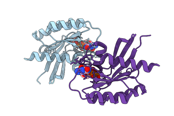
Crystal Structure Of Gdp-Bound Kras G12D/E62Q: Suppressing G12D Oncogenicity Via Second-Site E62Q Mutation
Organism: Homo sapiens
Method: X-RAY DIFFRACTION Resolution:1.94 Å Release Date: 2025-10-08 Classification: ONCOPROTEIN Ligands: MG, GDP, SO4 |
Organism: Homo sapiens
Method: X-RAY DIFFRACTION
Release Date: 2025-10-08
Ligands: MG, GDP, SO4
 |
Crystal Structure Of Gdp-Bound Kras E3K/G12D: Suppressing G12D Oncogenicity Via Second-Site E3K Mutation
Organism: Homo sapiens
Method: X-RAY DIFFRACTION Resolution:1.87 Å Release Date: 2025-10-08 Classification: ONCOPROTEIN Ligands: MG, GDP, GOL |
Organism: Homo sapiens
Method: X-RAY DIFFRACTION
Release Date: 2025-10-08
Ligands: MG, GDP, GOL
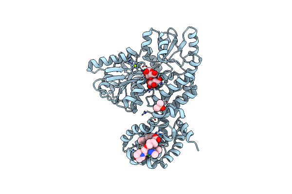 |
Crystal Structure Of Human Mbp-Myeloid Cell Leukemia 1 (Mcl-1) In Complex With Brd810 Inhibitor
Organism: Escherichia coli k-12, Homo sapiens
Method: X-RAY DIFFRACTION Resolution:1.56 Å Release Date: 2024-06-19 Classification: APOPTOSIS Ligands: MG, YI7, DMS, PGE |
Organism: Escherichia coli k-12, Homo sapiens
Method: X-RAY DIFFRACTION
Release Date: 2024-06-19
Ligands: MG, YI7, DMS, PGE
 |
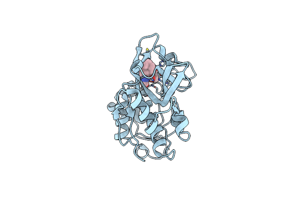
X-Ray Crystal Structure Of Jrd-Sik1/2I-3 Bound To A Mark2-Sik2 Chimera
Organism: Homo sapiens
Method: X-RAY DIFFRACTION Resolution:2.10 Å Release Date: 2024-01-10 Classification: BIOSYNTHETIC PROTEIN Ligands: PEG, SJ0, SO4 |
Organism: Homo sapiens
Method: X-RAY DIFFRACTION
Release Date: 2024-01-10
Ligands: PEG, SJ0, SO4
 |
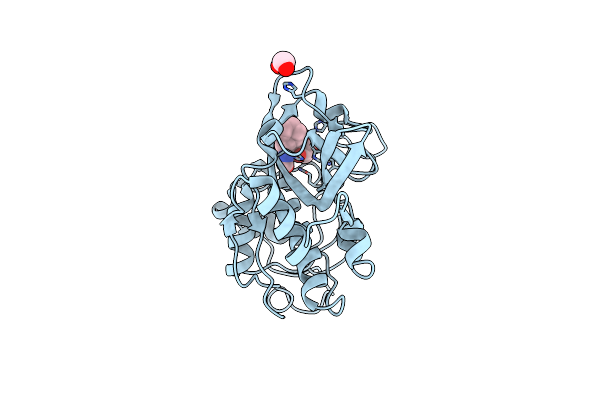
Human Cathepsin B In Complex With The Carbamate Inhibitor 7
Organism: Homo sapiens
Method: X-RAY DIFFRACTION Resolution:1.45 Å Release Date: 2023-10-04 Classification: HYDROLASE Ligands: P9F |
Organism: Homo sapiens
Method: X-RAY DIFFRACTION
Release Date: 2023-10-04
Ligands: P9F
 |
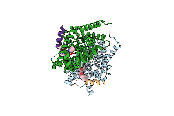
Human Cathepsin B In Complex With The Carbamate Inhibitor 31
Organism: Homo sapiens
Method: X-RAY DIFFRACTION Resolution:1.70 Å Release Date: 2023-10-04 Classification: HYDROLASE Ligands: P9U, EDO |
Organism: Homo sapiens
Method: X-RAY DIFFRACTION
Release Date: 2023-10-04
Ligands: P9U, EDO
 |
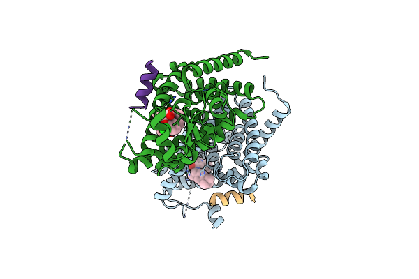
Crystal Structure Of Pparg And Ncor2 With An Inverse Agonist (Compound 7A)
Organism: Homo sapiens
Method: X-RAY DIFFRACTION Resolution:1.86 Å Release Date: 2022-12-28 Classification: TRANSCRIPTION Ligands: Q2X, GOL |
Organism: Homo sapiens
Method: X-RAY DIFFRACTION
Release Date: 2022-12-28
Ligands: Q2X, GOL
 |
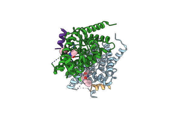
Crystal Structure Of Pparg And Ncor2 With Sr10221, An Inverse Agonist
Organism: Homo sapiens
Method: X-RAY DIFFRACTION Resolution:1.78 Å Release Date: 2022-12-28 Classification: TRANSCRIPTION Ligands: A8R |
Organism: Homo sapiens
Method: X-RAY DIFFRACTION
Release Date: 2022-12-28
Ligands: A8R
 |
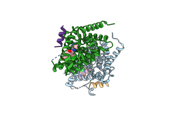
Crystal Structure Of Pparg And Ncor2 With An Inverse Agonist (Compound 7E)
Organism: Homo sapiens
Method: X-RAY DIFFRACTION Resolution:2.00 Å Release Date: 2022-12-28 Classification: TRANSCRIPTION Ligands: Q33 |
Organism: Homo sapiens
Method: X-RAY DIFFRACTION
Release Date: 2022-12-28
Ligands: Q33
 |
Crystal Structure Of Mutant Pparg (C313A) And Ncor2 With An Inverse Agonist (Compound 7E)
Organism: Homo sapiens
Method: X-RAY DIFFRACTION Resolution:2.22 Å Release Date: 2022-12-28 Classification: TRANSCRIPTION Ligands: Q33 |
Organism: Homo sapiens
Method: X-RAY DIFFRACTION
Release Date: 2022-12-28
Ligands: Q33