

Search Count: 11
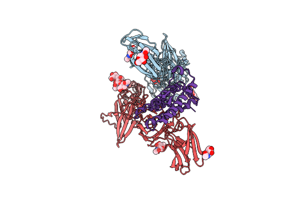 |
Cryo-Em Structure Of Murine Thrombopoietin Receptor Ectodomain In Complex With Tpo
Organism: Mus musculus, Saccharomyces cerevisiae
Method: ELECTRON MICROSCOPY Release Date: 2024-02-07 Classification: CYTOKINE/RECEPTOR Ligands: NAG, MAN |
Organism: Mus musculus, Saccharomyces cerevisiae
Method: ELECTRON MICROSCOPY
Release Date: 2024-02-07
Ligands: NAG, MAN
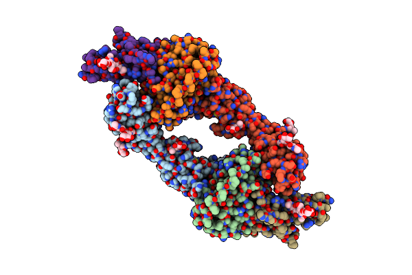 |
The Structure Of The Interleukin 11 Signalling Complex, Truncated Gp130
Organism: Homo sapiens
Method: ELECTRON MICROSCOPY Release Date: 2023-11-29 Classification: CYTOKINE Ligands: NAG |
Organism: Homo sapiens
Method: ELECTRON MICROSCOPY
Release Date: 2023-11-29
Ligands: NAG
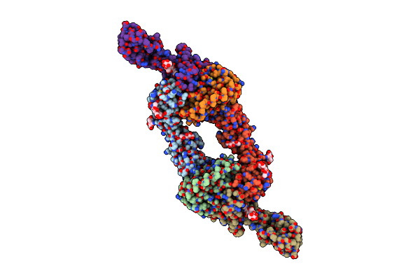 |
The Structure Of The Il-11 Signalling Complex, With Full-Length Extracellular Gp130
Organism: Homo sapiens
Method: ELECTRON MICROSCOPY Release Date: 2023-11-29 Classification: CYTOKINE Ligands: NAG |
Organism: Homo sapiens
Method: ELECTRON MICROSCOPY
Release Date: 2023-11-29
Ligands: NAG
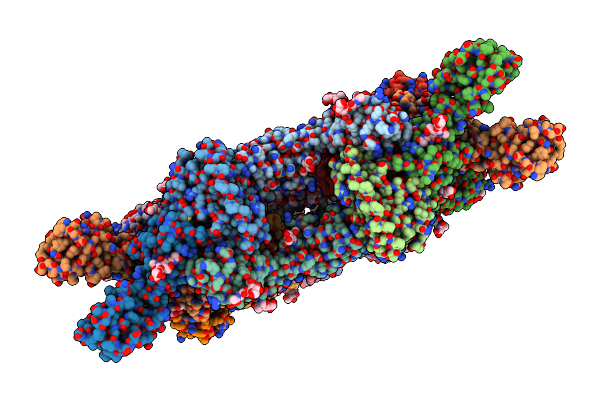 |
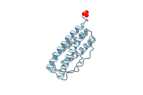
The Crystal Structure Of The Il-11 Signalling Complex
Organism: Homo sapiens
Method: X-RAY DIFFRACTION Resolution:3.78 Å Release Date: 2023-11-29 Classification: CYTOKINE Ligands: NAG |
Organism: Homo sapiens
Method: X-RAY DIFFRACTION
Release Date: 2023-11-29
Ligands: NAG
 |
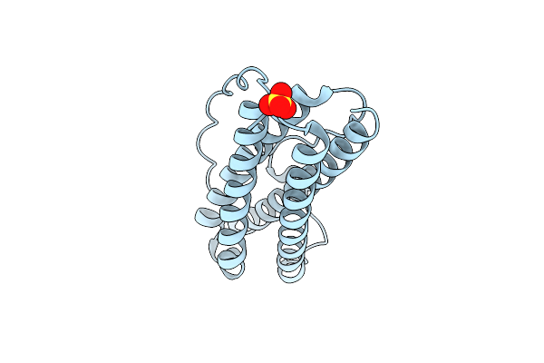
The Crystal Structure Of Interleukin-11, W147A Mutant
Organism: Homo sapiens
Method: X-RAY DIFFRACTION Resolution:1.48 Å Release Date: 2023-11-29 Classification: CYTOKINE Ligands: SO4, CL |
Organism: Homo sapiens
Method: X-RAY DIFFRACTION
Release Date: 2023-11-29
Ligands: SO4, CL
 |
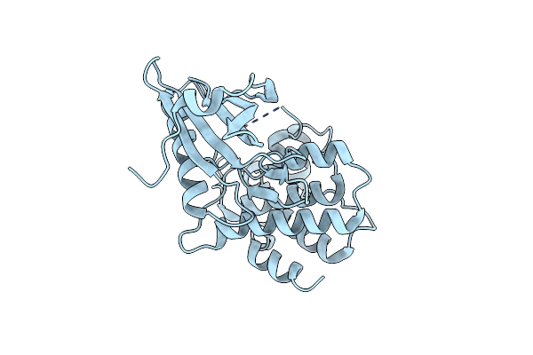
The Structure Of Interleukin-11 Mutein
Organism: Homo sapiens
Method: X-RAY DIFFRACTION Resolution:1.80 Å Release Date: 2023-11-29 Classification: CYTOKINE Ligands: SO4 |
Organism: Homo sapiens
Method: X-RAY DIFFRACTION
Release Date: 2023-11-29
Ligands: SO4
 |
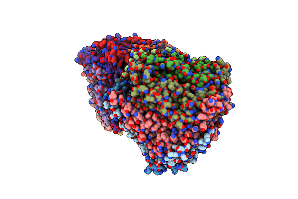
Crystal Structure Of Phosphorylated (T357/S358) Human Mlkl Pseudokinase Domain
Organism: Homo sapiens
Method: X-RAY DIFFRACTION Resolution:2.30 Å Release Date: 2023-11-08 Classification: TRANSFERASE |
Organism: Homo sapiens
Method: X-RAY DIFFRACTION
Release Date: 2023-11-08
 |
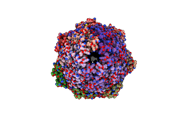
Crystal Structure Of The 11S Subunit Of The Plasmodium Falciparum Proteasome, Pa28
Organism: Plasmodium falciparum
Method: X-RAY DIFFRACTION Resolution:3.10 Å Release Date: 2019-08-07 Classification: PROTEIN BINDING Ligands: SO4 |
Organism: Plasmodium falciparum
Method: X-RAY DIFFRACTION
Release Date: 2019-08-07
Ligands: SO4
 |
The Structure Of The Plasmodium Falciparum 20S Proteasome In Complex With Two Pa28 Activators
Organism: Plasmodium falciparum (isolate 3d7)
Method: ELECTRON MICROSCOPY Release Date: 2019-08-07 Classification: HYDROLASE |
Organism: Plasmodium falciparum (isolate 3d7)
Method: ELECTRON MICROSCOPY
Release Date: 2019-08-07
 |
The Structure Of The Plasmodium Falciparum 20S Proteasome.
Organism: Plasmodium falciparum (isolate 3d7)
Method: ELECTRON MICROSCOPY Release Date: 2019-08-07 Classification: HYDROLASE |
Organism: Plasmodium falciparum (isolate 3d7)
Method: ELECTRON MICROSCOPY
Release Date: 2019-08-07
 |
The Structure Of The Plasmodium Falciparum 20S Proteasome In Complex With One Pa28 Activator
Organism: Plasmodium falciparum 3d7
Method: ELECTRON MICROSCOPY Release Date: 2019-08-07 Classification: HYDROLASE |
Organism: Plasmodium falciparum 3d7
Method: ELECTRON MICROSCOPY
Release Date: 2019-08-07