

Search Count: 22
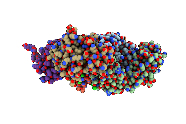 |
Pfs230 Domain 1 Bound By Rupa-32 Fab
Organism: Homo sapiens, Plasmodium falciparum
Method: X-RAY DIFFRACTION Resolution:2.59 Å Release Date: 2023-02-15 Classification: CELL INVASION/IMMUNE SYSTEM Ligands: NH4, CL, EDO, TRS |
Organism: Homo sapiens, Plasmodium falciparum
Method: X-RAY DIFFRACTION
Release Date: 2023-02-15
Ligands: NH4, CL, EDO, TRS
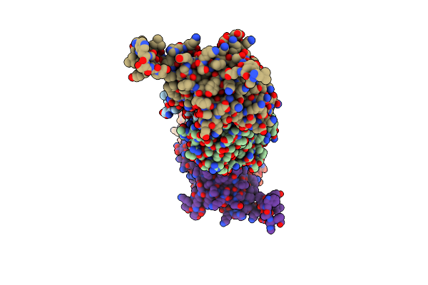 |
Pfs230 Domain 1 Bound By Rupa-55 Fab
Organism: Homo sapiens, Plasmodium falciparum
Method: X-RAY DIFFRACTION Resolution:2.92 Å Release Date: 2023-02-15 Classification: CELL INVASION/IMMUNE SYSTEM |
Organism: Homo sapiens, Plasmodium falciparum
Method: X-RAY DIFFRACTION
Release Date: 2023-02-15
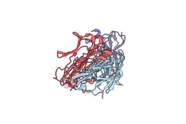 |
Pfs230 Domain 1 Bound By Rupa-38 Fab
Organism: Homo sapiens, Plasmodium falciparum
Method: X-RAY DIFFRACTION Resolution:2.09 Å Release Date: 2023-02-15 Classification: CELL INVASION/IMMUNE SYSTEM Ligands: CL, IPA |
Organism: Homo sapiens, Plasmodium falciparum
Method: X-RAY DIFFRACTION
Release Date: 2023-02-15
Ligands: CL, IPA
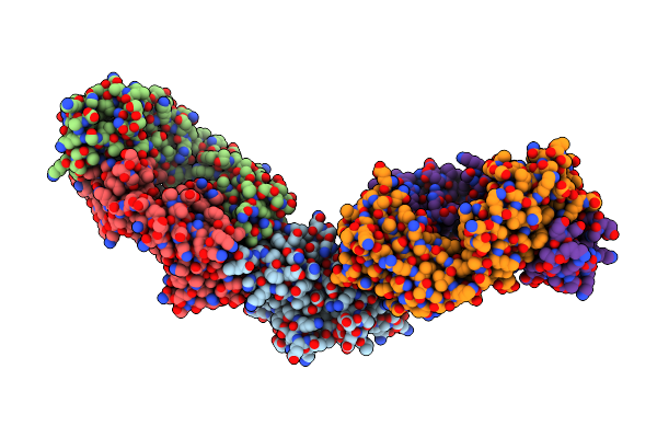 |
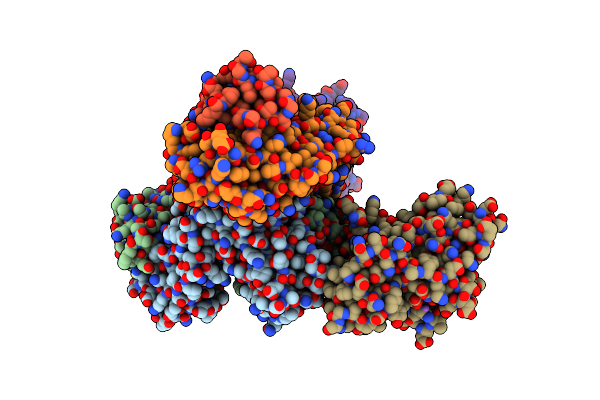
Pfs230 Domain 1 Bound By Rupa-97 And 15C5 Fabs
Organism: Plasmodium falciparum, Homo sapiens
Method: X-RAY DIFFRACTION Resolution:3.29 Å Release Date: 2023-02-15 Classification: CELL INVASION/IMMUNE SYSTEM |
Organism: Plasmodium falciparum, Homo sapiens
Method: X-RAY DIFFRACTION
Release Date: 2023-02-15
 |
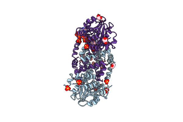
Pfs230 Domain 1 Bound By Lmiv230-02 Fab
Organism: Homo sapiens, Plasmodium falciparum
Method: X-RAY DIFFRACTION Resolution:2.06 Å Release Date: 2023-02-15 Classification: CELL INVASION/IMMUNE SYSTEM |
Organism: Homo sapiens, Plasmodium falciparum
Method: X-RAY DIFFRACTION
Release Date: 2023-02-15
 |
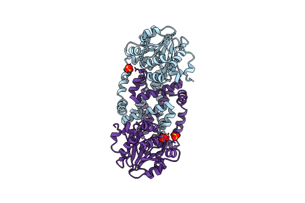
Ilvc, A Ketol-Acid Reductoisomerase, From Streptococcus Pneumoniae_Wt
Organism: Streptococcus pneumoniae d39
Method: X-RAY DIFFRACTION Resolution:1.69 Å Release Date: 2020-08-12 Classification: ISOMERASE Ligands: GOL, SO4, NAP, MG |
Organism: Streptococcus pneumoniae d39
Method: X-RAY DIFFRACTION
Release Date: 2020-08-12
Ligands: GOL, SO4, NAP, MG
 |
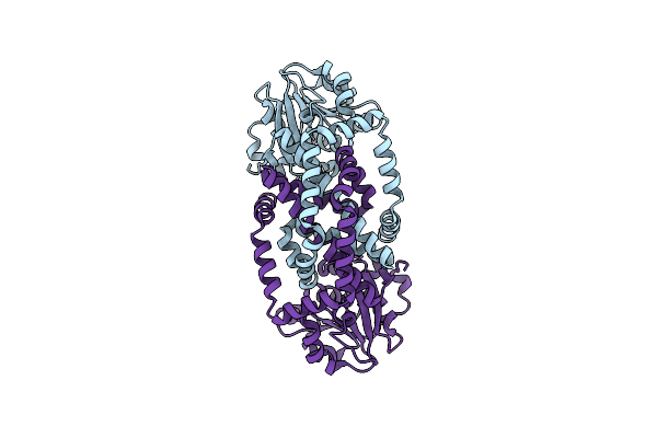
Ilvc, A Ketol-Acid Reductoisomerase, From Streptococcus Pneumoniae_R49E
Organism: Streptococcus pneumoniae d39
Method: X-RAY DIFFRACTION Resolution:1.95 Å Release Date: 2020-08-12 Classification: ISOMERASE Ligands: SO4, GOL |
Organism: Streptococcus pneumoniae d39
Method: X-RAY DIFFRACTION
Release Date: 2020-08-12
Ligands: SO4, GOL
 |
Ilvc, A Ketol-Acid Reductoisomerase, From Streptococcus Pneumoniae_E195S
Organism: Streptococcus pneumoniae d39
Method: X-RAY DIFFRACTION Resolution:2.02 Å Release Date: 2020-08-12 Classification: ISOMERASE |
Organism: Streptococcus pneumoniae d39
Method: X-RAY DIFFRACTION
Release Date: 2020-08-12
 |
Ilvc, A Ketol-Acid Reductoisomerase, From Streptococcus Pneumoniae_D83G
Organism: Streptococcus pneumoniae d39
Method: X-RAY DIFFRACTION Resolution:2.29 Å Release Date: 2020-08-12 Classification: ISOMERASE |
Organism: Streptococcus pneumoniae d39
Method: X-RAY DIFFRACTION
Release Date: 2020-08-12
 |
Ilvc, A Ketol-Acid Reductoisomerase, From Streptococcus Pnuemoniae_D191G
Organism: Streptococcus pneumoniae d39
Method: X-RAY DIFFRACTION Resolution:2.02 Å Release Date: 2020-08-12 Classification: ISOMERASE Ligands: NAP, SO4, GOL, NH4 |
Organism: Streptococcus pneumoniae d39
Method: X-RAY DIFFRACTION
Release Date: 2020-08-12
Ligands: NAP, SO4, GOL, NH4
 |
Crystal Structure Of N-Glycosylated Human Calcitonin Receptor Extracellular Domain In Complex With Salmon Calcitonin (16-32)
Organism: Escherichia coli, Homo sapiens, Oncorhynchus sp.
Method: X-RAY DIFFRACTION Resolution:1.78 Å Release Date: 2020-02-12 Classification: SIGNALING PROTEIN Ligands: NAG, SO4 |
Organism: Escherichia coli, Homo sapiens, Oncorhynchus sp.
Method: X-RAY DIFFRACTION
Release Date: 2020-02-12
Ligands: NAG, SO4
 |
Crystal Structure Of N-Glycosylated Human Calcitonin Receptor Extracellular Domain In Complex With Salmon Calcitonin (22-32)
Organism: Escherichia coli, Homo sapiens, Oncorhynchus sp.
Method: X-RAY DIFFRACTION Resolution:2.85 Å Release Date: 2020-02-12 Classification: SIGNALING PROTEIN Ligands: NAG, ACT |
Organism: Escherichia coli, Homo sapiens, Oncorhynchus sp.
Method: X-RAY DIFFRACTION
Release Date: 2020-02-12
Ligands: NAG, ACT
 |
Crystal Structure Of D-Xylose Reductase From Scheffersomyces Stipitis In Complex With Nadph
Organism: Scheffersomyces stipitis (strain atcc 58785 / cbs 6054 / nbrc 10063 / nrrl y-11545)
Method: X-RAY DIFFRACTION Resolution:2.00 Å Release Date: 2018-12-05 Classification: OXIDOREDUCTASE Ligands: NAP |
Organism: Scheffersomyces stipitis (strain atcc 58785 / cbs 6054 / nbrc 10063 / nrrl y-11545)
Method: X-RAY DIFFRACTION
Release Date: 2018-12-05
Ligands: NAP
 |
Crystal Structure Of D-Xylose Reductase From Scheffersomyces Stipitis
Organism: Scheffersomyces stipitis (strain atcc 58785 / cbs 6054 / nbrc 10063 / nrrl y-11545)
Method: X-RAY DIFFRACTION Resolution:1.95 Å Release Date: 2018-12-05 Classification: OXIDOREDUCTASE Ligands: GOL |
Organism: Scheffersomyces stipitis (strain atcc 58785 / cbs 6054 / nbrc 10063 / nrrl y-11545)
Method: X-RAY DIFFRACTION
Release Date: 2018-12-05
Ligands: GOL
 |
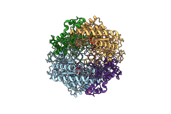
Crystal Structure Of The Human Clr:Ramp1 Extracellular Domain Heterodimer In Complex With Adrenomedullin 2/Intermedin
Organism: Escherichia coli o157:h7, Homo sapiens
Method: X-RAY DIFFRACTION Resolution:2.05 Å Release Date: 2018-09-05 Classification: PEPTIDE BINDING PROTEIN |
Organism: Escherichia coli o157:h7, Homo sapiens
Method: X-RAY DIFFRACTION
Release Date: 2018-09-05
 |
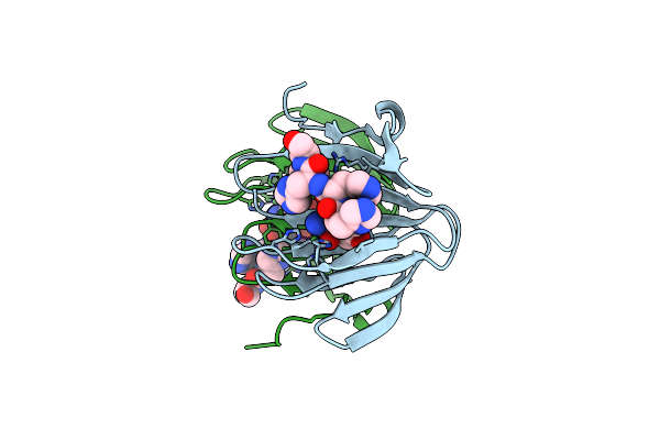
Crystal Structure Of Xylose Isomerase From Piromyces Sp. E2
Organism: Piromyces sp. (strain e2)
Method: X-RAY DIFFRACTION Resolution:2.70 Å Release Date: 2018-05-09 Classification: ISOMERASE Ligands: MN, GOL |
Organism: Piromyces sp. (strain e2)
Method: X-RAY DIFFRACTION
Release Date: 2018-05-09
Ligands: MN, GOL
 |
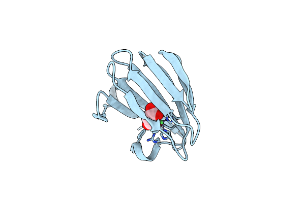
The Structure Of Kdgf From Yersinia Enterocolitica.
Organism: Yersinia enterocolitica
Method: X-RAY DIFFRACTION Resolution:1.50 Å Release Date: 2016-05-04 Classification: HYDROLASE Ligands: NI |
Organism: Yersinia enterocolitica
Method: X-RAY DIFFRACTION
Release Date: 2016-05-04
Ligands: NI
 |
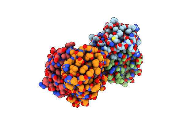
The Structure Of Kdgf From Yersinia Enterocolitica With Malonate Bound In The Active Site.
Organism: Yersinia enterocolitica
Method: X-RAY DIFFRACTION Resolution:1.50 Å Release Date: 2016-05-04 Classification: HYDROLASE Ligands: NI, MLA |
Organism: Yersinia enterocolitica
Method: X-RAY DIFFRACTION
Release Date: 2016-05-04
Ligands: NI, MLA
 |
The Structure Of Kdgf From Halomonas Sp.
Organism: Halomonas sp.
Method: X-RAY DIFFRACTION Resolution:2.00 Å Release Date: 2016-05-04 Classification: LYASE Ligands: NI, FLC |
Organism: Halomonas sp.
Method: X-RAY DIFFRACTION
Release Date: 2016-05-04
Ligands: NI, FLC
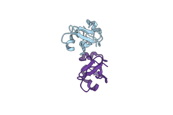 |
Crystal Structure Of Cide-N Domain Of Fsp27
Organism: Mus musculus
Method: X-RAY DIFFRACTION Resolution:2.00 Å Release Date: 2014-07-02 Classification: APOPTOSIS |
Organism: Mus musculus
Method: X-RAY DIFFRACTION
Release Date: 2014-07-02