

Search Count: 32
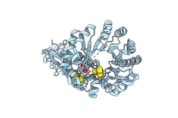 |
X-Ray Crystal Structure Of Tige From Paramaledivibacter Caminithermalis
Organism: Paramaledivibacter caminithermalis
Method: X-RAY DIFFRACTION Resolution:1.66 Å Release Date: 2024-02-07 Classification: OXIDOREDUCTASE Ligands: SF4, GOL |
Organism: Paramaledivibacter caminithermalis
Method: X-RAY DIFFRACTION
Release Date: 2024-02-07
Ligands: SF4, GOL
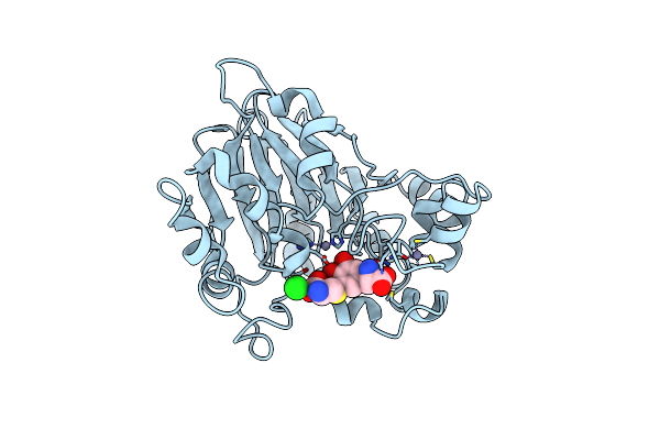 |
Pseudomonas Putida Pqqb With A Non-Physiological Zinc At The Active Site Binds The Substrate Mimic, 5-Cysteinyl-3,4-Dihydroxyphenylalanine (5-Cys-Dopa), Non-Specifically But Supports The Proposed Function Of The Enzyme In Pyrroloquinoline Quinone Biosynthesis.
Organism: Pseudomonas putida (strain atcc 47054 / dsm 6125 / ncimb 11950 / kt2440)
Method: X-RAY DIFFRACTION Resolution:2.35 Å Release Date: 2019-05-22 Classification: OXIDOREDUCTASE Ligands: ZN, CL, HKS |
Organism: Pseudomonas putida (strain atcc 47054 / dsm 6125 / ncimb 11950 / kt2440)
Method: X-RAY DIFFRACTION
Release Date: 2019-05-22
Ligands: ZN, CL, HKS
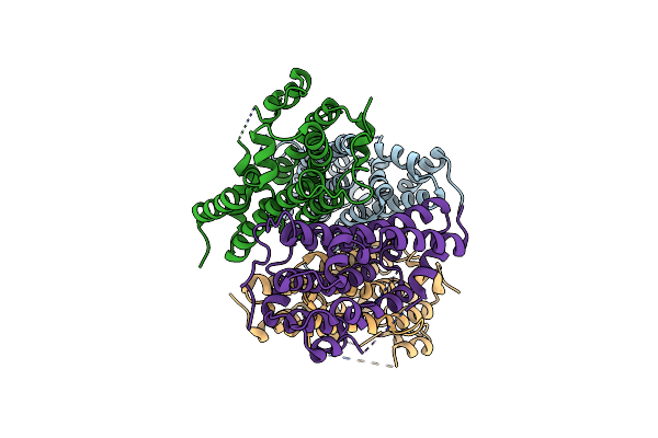 |
Crystal Structure For Methylobacterium Extorquens Pqqc (Truncation Of Natural Cd Fusion)
Organism: Methylobacterium extorquens (strain atcc 14718 / dsm 1338 / jcm 2805 / ncimb 9133 / am1)
Method: X-RAY DIFFRACTION Resolution:2.00 Å Release Date: 2018-05-16 Classification: OXIDOREDUCTASE |
 |
Crystal Structure For Methylobacterium Extorquens Pqqcd (Natural Fusion)
Organism: Methylobacterium extorquens (strain atcc 14718 / dsm 1338 / jcm 2805 / ncimb 9133 / am1)
Method: X-RAY DIFFRACTION Resolution:2.85 Å Release Date: 2018-05-16 Classification: OXIDOREDUCTASE |
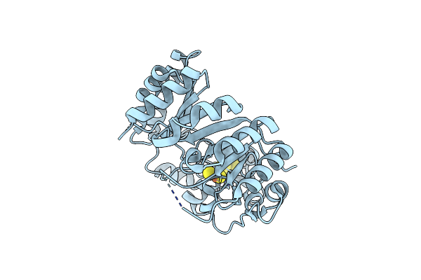 |
X-Ray Structure Of Pqqe From Methylobacterium Extorquens
Organism: Methylobacterium extorquens
Method: X-RAY DIFFRACTION Resolution:3.20 Å Release Date: 2018-02-14 Classification: OXIDOREDUCTASE Ligands: FES, SF4 |
Organism: Methylobacterium extorquens
Method: X-RAY DIFFRACTION
Release Date: 2018-02-14
Ligands: FES, SF4
 |
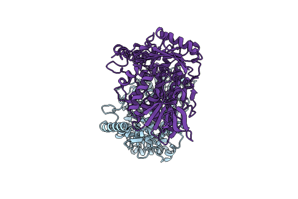
The Solution Nmr Structure For The Pqqd Truncation Of Methylobacterium Extorquens Pqqcd Representing A Functional And Stand-Alone Ribosomally Synthesized And Post-Translational Modified (Ripp) Recognition Element (Rre)
Organism: Methylobacterium extorquens (strain atcc 14718 / dsm 1338 / am1)
Method: SOLUTION NMR Release Date: 2017-05-24 Classification: CHAPERONE |
Organism: Methylobacterium extorquens (strain atcc 14718 / dsm 1338 / am1)
Method: SOLUTION NMR
Release Date: 2017-05-24
 |
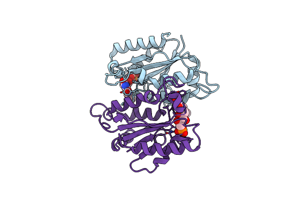
Crystal Structure Of Thermophilic Tryptophan Halogenase (Th-Hal) Enzyme From Streptomycin Violaceusniger.
Organism: Streptomyces violaceusniger
Method: X-RAY DIFFRACTION Resolution:2.33 Å Release Date: 2016-10-19 Classification: HYDROLASE |
Organism: Streptomyces violaceusniger
Method: X-RAY DIFFRACTION
Release Date: 2016-10-19
 |
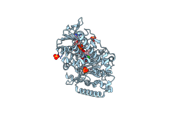
Crystal Structure Of Thermophilic Tryptophan Halogenase (Th-Hal) Enzyme From Streptomycin Violaceusniger.
Organism: Bacillus subtilis
Method: X-RAY DIFFRACTION Resolution:2.53 Å Release Date: 2016-10-19 Classification: OXIDOREDUCTASE Ligands: FMN |
Organism: Bacillus subtilis
Method: X-RAY DIFFRACTION
Release Date: 2016-10-19
Ligands: FMN
 |
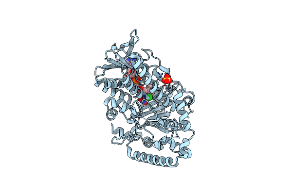
F454K Mutant Of Tryptophan 7-Halogenase Prna
Organism: Pseudomonas fluorescens
Method: X-RAY DIFFRACTION Resolution:2.20 Å Release Date: 2016-04-13 Classification: OXIDOREDUCTASE Ligands: FAD, CL, PO4, SO4 |
Organism: Pseudomonas fluorescens
Method: X-RAY DIFFRACTION
Release Date: 2016-04-13
Ligands: FAD, CL, PO4, SO4
 |
Crystal Structure Of Tryptophan 7-Halogenase (Prna) Mutant E450K
Organism: Pseudomonas fluorescens
Method: X-RAY DIFFRACTION Resolution:2.29 Å Release Date: 2016-01-20 Classification: OXIDOREDUCTASE Ligands: FAD, SO4, CL |
Organism: Pseudomonas fluorescens
Method: X-RAY DIFFRACTION
Release Date: 2016-01-20
Ligands: FAD, SO4, CL
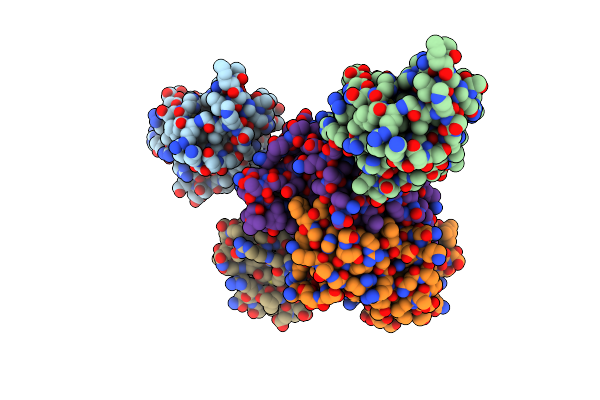 |
Crystal Structure Of Thioesterase Pa1618 From Pseudomonas Aeruginosa
Organism: Pseudomonas aeruginosa
Method: X-RAY DIFFRACTION Resolution:1.77 Å Release Date: 2015-05-13 Classification: HYDROLASE |
Organism: Pseudomonas aeruginosa
Method: X-RAY DIFFRACTION
Release Date: 2015-05-13
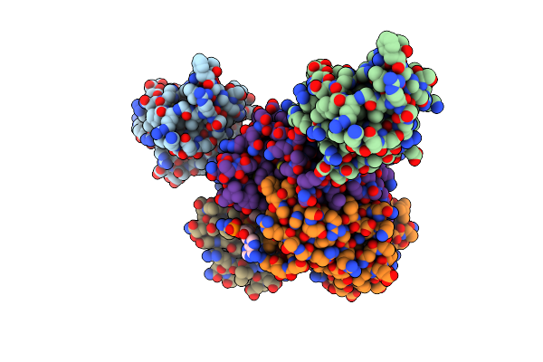 |
Crystal Structure Of Thioesterase Pa1618 From Pseudomonas Aeruginosa In Complex With Phenacyl-Coa
Organism: Pseudomonas aeruginosa
Method: X-RAY DIFFRACTION Resolution:1.62 Å Release Date: 2015-05-13 Classification: HYDROLASE Ligands: 0FQ |
Organism: Pseudomonas aeruginosa
Method: X-RAY DIFFRACTION
Release Date: 2015-05-13
Ligands: 0FQ
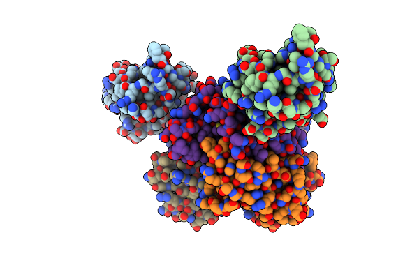 |
Crystal Structure Of Thioesterase Pa1618 From Pseudomonas Aeruginosa In Complex With Benzoyl-Do-Coa
Organism: Pseudomonas aeruginosa
Method: X-RAY DIFFRACTION Resolution:1.77 Å Release Date: 2015-05-13 Classification: HYDROLASE Ligands: 31B |
Organism: Pseudomonas aeruginosa
Method: X-RAY DIFFRACTION
Release Date: 2015-05-13
Ligands: 31B
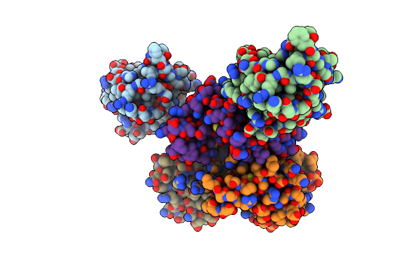 |
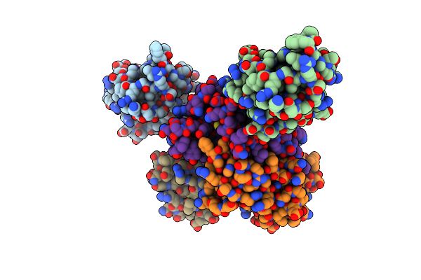
Crystal Structure Of Mutant Thioesterase Pa1618 (E64A) From Pseudomonas Aeruginosa
Organism: Pseudomonas aeruginosa
Method: X-RAY DIFFRACTION Resolution:2.30 Å Release Date: 2015-05-13 Classification: HYDROLASE |
Organism: Pseudomonas aeruginosa
Method: X-RAY DIFFRACTION
Release Date: 2015-05-13
 |
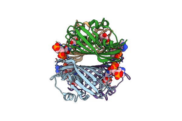
Crystal Structure Of Mutant Thioesterase Pa1618 (Q49A) From Pseudomonas Aeruginosa
Organism: Pseudomonas aeruginosa
Method: X-RAY DIFFRACTION Resolution:2.02 Å Release Date: 2015-05-13 Classification: HYDROLASE |
Organism: Pseudomonas aeruginosa
Method: X-RAY DIFFRACTION
Release Date: 2015-05-13
 |
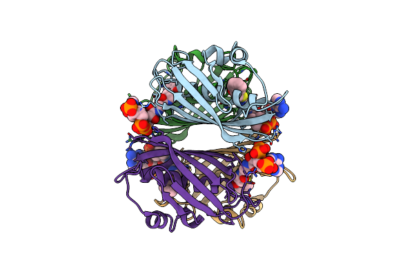
X-Ray Crystal Structure Of E. Coli Ydii Complexed With 2,4-Dihydroxyphenacyl Coa
Organism: Escherichia coli
Method: X-RAY DIFFRACTION Resolution:1.89 Å Release Date: 2014-07-30 Classification: HYDROLASE Ligands: HFQ |
Organism: Escherichia coli
Method: X-RAY DIFFRACTION
Release Date: 2014-07-30
Ligands: HFQ
 |
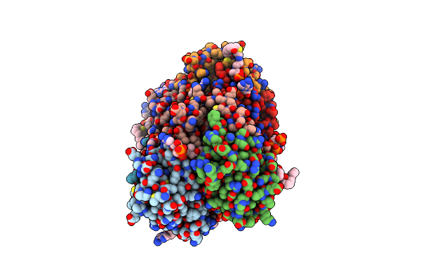
X-Ray Crystal Structure Of E. Coli Ydii Complexed With Phenacyl-Coa
Organism: Escherichia coli
Method: X-RAY DIFFRACTION Resolution:1.89 Å Release Date: 2014-07-30 Classification: HYDROLASE Ligands: 0FQ |
Organism: Escherichia coli
Method: X-RAY DIFFRACTION
Release Date: 2014-07-30
Ligands: 0FQ
 |
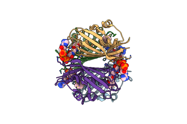
X-Ray Crystal Structure Of E. Coli Ydii Complexed With Undeca-2-One-Coa
Organism: Escherichia coli
Method: X-RAY DIFFRACTION Resolution:1.90 Å Release Date: 2014-07-30 Classification: HYDROLASE Ligands: UOQ, CL |
Organism: Escherichia coli
Method: X-RAY DIFFRACTION
Release Date: 2014-07-30
Ligands: UOQ, CL
 |
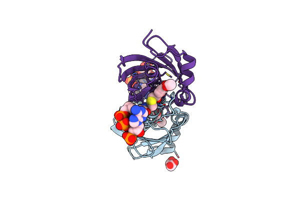
X-Ray Crystal Structure Of E. Coli Ybdb Complexed With Phenacyl-Coa
Organism: Escherichia coli
Method: X-RAY DIFFRACTION Resolution:1.85 Å Release Date: 2014-07-30 Classification: HYDROLASE Ligands: 0FQ |
Organism: Escherichia coli
Method: X-RAY DIFFRACTION
Release Date: 2014-07-30
Ligands: 0FQ
 |
X-Ray Crystal Structure Of E. Coli Ybdb Complexed With 2,4-Dihydroxyphenacyl-Coa
Organism: Escherichia coli
Method: X-RAY DIFFRACTION Resolution:2.17 Å Release Date: 2014-07-30 Classification: HYDROLASE Ligands: HFQ, MLI, ACT |
Organism: Escherichia coli
Method: X-RAY DIFFRACTION
Release Date: 2014-07-30
Ligands: HFQ, MLI, ACT