

Search Count: 22
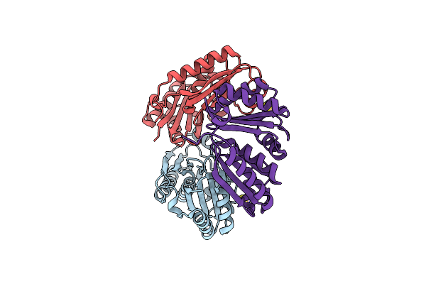 |
Carboxysome Shell Protein Ccmp From Synechococcus Elongatus Pcc 7942
Organism: Synechococcus elongatus pcc 7942
Method: X-RAY DIFFRACTION Resolution:1.65 Å Release Date: 2017-04-12 Classification: TRANSPORT PROTEIN Ligands: SCN |
Organism: Synechococcus elongatus pcc 7942
Method: X-RAY DIFFRACTION
Release Date: 2017-04-12
Ligands: SCN
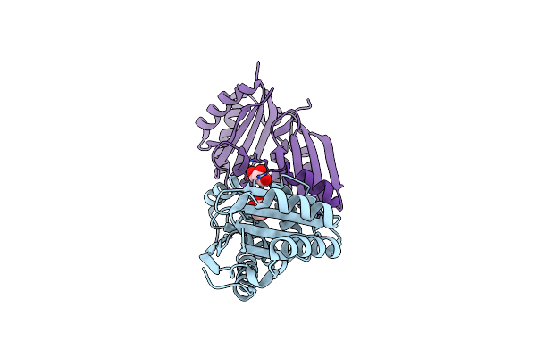 |
Carboxysome Shell Protein Ccmp From Synechococcus Elongatus Pcc 7942
Organism: Synechococcus elongatus pcc 7942
Method: X-RAY DIFFRACTION Resolution:1.45 Å Release Date: 2017-04-12 Classification: TRANSPORT PROTEIN Ligands: GOL, CL |
Organism: Synechococcus elongatus pcc 7942
Method: X-RAY DIFFRACTION
Release Date: 2017-04-12
Ligands: GOL, CL
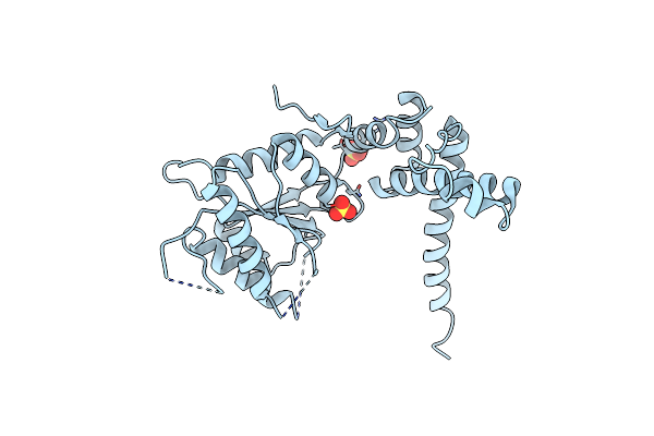 |
Rubisco Activase From Arabidopsis Thaliana
Organism: Arabidopsis thaliana
Method: X-RAY DIFFRACTION Resolution:2.90 Å Release Date: 2015-04-08 Classification: CHAPERONE Ligands: SO4 |
Organism: Arabidopsis thaliana
Method: X-RAY DIFFRACTION
Release Date: 2015-04-08
Ligands: SO4
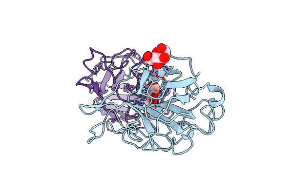 |
The Lytic Polysaccharide Monooxygenase Gh61D Structure From The Basidiomycota Fungus Phanerochaete Chrysosporium
Organism: Phanerochaete chrysosporium
Method: X-RAY DIFFRACTION Resolution:1.75 Å Release Date: 2013-04-03 Classification: HYDROLASE Ligands: CU, GOL, MAN |
Organism: Phanerochaete chrysosporium
Method: X-RAY DIFFRACTION
Release Date: 2013-04-03
Ligands: CU, GOL, MAN
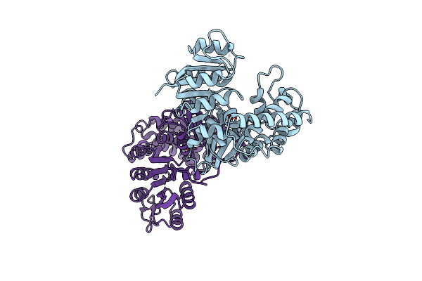 |
X-Ray Structure Of 1-Deoxy-D-Xylulose 5-Phosphate Reductoisomerase, Dxr, Rv2870C, From Mycobacterium Tuberculosis, In Complex With Manganese.
Organism: Mycobacterium tuberculosis
Method: X-RAY DIFFRACTION Resolution:1.90 Å Release Date: 2011-06-29 Classification: OXIDOREDUCTASE Ligands: MN |
Organism: Mycobacterium tuberculosis
Method: X-RAY DIFFRACTION
Release Date: 2011-06-29
Ligands: MN
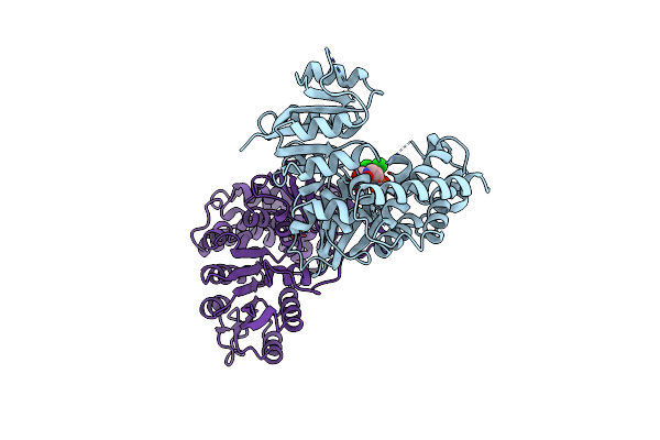 |
X-Ray Structure Of 1-Deoxy-D-Xylulose 5-Phosphate Reductoisomerase, Dxr, Rv2870C, From Mycobacterium Tuberculosis, In Complex With A 3,4- Dichlorophenyl-Substituted Fosmidomycin Analogue And Manganese.
Organism: Mycobacterium tuberculosis
Method: X-RAY DIFFRACTION Resolution:2.05 Å Release Date: 2011-06-29 Classification: OXIDOREDUCTASE Ligands: 34F, MN |
Organism: Mycobacterium tuberculosis
Method: X-RAY DIFFRACTION
Release Date: 2011-06-29
Ligands: 34F, MN
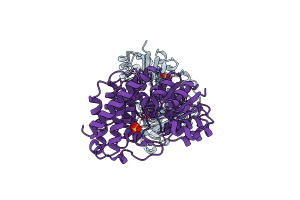 |
X-Ray Structure Of 1-Deoxy-D-Xylulose 5-Phosphate Reductoisomerase, Dxr, Rv2870C, From Mycobacterium Tuberculosis, In Complex With Manganese.
Organism: Mycobacterium tuberculosis
Method: X-RAY DIFFRACTION Resolution:1.65 Å Release Date: 2011-06-29 Classification: OXIDOREDUCTASE Ligands: MN, SO4 |
Organism: Mycobacterium tuberculosis
Method: X-RAY DIFFRACTION
Release Date: 2011-06-29
Ligands: MN, SO4
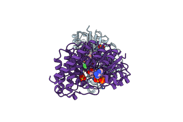 |
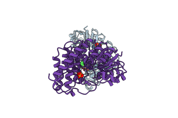
X-Ray Structure Of 1-Deoxy-D-Xylulose 5-Phosphate Reductoisomerase, Dxr, Rv2870C, From Mycobacterium Tuberculosis, In Complex With A 3,4- Dichlorophenyl-Substituted Fosmidomycin Analogue, Manganese And Nadph.
Organism: Mycobacterium tuberculosis
Method: X-RAY DIFFRACTION Resolution:1.96 Å Release Date: 2011-06-29 Classification: OXIDOREDUCTASE Ligands: 34F, NDP, MN, SO4 |
Organism: Mycobacterium tuberculosis
Method: X-RAY DIFFRACTION
Release Date: 2011-06-29
Ligands: 34F, NDP, MN, SO4
 |
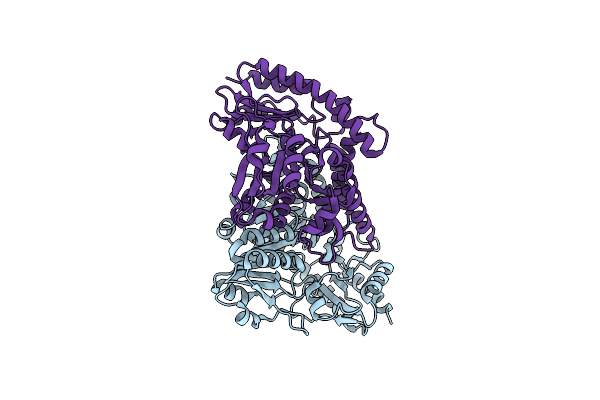
X-Ray Structure Of 1-Deoxy-D-Xylulose 5-Phosphate Reductoisomerase, Dxr, Rv2870C, From Mycobacterium Tuberculosis, In Complex With A 3,4- Dichlorophenyl-Substituted Fr900098 Analogue And Manganese.
Organism: Mycobacterium tuberculosis
Method: X-RAY DIFFRACTION Resolution:1.95 Å Release Date: 2011-06-29 Classification: OXIDOREDUCTASE Ligands: FM5, MN, SO4 |
Organism: Mycobacterium tuberculosis
Method: X-RAY DIFFRACTION
Release Date: 2011-06-29
Ligands: FM5, MN, SO4
 |
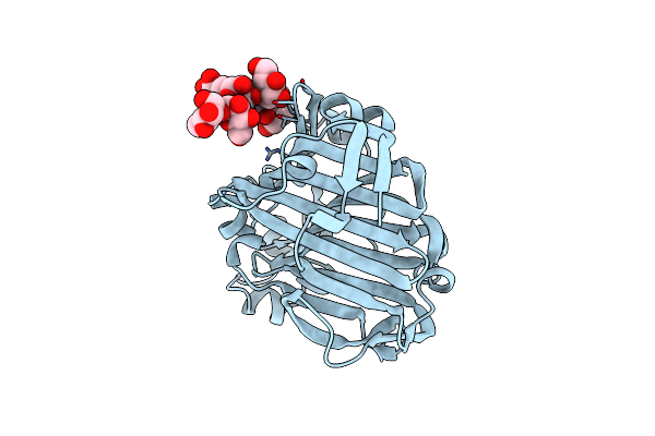
Crystal Structure Of Mycobacterium Tuberculosis Rv3778C Protein
Organism: Mycobacterium tuberculosis
Method: X-RAY DIFFRACTION Resolution:1.80 Å Release Date: 2008-03-11 Classification: TRANSFERASE |
Organism: Mycobacterium tuberculosis
Method: X-RAY DIFFRACTION
Release Date: 2008-03-11
 |
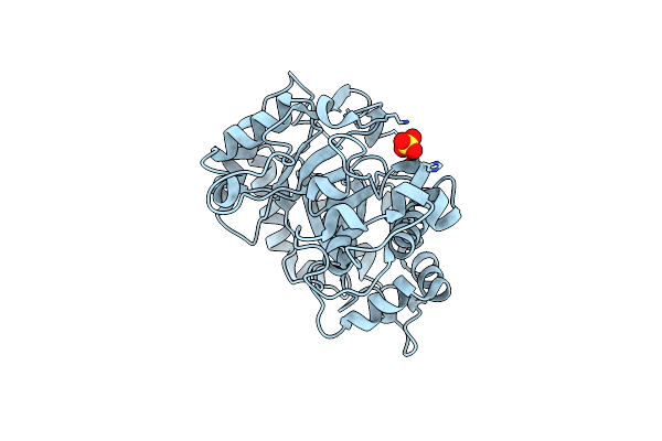
Endo-1,3(4)-Beta-Glucanase From Phanerochaete Chrysosporium, Solved Using Native Sulfur Sad, Exhibiting Intact Heptasaccharide Glycosylation
Organism: Phanerochaete chrysosporium
Method: X-RAY DIFFRACTION Resolution:1.35 Å Release Date: 2006-10-25 Classification: HYDROLASE |
Organism: Phanerochaete chrysosporium
Method: X-RAY DIFFRACTION
Release Date: 2006-10-25
 |
X-Ray Structure Of Beta-Mannanase From Blue Mussel Mytilus Edulis
Organism: Mytilus edulis
Method: X-RAY DIFFRACTION Resolution:1.60 Å Release Date: 2006-02-22 Classification: HYDROLASE Ligands: SO4 |
Organism: Mytilus edulis
Method: X-RAY DIFFRACTION
Release Date: 2006-02-22
Ligands: SO4
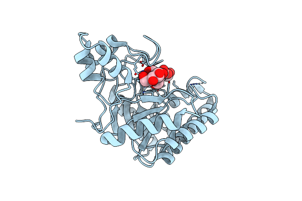 |
Catalytic Domain Of Endo-1,4-Glucanase Cel6A From Thermobifida Fusca In Complex With Methyl Cellobiosyl-4-Thio-Beta-Cellobioside
Organism: Thermomonospora fusca
Method: X-RAY DIFFRACTION Resolution:1.50 Å Release Date: 2005-10-05 Classification: HYDROLASE |
Organism: Thermomonospora fusca
Method: X-RAY DIFFRACTION
Release Date: 2005-10-05
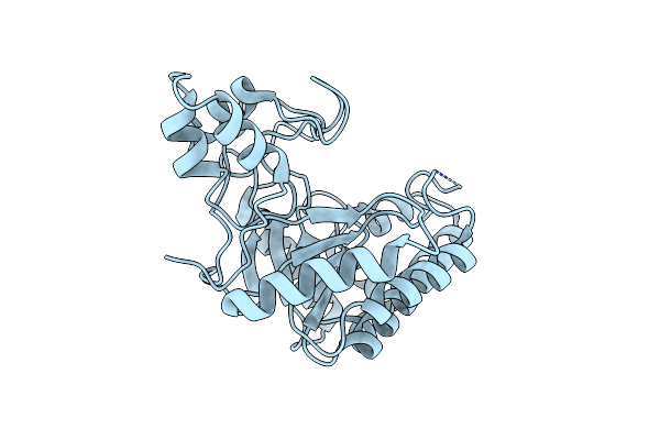 |
Catalytic Domain Of Endo-1,4-Glucanase Cel6A Mutant Y73S From Thermobifida Fusca
Organism: Thermomonospora fusca
Method: X-RAY DIFFRACTION Resolution:1.15 Å Release Date: 2005-10-05 Classification: HYDROLASE |
Organism: Thermomonospora fusca
Method: X-RAY DIFFRACTION
Release Date: 2005-10-05
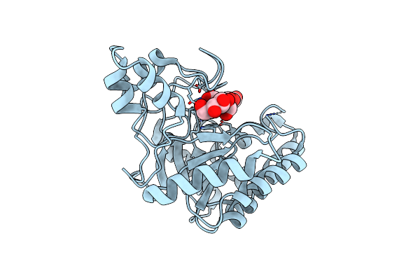 |
Catalytic Domain Of Endo-1,4-Glucanase Cel6A Mutant Y73S From Thermobifida Fusca In Complex With Cellotetrose
Organism: Thermomonospora fusca
Method: X-RAY DIFFRACTION Resolution:1.64 Å Release Date: 2005-10-05 Classification: HYDROLASE |
Organism: Thermomonospora fusca
Method: X-RAY DIFFRACTION
Release Date: 2005-10-05
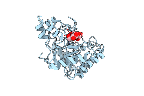 |
Catalytic Domain Of Endo-1,4-Glucanase Cel6A Mutant Y73S From Thermobifida Fusca In Complex With Methyl Cellobiosyl-4-Thio-Beta- Cellobioside
Organism: Thermomonospora fusca
Method: X-RAY DIFFRACTION Resolution:1.04 Å Release Date: 2005-10-05 Classification: HYDROLASE |
Organism: Thermomonospora fusca
Method: X-RAY DIFFRACTION
Release Date: 2005-10-05
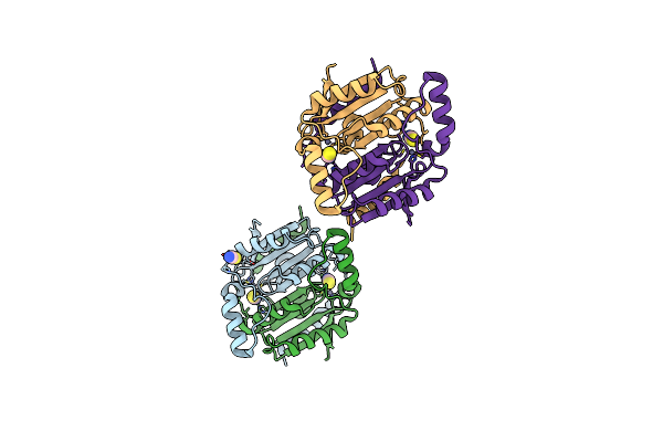 |
Crystal Structure Of Rv1284 From Mycobacterium Tuberculosis In Complex With Thiocyanate
Organism: Mycobacterium tuberculosis
Method: X-RAY DIFFRACTION Resolution:2.00 Å Release Date: 2005-03-08 Classification: UNKNOWN FUNCTION Ligands: ZN, SCN |
Organism: Mycobacterium tuberculosis
Method: X-RAY DIFFRACTION
Release Date: 2005-03-08
Ligands: ZN, SCN
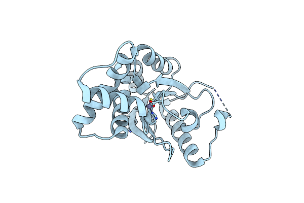 |
Crystal Structure Of Carbonic Anhydrase Rv3588C From Mycobacterium Tuberculosis
Organism: Mycobacterium tuberculosis
Method: X-RAY DIFFRACTION Resolution:1.75 Å Release Date: 2005-03-08 Classification: LYASE Ligands: ZN, MG |
Organism: Mycobacterium tuberculosis
Method: X-RAY DIFFRACTION
Release Date: 2005-03-08
Ligands: ZN, MG
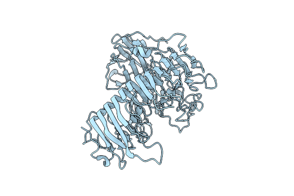 |
Dex49A From Penicillium Minioluteum
Organism: Penicillium minioluteum
Method: X-RAY DIFFRACTION Resolution:1.80 Å Release Date: 2003-09-11 Classification: HYDROLASE |
Organism: Penicillium minioluteum
Method: X-RAY DIFFRACTION
Release Date: 2003-09-11
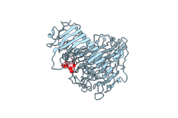 |
Dex49A From Penicillium Minioluteum Complex With Isomaltose
Organism: Penicillium minioluteum
Method: X-RAY DIFFRACTION Resolution:1.65 Å Release Date: 2003-09-11 Classification: HYDROLASE |
Organism: Penicillium minioluteum
Method: X-RAY DIFFRACTION
Release Date: 2003-09-11