
Search Count: 170
All
Selected
 |
Organism: Homo sapiens, Severe acute respiratory syndrome coronavirus 2, Mus musculus
Method: ELECTRON MICROSCOPY Release Date: 2026-03-18 Classification: VIRAL PROTEIN/HYDROLASE Ligands: NAG |
 |
Organism: Severe acute respiratory syndrome coronavirus 2, Mus musculus
Method: ELECTRON MICROSCOPY Resolution:2.33 Å Release Date: 2026-03-18 Classification: VIRAL PROTEIN |
 |
Organism: Bacillus phage spbetal1
Method: X-RAY DIFFRACTION Resolution:2.30 Å Release Date: 2025-12-24 Classification: VIRAL PROTEIN Ligands: NAD, SO4, GOL |
 |
Organism: Bacillus phage spbetal1
Method: X-RAY DIFFRACTION Resolution:2.40 Å Release Date: 2025-12-24 Classification: VIRAL PROTEIN Ligands: GOL, NCA, SO4 |
 |
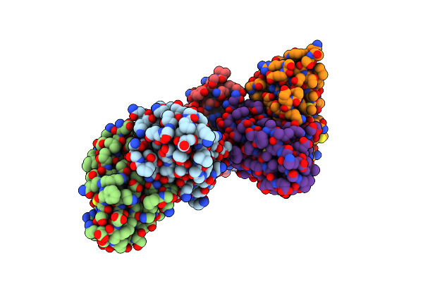
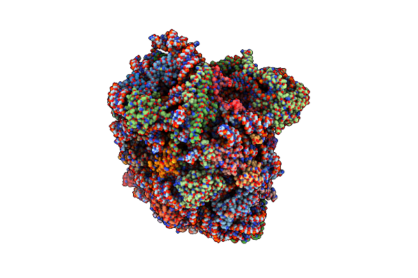
Cryo-Em Structure Of Sars-Cov-2 Ba.5 Spike Protein In Complex With Nab 1C4 (Local Refinement)
Organism: Severe acute respiratory syndrome coronavirus 2, Mus musculus
Method: ELECTRON MICROSCOPY Release Date: 2025-12-10 Classification: VIRAL PROTEIN/IMMUNE SYSTEM |
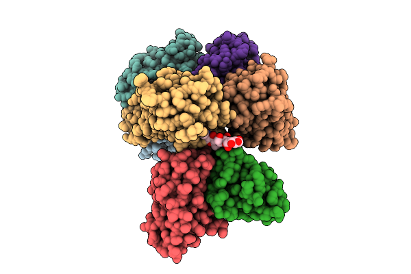 |
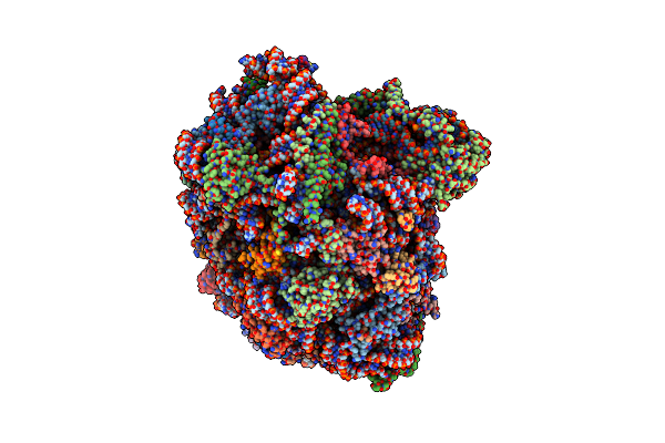
Cryo-Em Structure Of Sars-Cov-2 Spike Protein In Complex With Three-Nab 8H12, 3E2 And 1C4
Organism: Severe acute respiratory syndrome coronavirus 2, Mus musculus
Method: ELECTRON MICROSCOPY Release Date: 2025-12-10 Classification: VIRAL PROTEIN/IMMUNE SYSTEM |
 |
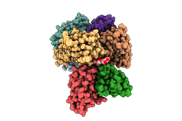
Cryo-Em Structure Of Sars-Cov-2 Ba.1 Spike Protein In Complex With Three-Nab 8H12, 3E2 And 1C4
Organism: Severe acute respiratory syndrome coronavirus 2, Mus musculus
Method: ELECTRON MICROSCOPY Release Date: 2025-12-10 Classification: VIRAL PROTEIN/IMMUNE SYSTEM |
 |
Cryo-Em Structure Of Sars-Cov-2 Ba.2 Spike Protein In Complex With Triple-Nab 8H12, 3E2 And 1C4 (Local Refinement)
Organism: Severe acute respiratory syndrome coronavirus 2, Mus musculus
Method: ELECTRON MICROSCOPY Release Date: 2025-12-10 Classification: VIRAL PROTEIN/IMMUNE SYSTEM |
 |
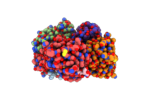
Organism: Homo sapiens, Sus scrofa
Method: ELECTRON MICROSCOPY Release Date: 2025-12-03 Classification: MOTOR PROTEIN Ligands: ADP, ATP, ZN |
 |
Organism: Trypanosoma cruzi
Method: ELECTRON MICROSCOPY Release Date: 2025-10-01 Classification: HYDROLASE |
 |
Organism: Trypanosoma cruzi
Method: ELECTRON MICROSCOPY Resolution:3.82 Å Release Date: 2025-10-01 Classification: HYDROLASE |
 |
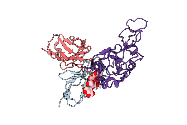
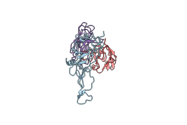
Locally Refined Region Of Sars-Cov-2 Spike In Complex With Antibodies 9G11 And 3E2.
Organism: Severe acute respiratory syndrome coronavirus 2, Mus musculus
Method: ELECTRON MICROSCOPY Release Date: 2025-02-12 Classification: VIRAL PROTEIN/IMMUNE SYSTEM Ligands: NAG |
 |
Organism: Mus musculus, Saccharomyces cerevisiae
Method: ELECTRON MICROSCOPY Release Date: 2024-06-26 Classification: RIBOSOME Ligands: MG, CL, SPM, SPD, ZN |
 |
Organism: Saccharomyces cerevisiae
Method: ELECTRON MICROSCOPY Release Date: 2024-06-12 Classification: RIBOSOME Ligands: MG, SPM, SPD, ZN |
 |
Organism: Mus musculus, Saccharomyces cerevisiae
Method: ELECTRON MICROSCOPY Release Date: 2024-06-12 Classification: RIBOSOME Ligands: MG, CL, SPD, SPM, ZN |
 |
Organism: Saccharomyces cerevisiae
Method: ELECTRON MICROSCOPY Release Date: 2024-06-12 Classification: RIBOSOME Ligands: MG, CL, SPM, SPD, ZN |
 |
Cryo-Em Structure Of Sars-Cov-2 Ba.4/5 Spike Protein In Complex With 1G11 (Local Refinement)
Organism: Homo sapiens, Severe acute respiratory syndrome coronavirus 2
Method: ELECTRON MICROSCOPY Release Date: 2023-11-15 Classification: VIRAL PROTEIN/IMMUNE SYSTEM |
 |
Cryo-Em Structure Of Sars-Cov-2 Spike Protein In Complex With Double Nabs 8H12 And 3E2 (Local Refinement)
Organism: Severe acute respiratory syndrome coronavirus 2, Mus musculus
Method: ELECTRON MICROSCOPY Release Date: 2023-08-16 Classification: VIRAL PROTEIN/IMMUNE SYSTEM |
 |
Cryo-Em Structure Of Sars-Cov-2 Spike Protein In Complex With Double Nabs 8H12 And 1C4 (Local Refinement)
Organism: Severe acute respiratory syndrome coronavirus 2, Mus musculus
Method: ELECTRON MICROSCOPY Release Date: 2023-08-16 Classification: VIRAL PROTEIN/IMMUNE SYSTEM |
 |
Cryo-Em Structure Of Sars-Cov-2 Spike Protein In Complex With Double Nabs 3E2 And 1C4 (Local Refinement)
Organism: Severe acute respiratory syndrome coronavirus 2, Mus musculus
Method: ELECTRON MICROSCOPY Release Date: 2023-08-16 Classification: VIRAL PROTEIN/IMMUNE SYSTEM |