

Search Count: 11
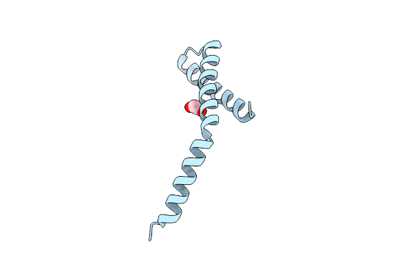 |
Crystal Structure Of Periplasmic Domain Of Antiholin Ri From T4 Phage
Organism: Enterobacteria phage t4
Method: X-RAY DIFFRACTION Resolution:2.21 Å Release Date: 2020-06-24 Classification: VIRAL PROTEIN Ligands: EDO |
Organism: Enterobacteria phage t4
Method: X-RAY DIFFRACTION
Release Date: 2020-06-24
Ligands: EDO
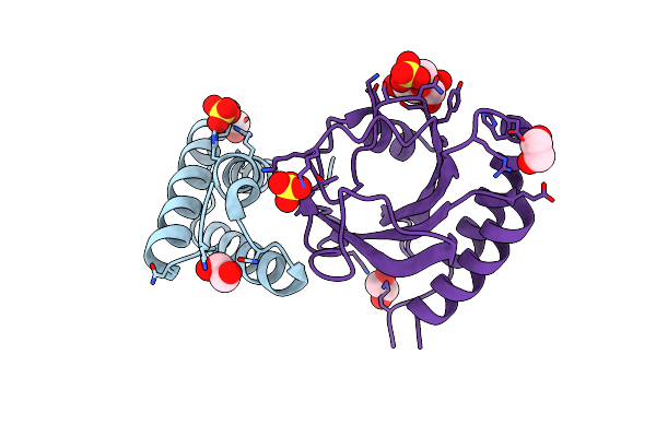 |
Crystal Structure Of The Complex Between Periplasmic Domains Of Antiholin Ri And Holin T From T4 Phage, In P6522
Organism: Escherichia phage ecml-134, Escherichia phage vb_ecom_nbg2
Method: X-RAY DIFFRACTION Resolution:2.20 Å Release Date: 2020-06-24 Classification: VIRAL PROTEIN Ligands: SO4, EDO, BTB, CL |
Organism: Escherichia phage ecml-134, Escherichia phage vb_ecom_nbg2
Method: X-RAY DIFFRACTION
Release Date: 2020-06-24
Ligands: SO4, EDO, BTB, CL
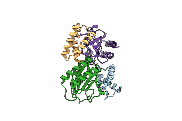 |
Crystal Structure Of The Complex Between Periplasmic Domains Of Antiholin Ri And Holin T From T4 Phage, In H32
Organism: Escherichia phage ecml-134, Escherichia phage vb_ecom_nbg2
Method: X-RAY DIFFRACTION Resolution:1.65 Å Release Date: 2020-06-24 Classification: VIRAL PROTEIN |
Organism: Escherichia phage ecml-134, Escherichia phage vb_ecom_nbg2
Method: X-RAY DIFFRACTION
Release Date: 2020-06-24
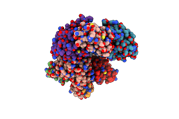 |
Crystal Structure Of The Complex Between Periplasmic Domains Of Antiholin Ri And Holin T From T4 Phage, In P21
Organism: Enterobacteria phage t4, Escherichia phage vb_ecom_nbg2
Method: X-RAY DIFFRACTION Resolution:2.30 Å Release Date: 2020-06-24 Classification: VIRAL PROTEIN |
Organism: Enterobacteria phage t4, Escherichia phage vb_ecom_nbg2
Method: X-RAY DIFFRACTION
Release Date: 2020-06-24
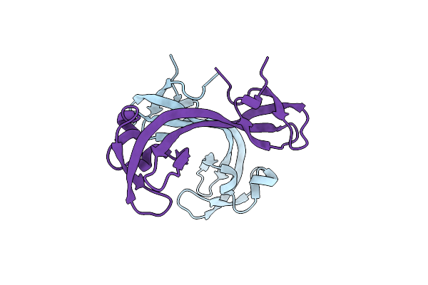 |
Crystal Structure Of Hybrid Tudor Domain Of Human Lysine Demethylase Kdm4B
Organism: Homo sapiens
Method: X-RAY DIFFRACTION Resolution:2.56 Å Release Date: 2016-03-16 Classification: REPLICATION |
Organism: Homo sapiens
Method: X-RAY DIFFRACTION
Release Date: 2016-03-16
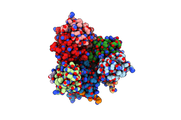 |
Crystal Structure Of Double Tudor Domain Of Human Lysine Demethylase Kdm4A Complexed With Histone H3K23Me3
Organism: Homo sapiens
Method: X-RAY DIFFRACTION Resolution:2.29 Å Release Date: 2016-02-10 Classification: OXIDOREDUCTASE |
Organism: Homo sapiens
Method: X-RAY DIFFRACTION
Release Date: 2016-02-10
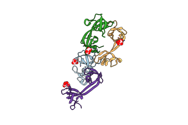 |
Crystal Structure Of Double Tudor Domain Of Human Lysine Demethylase Kdm4A
Organism: Homo sapiens
Method: X-RAY DIFFRACTION Resolution:1.99 Å Release Date: 2015-11-25 Classification: OXIDOREDUCTASE Ligands: SRT |
Organism: Homo sapiens
Method: X-RAY DIFFRACTION
Release Date: 2015-11-25
Ligands: SRT
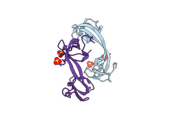 |
Crystal Structure Of Double Tudor Domain Of Human Lysine Demethylase Kdm4A
Organism: Homo sapiens
Method: X-RAY DIFFRACTION Resolution:2.15 Å Release Date: 2015-11-25 Classification: OXIDOREDUCTASE Ligands: SO4 |
Organism: Homo sapiens
Method: X-RAY DIFFRACTION
Release Date: 2015-11-25
Ligands: SO4
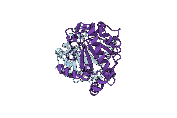 |
Structure Of The Salmonella Plasmid Virulence C Protein (Spvc) H106N Mutant.
Organism: Salmonella enterica subsp. enterica serovar typhimurium
Method: X-RAY DIFFRACTION Resolution:2.30 Å Release Date: 2013-08-07 Classification: LYASE |
Organism: Salmonella enterica subsp. enterica serovar typhimurium
Method: X-RAY DIFFRACTION
Release Date: 2013-08-07
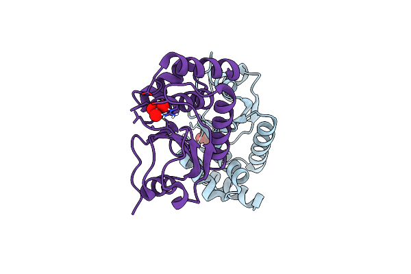 |
Structure Of Vhz Bound To Metavanadate
Organism: Homo sapiens
Method: X-RAY DIFFRACTION Resolution:1.15 Å Release Date: 2012-12-19 Classification: HYDROLASE Ligands: VN4 |
Organism: Homo sapiens
Method: X-RAY DIFFRACTION
Release Date: 2012-12-19
Ligands: VN4
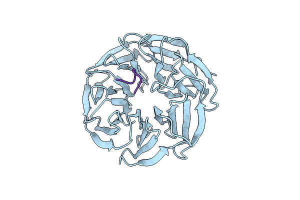 |
Symmetric Dimethylation Of H3 Arginine 2 Is A Novel Histone Mark That Supports Euchromatin Maintenance
Organism: Homo sapiens
Method: X-RAY DIFFRACTION Resolution:1.90 Å Release Date: 2012-01-11 Classification: TRANSCRIPTION |
Organism: Homo sapiens
Method: X-RAY DIFFRACTION
Release Date: 2012-01-11