

Search Count: 18
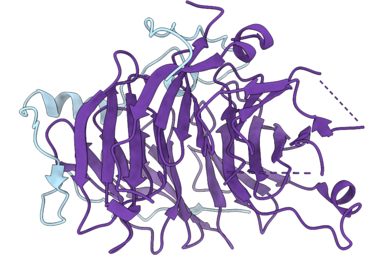 |
Structure Of S. Pombe Pnuts (565 - 644) Bound To Swd2.2 - Crystal Form 2
Organism: Schizosaccharomyces pombe
Method: X-RAY DIFFRACTION Resolution:1.53 Å Release Date: 2026-08-05 Classification: TRANSCRIPTION |
Organism: Schizosaccharomyces pombe
Method: X-RAY DIFFRACTION
Release Date: 2026-08-05
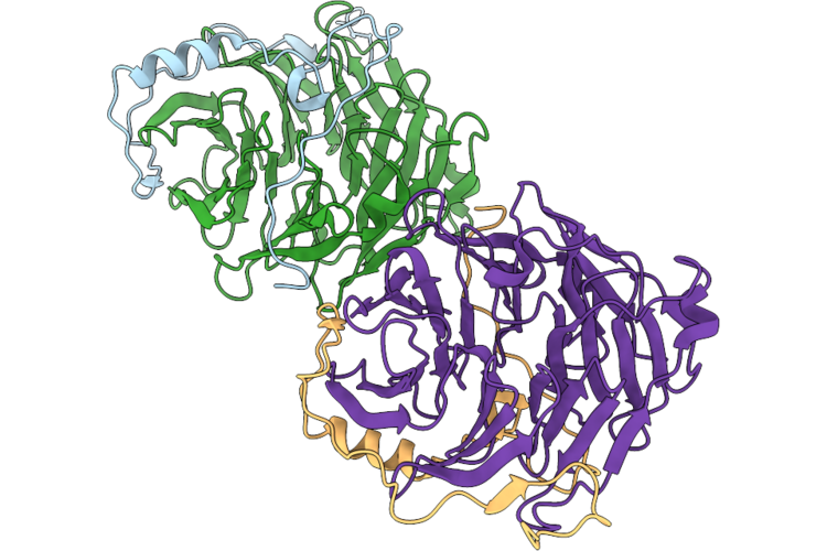 |
Structure Of S. Pombe Pnuts (565 - 644) Bound To Swd2.2 - Crystal Form 1
Organism: Schizosaccharomyces pombe
Method: X-RAY DIFFRACTION Resolution:1.61 Å Release Date: 2026-08-05 Classification: TRANSCRIPTION |
Organism: Schizosaccharomyces pombe
Method: X-RAY DIFFRACTION
Release Date: 2026-08-05
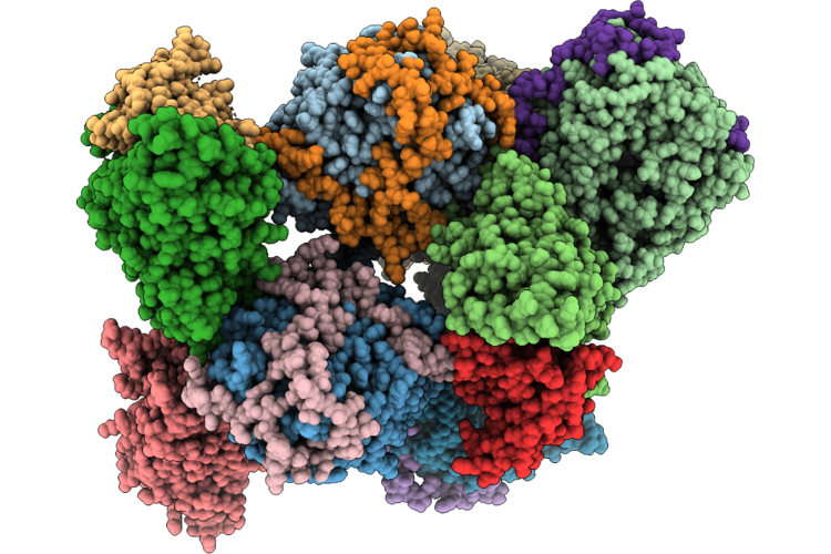 |
Structure Of S. Pombe Pta1 - Ssu72 - Pnuts - Swd2.2 Complex - Crystal Form 1
Organism: Schizosaccharomyces pombe, Schizosaccharomyces pombe (strain 972 / atcc 24843)
Method: X-RAY DIFFRACTION Resolution:3.79 Å Release Date: 2026-08-05 Classification: TRANSCRIPTION |
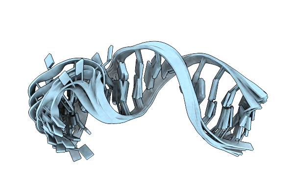 |
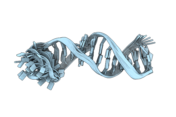
5_Sl5B_Gc Of The 5_Sl5 Rna
Organism: Severe acute respiratory syndrome coronavirus
Method: SOLUTION NMR Release Date: 2024-11-06 Classification: RNA |
Organism: Severe acute respiratory syndrome coronavirus
Method: SOLUTION NMR
Release Date: 2024-11-06
 |
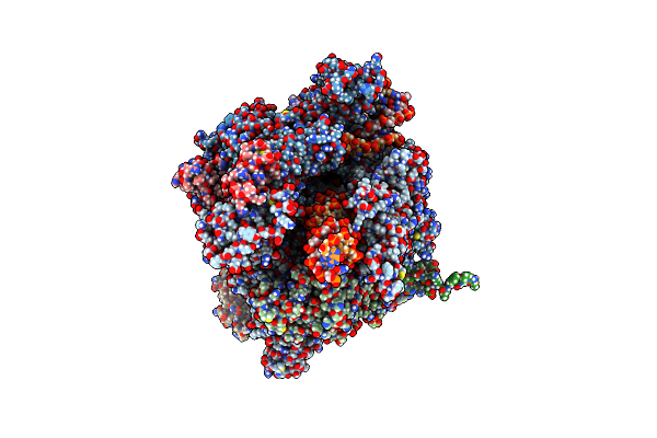
5_Sl5A Of S_Sl5 Of Sars-Cov-2
Organism: Severe acute respiratory syndrome coronavirus
Method: SOLUTION NMR Release Date: 2024-11-06 Classification: RNA |
Organism: Severe acute respiratory syndrome coronavirus
Method: SOLUTION NMR
Release Date: 2024-11-06
 |
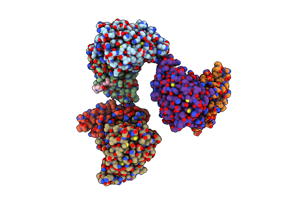
Structure Of S. Pombe Rna Polymerase Ii In Complex With Dsif And Rat1/Rai1
Method: ELECTRON MICROSCOPY
Release Date: 2024-10-23 Classification: RNA BINDING PROTEIN Ligands: ZN |
Method: ELECTRON MICROSCOPY
Release Date: 2024-10-23
Ligands: ZN
 |
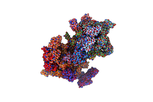
Crystal Structure Of Procaspase-8 In Complex With Covalent Small Molecule Inhibitor 63-R
Organism: Homo sapiens
Method: X-RAY DIFFRACTION Resolution:2.88 Å Release Date: 2020-01-29 Classification: HYDROLASE/HYDROLASE INHIBITOR Ligands: 63R |
Organism: Homo sapiens
Method: X-RAY DIFFRACTION
Release Date: 2020-01-29
Ligands: 63R
 |
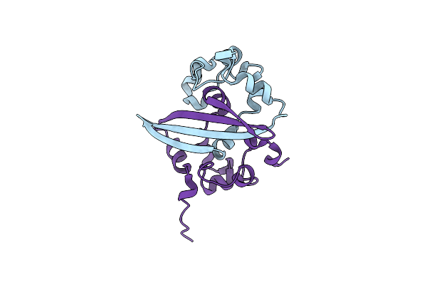
X-Ray Crystal Structure Of Escherichia Coli Rna Polymerase (Rpob-H526Y) And Ppapp Complex
Organism: Escherichia coli
Method: X-RAY DIFFRACTION Resolution:3.60 Å Release Date: 2018-01-17 Classification: transferase/transferase inhibitor Ligands: ECJ, MG, ZN |
Organism: Escherichia coli
Method: X-RAY DIFFRACTION
Release Date: 2018-01-17
Ligands: ECJ, MG, ZN
 |
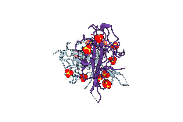
C-Terminal Domain Of Human Coronavirus Nl63 Nucleocapsid Protein
Organism: Human coronavirus nl63
Method: X-RAY DIFFRACTION Resolution:1.50 Å Release Date: 2017-02-22 Classification: VIRAL PROTEIN |
Organism: Human coronavirus nl63
Method: X-RAY DIFFRACTION
Release Date: 2017-02-22
 |
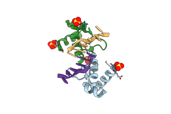
N-Terminal Domain Of A Human Coronavirus Nl63 Nucleocapsid Protein
Organism: Human coronavirus nl63
Method: X-RAY DIFFRACTION Resolution:1.49 Å Release Date: 2017-02-22 Classification: RNA BINDING PROTEIN Ligands: SO4 |
Organism: Human coronavirus nl63
Method: X-RAY DIFFRACTION
Release Date: 2017-02-22
Ligands: SO4
 |
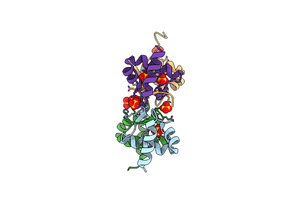
The Binding Mode Of Cyprinid Herpesvirus3 Orf112-Zalpha To Z-Dna
Organism: Cyprinid herpesvirus 3, Synthetic construct
Method: X-RAY DIFFRACTION Resolution:1.50 Å Release Date: 2015-11-18 Classification: DNA BINDING PROTEIN Ligands: SO4 |
Organism: Cyprinid herpesvirus 3, Synthetic construct
Method: X-RAY DIFFRACTION
Release Date: 2015-11-18
Ligands: SO4
 |
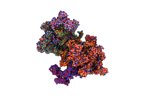
The Crystal Structure Of The Zalpha Domain From Cyprinid Herpes Virus 3
Organism: Cyprinid herpesvirus 3
Method: X-RAY DIFFRACTION Resolution:1.76 Å Release Date: 2013-09-11 Classification: DNA BINDING PROTEIN Ligands: SO4 |
Organism: Cyprinid herpesvirus 3
Method: X-RAY DIFFRACTION
Release Date: 2013-09-11
Ligands: SO4
 |
X-Ray Crystal Structure Of Escherichia Coli Sigma70 Holoenzyme In Complex With Guanosine Pentaphosphate (Pppgpp)
Organism: Escherichia coli
Method: X-RAY DIFFRACTION Resolution:4.20 Å Release Date: 2013-04-17 Classification: TRANSCRIPTION, TRANSFERASE Ligands: ZN, 0O2 |
Organism: Escherichia coli
Method: X-RAY DIFFRACTION
Release Date: 2013-04-17
Ligands: ZN, 0O2
 |
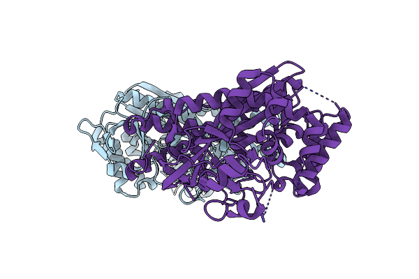
X-Ray Crystal Structure Of Escherichia Coli Sigma70 Holoenzyme In Complex With Guanosine Tetraphosphate (Ppgpp)
Organism: Escherichia coli
Method: X-RAY DIFFRACTION Resolution:3.90 Å Release Date: 2013-04-10 Classification: TRANSCRIPTION, TRANSFERASE Ligands: ZN, G4P |
Organism: Escherichia coli
Method: X-RAY DIFFRACTION
Release Date: 2013-04-10
Ligands: ZN, G4P
 |
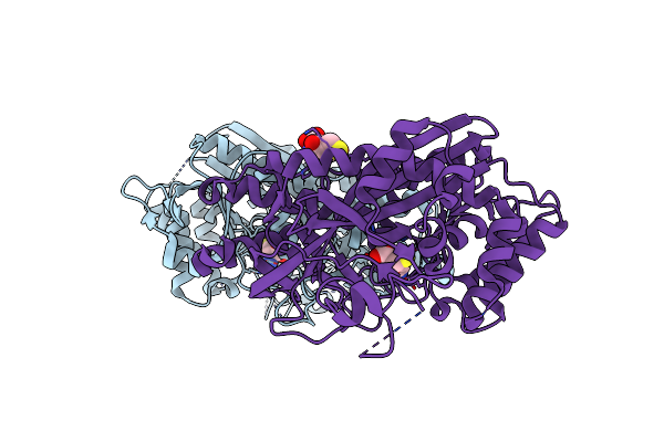
Apoenzyme Structure Of Homoglutathione Synthetase From Glycine Max In Open Conformation
Organism: Glycine max
Method: X-RAY DIFFRACTION Resolution:2.00 Å Release Date: 2009-12-22 Classification: LIGASE |
Organism: Glycine max
Method: X-RAY DIFFRACTION
Release Date: 2009-12-22
 |
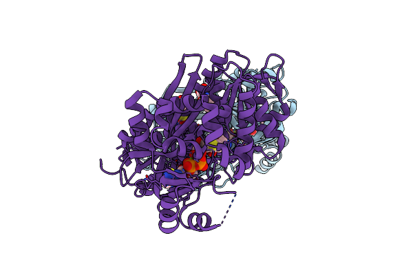
Structure Of Homoglutathione Synthetase From Glycine Max In Open Conformation With Gamma-Glutamyl-Cysteine Bound.
Organism: Glycine max
Method: X-RAY DIFFRACTION Resolution:2.11 Å Release Date: 2009-12-22 Classification: LIGASE Ligands: 3GC |
Organism: Glycine max
Method: X-RAY DIFFRACTION
Release Date: 2009-12-22
Ligands: 3GC
 |
Structure Of Homoglutathione Synthetase From Glycine Max In Closed Conformation With Homoglutathione, Adp, A Sulfate Ion, And Three Magnesium Ions Bound
Organism: Glycine max
Method: X-RAY DIFFRACTION Resolution:1.90 Å Release Date: 2009-12-22 Classification: LIGASE Ligands: ADP, HGS, MG, SO4 |
Organism: Glycine max
Method: X-RAY DIFFRACTION
Release Date: 2009-12-22
Ligands: ADP, HGS, MG, SO4
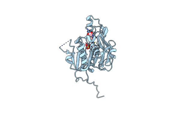 |
Crystal Structure Of Homoserine O-Acetyltransferase (Meta) From Bacillus Cereus With Homoserine
Organism: Bacillus cereus
Method: X-RAY DIFFRACTION Resolution:2.00 Å Release Date: 2008-01-22 Classification: TRANSFERASE Ligands: SO4, HSE |
Organism: Bacillus cereus
Method: X-RAY DIFFRACTION
Release Date: 2008-01-22
Ligands: SO4, HSE