

Search Count: 31
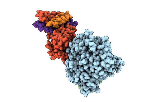 |
Symmetry-Expanded Reconstruction Of Augmin T-Ii Bonsai On The Gtpgammas Microtubule
Organism: Xenopus laevis, Bos taurus
Method: ELECTRON MICROSCOPY Release Date: 2025-11-12 Classification: CELL CYCLE Ligands: GTP, MG, GSP |
Organism: Xenopus laevis, Bos taurus
Method: ELECTRON MICROSCOPY
Release Date: 2025-11-12
Ligands: GTP, MG, GSP
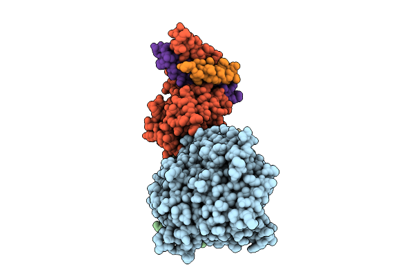 |
Symmetry-Expanded Reconstruction Of Augmin T-Ii Bonsai On The Microtubule
Organism: Xenopus laevis, Bos taurus
Method: ELECTRON MICROSCOPY Release Date: 2025-08-13 Classification: CELL CYCLE Ligands: GTP, MG, G2P |
Organism: Xenopus laevis, Bos taurus
Method: ELECTRON MICROSCOPY
Release Date: 2025-08-13
Ligands: GTP, MG, G2P
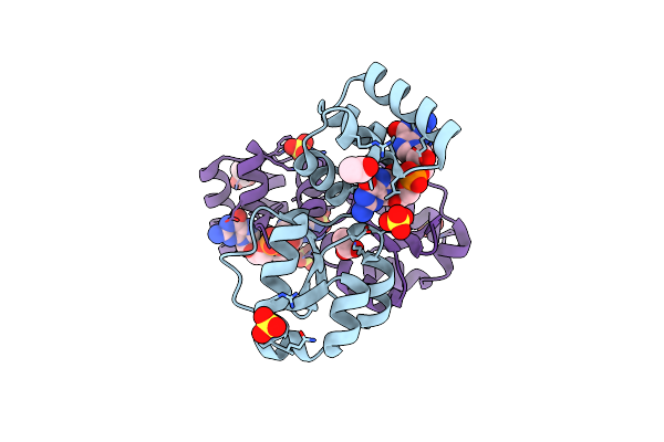 |
X-Ray Structure Of The Thermus Thermophilus Q190E Mutant Of The Pilf-Gspiib Domain In The C-Di-Gmp Bound State
Organism: Thermus thermophilus hb27
Method: X-RAY DIFFRACTION Resolution:1.90 Å Release Date: 2025-06-25 Classification: MOTOR PROTEIN Ligands: C2E, SO4, ACY |
Organism: Thermus thermophilus hb27
Method: X-RAY DIFFRACTION
Release Date: 2025-06-25
Ligands: C2E, SO4, ACY
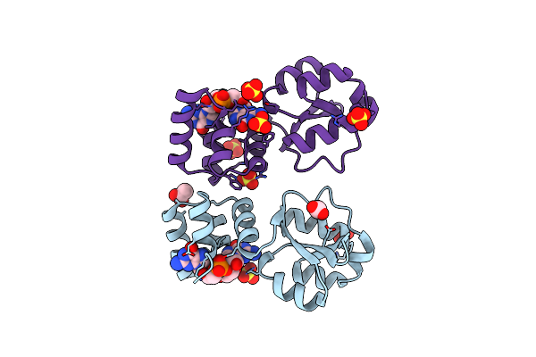 |
X-Ray Structure Of The Thermus Thermophilus Q218E Mutant Of The Pilf-Gspiib Domain In The C-Di-Gmp Bound State
Organism: Thermus thermophilus hb27
Method: X-RAY DIFFRACTION Resolution:1.55 Å Release Date: 2025-06-25 Classification: MOTOR PROTEIN Ligands: C2E, SO4, ACT |
Organism: Thermus thermophilus hb27
Method: X-RAY DIFFRACTION
Release Date: 2025-06-25
Ligands: C2E, SO4, ACT
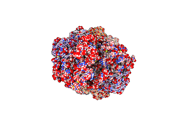 |
Magic Angle Spinning Nmr Structure Of Human Cofilin-2 Assembled On Actin Filaments
Organism: Oryctolagus cuniculus, Homo sapiens
Method: SOLID-STATE NMR Release Date: 2023-08-16 Classification: CONTRACTILE PROTEIN, CELL CYCLE |
Organism: Oryctolagus cuniculus, Homo sapiens
Method: SOLID-STATE NMR
Release Date: 2023-08-16
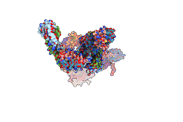 |
Structure Of The Vertebrate Augmin Complex
Organism: Xenopus laevis
Method: ELECTRON MICROSCOPY Release Date: 2023-04-19 Classification: CELL CYCLE |
Organism: Xenopus laevis
Method: ELECTRON MICROSCOPY
Release Date: 2023-04-19
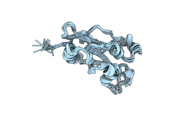 |
Magic Angle Spinning Nmr Structure Of Human Cofilin-2 Assembled On Actin Filaments
Organism: Homo sapiens
Method: SOLID-STATE NMR Release Date: 2022-04-20 Classification: CONTRACTILE PROTEIN |
Organism: Homo sapiens
Method: SOLID-STATE NMR
Release Date: 2022-04-20
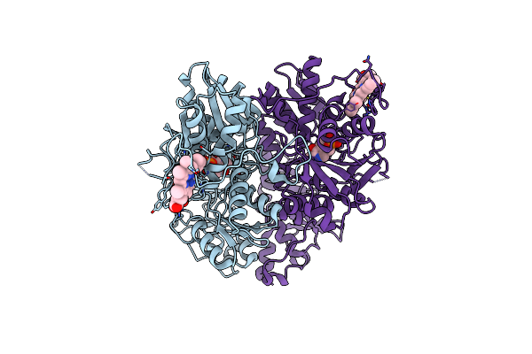 |
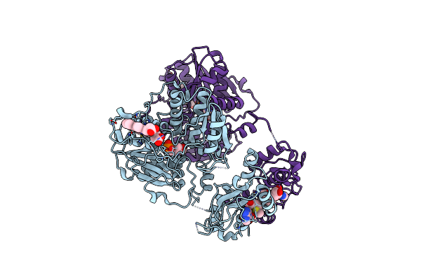
Structure Of Active Cystathionine B-Synthase From Apis Mellifera
Organism: Apis mellifera
Method: X-RAY DIFFRACTION Resolution:3.20 Å Release Date: 2018-01-03 Classification: LYASE Ligands: HEM, PLP |
Organism: Apis mellifera
Method: X-RAY DIFFRACTION
Release Date: 2018-01-03
Ligands: HEM, PLP
 |
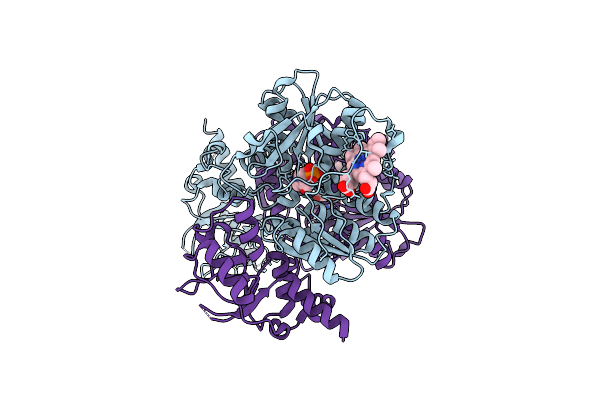
Crystal Structure Of Delta516-525 E201S Human Cystathionine Beta-Synthase With Adomet
Organism: Homo sapiens
Method: X-RAY DIFFRACTION Resolution:3.58 Å Release Date: 2014-09-24 Classification: LYASE Ligands: HEM, PLP, SAM |
Organism: Homo sapiens
Method: X-RAY DIFFRACTION
Release Date: 2014-09-24
Ligands: HEM, PLP, SAM
 |
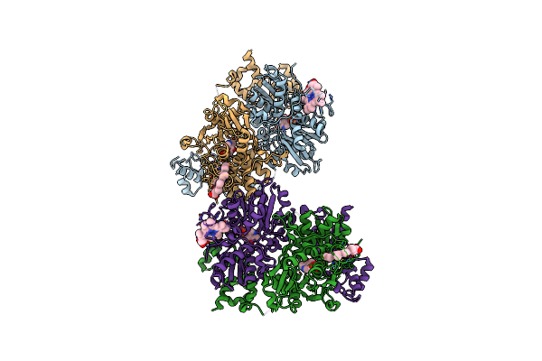
Crystal Structure Of Delta516-525 Human Cystathionine Beta-Synthase Containing C-Terminal 6Xhis-Tag
Organism: Homo sapiens
Method: X-RAY DIFFRACTION Resolution:2.97 Å Release Date: 2013-09-18 Classification: LYASE Ligands: PLP, HEM |
Organism: Homo sapiens
Method: X-RAY DIFFRACTION
Release Date: 2013-09-18
Ligands: PLP, HEM
 |
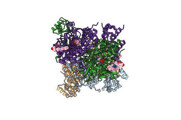
Crystal Structure Of Delta1-39 And Delta516-525 Human Cystathionine Beta-Synthase D444N Mutant Containing C-Terminal 6Xhis Tag
Organism: Homo sapiens
Method: X-RAY DIFFRACTION Resolution:3.39 Å Release Date: 2013-09-18 Classification: LYASE Ligands: PLP, HEM |
Organism: Homo sapiens
Method: X-RAY DIFFRACTION
Release Date: 2013-09-18
Ligands: PLP, HEM
 |
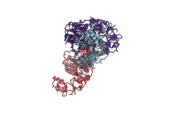
Crystal Structure Of Delta516-525 Human Cystathionine Beta-Synthase D444N Mutant Containing C-Terminal 6Xhis Tag
Organism: Homo sapiens
Method: X-RAY DIFFRACTION Resolution:2.63 Å Release Date: 2013-09-18 Classification: LYASE Ligands: PLP, HEM |
Organism: Homo sapiens
Method: X-RAY DIFFRACTION
Release Date: 2013-09-18
Ligands: PLP, HEM
 |
Crystal Structure Of Delta516-525 Human Cystathionine Beta-Synthase
Organism: Homo sapiens
Method: X-RAY DIFFRACTION Resolution:3.63 Å Release Date: 2013-09-18 Classification: LYASE Ligands: PLP, HEM |
Organism: Homo sapiens
Method: X-RAY DIFFRACTION
Release Date: 2013-09-18
Ligands: PLP, HEM
 |
Structure Of The Neuronal Nitric Oxide Synthase Heme Domain In Complex With 6-(((3R,4R)-4-(2-((1S,2R)-2-(3-Fluorophenyl)Cyclopropylamino)Ethoxy)Pyrrolidin-3-Yl)Methyl)-4-Methylpyridin-2-Amine
Organism: Rattus norvegicus
Method: X-RAY DIFFRACTION Resolution:1.84 Å Release Date: 2012-05-02 Classification: Oxidoreductase/Inhibitor Ligands: HEM, H4B, ACT, Y2B, ZN |
Organism: Rattus norvegicus
Method: X-RAY DIFFRACTION
Release Date: 2012-05-02
Ligands: HEM, H4B, ACT, Y2B, ZN
 |
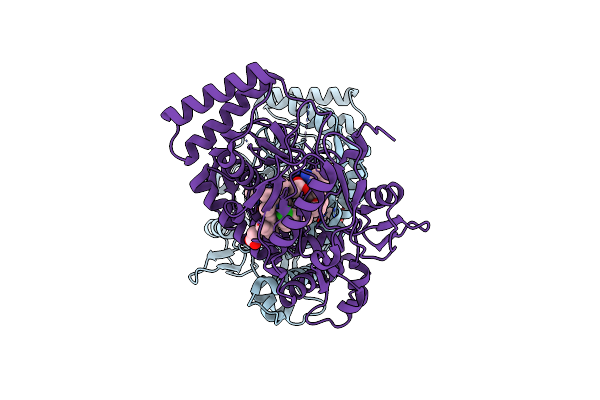
Structure Of The Neuronal Nitric Oxide Synthase Heme Domain In Complex With 4-Methyl-6-{[(3R,4R)-4-(2-{[(1R,2S)-2-(3-Methylphenyl)Cyclopropyl]Amino}Ethoxy)Pyrrolidin-3-Yl]Methyl}Pyridin-2-Amine And Its Isomer
Organism: Rattus norvegicus
Method: X-RAY DIFFRACTION Resolution:2.21 Å Release Date: 2012-05-02 Classification: Oxidoreductase/Inhibitor Ligands: HEM, H4B, X2C, ACT, ZN |
Organism: Rattus norvegicus
Method: X-RAY DIFFRACTION
Release Date: 2012-05-02
Ligands: HEM, H4B, X2C, ACT, ZN
 |
Structure Of The Neuronal Nitric Oxide Synthase Heme Domain In Complex With 6-(((3R,4R)-4-(2-((1S,2R/1R,2S)-2-(3-Clorophenyl)Cyclopropylamino)Ethoxy)Pyrrolidin-3-Yl)Methyl)-4-Methylpyridin-2-Amine
Organism: Rattus norvegicus
Method: X-RAY DIFFRACTION Resolution:1.93 Å Release Date: 2012-05-02 Classification: OXIDOREDUCTASE/INHIBITOR Ligands: HEM, H4B, ACT, X2D, ZN |
Organism: Rattus norvegicus
Method: X-RAY DIFFRACTION
Release Date: 2012-05-02
Ligands: HEM, H4B, ACT, X2D, ZN
 |
Structure Of The Neuronal Nitric Oxide Synthase Heme Domain In Complex With 6-{[(3R,4R)-4-(2-{[(2R/2S)-1-(3-Fluorophenyl)Propan-2-Yl]Amino}Ethoxy)Pyrrolidin-3-Yl]Methyl}-4-Methylpyridin-2-Amine
Organism: Rattus norvegicus
Method: X-RAY DIFFRACTION Resolution:1.95 Å Release Date: 2012-05-02 Classification: OXIDOREDUCTASE/INHIBITOR Ligands: HEM, H4B, X2E, ACT, ZN |
Organism: Rattus norvegicus
Method: X-RAY DIFFRACTION
Release Date: 2012-05-02
Ligands: HEM, H4B, X2E, ACT, ZN
 |
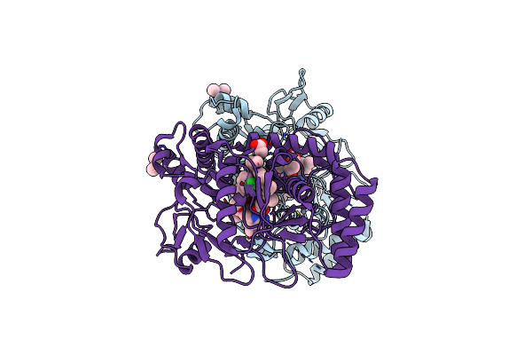
Structure Of The Neuronal Nitric Oxide Synthase Heme Domain In Complex With 6-(((3*R*,4*R*)-4-(2-(((*S* )-1-(3-Fluorophenyl)Propan-2-Yl)Amino)Ethoxy)Pyrrolidin-3-Yl)Methyl)-4-Methylpyridin-2-Amine
Organism: Rattus norvegicus
Method: X-RAY DIFFRACTION Resolution:1.95 Å Release Date: 2012-05-02 Classification: OXIDOREDUCTASE/INHIBITOR Ligands: HEM, H4B, Y2F, ACT, ZN |
Organism: Rattus norvegicus
Method: X-RAY DIFFRACTION
Release Date: 2012-05-02
Ligands: HEM, H4B, Y2F, ACT, ZN
 |
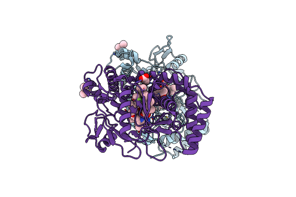
Structure Of The Endothelial Nitric Oxide Synthase Heme Domain In Complex With 6-(((3R,4R)-4-(2-((1S,2R/1R,2S)-2-(3-Clorophenyl)Cyclopropylamino)Ethoxy)Pyrrolidin-3-Yl)Methyl)-4-Methylpyridin-2-Amine
Organism: Bos taurus
Method: X-RAY DIFFRACTION Resolution:2.08 Å Release Date: 2012-05-02 Classification: OXIDOREDUCTASE/INHIBITOR Ligands: HEM, H4B, X2D, ACT, GOL, CAD, ZN |
Organism: Bos taurus
Method: X-RAY DIFFRACTION
Release Date: 2012-05-02
Ligands: HEM, H4B, X2D, ACT, GOL, CAD, ZN
 |
Structure Of The Endothelial Nitric Oxide Synthase Heme Domain In Complex With 6-{[(3R,4R)-4-(2-{[(2R/2S)-1-(3-Fluorophenyl)Propan-2-Yl]Amino}Ethoxy)Pyrrolidin-3-Yl]Methyl}-4-Methylpyridin-2-Amine
Organism: Bos taurus
Method: X-RAY DIFFRACTION Resolution:2.35 Å Release Date: 2012-05-02 Classification: OXIDOREDUCTASE/INHIBITOR Ligands: HEM, H4B, X2E, ACT, GOL, CAD, ZN |
Organism: Bos taurus
Method: X-RAY DIFFRACTION
Release Date: 2012-05-02
Ligands: HEM, H4B, X2E, ACT, GOL, CAD, ZN