

Search Count: 12
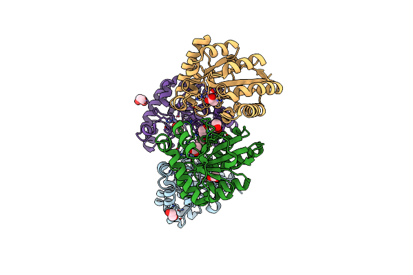 |
Crystal Structure Of The Polysaccharide Deacetylase Bc1974 From Bacillus Cereus
Organism: Bacillus cereus
Method: X-RAY DIFFRACTION Resolution:1.80 Å Release Date: 2018-02-21 Classification: HYDROLASE Ligands: ZN, ACT, EDO |
Organism: Bacillus cereus
Method: X-RAY DIFFRACTION
Release Date: 2018-02-21
Ligands: ZN, ACT, EDO
 |
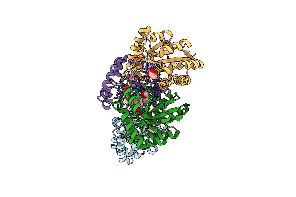
Crystal Structure Of The Polysaccharide Deacetylase Bc1974 From Bacillus Cereus In Complex With N-Hydroxynaphthalene-1-Carboxamide
Organism: Bacillus cereus
Method: X-RAY DIFFRACTION Resolution:1.45 Å Release Date: 2018-02-21 Classification: HYDROLASE Ligands: 8GK, ZN, EDO, NA, PGE |
Organism: Bacillus cereus
Method: X-RAY DIFFRACTION
Release Date: 2018-02-21
Ligands: 8GK, ZN, EDO, NA, PGE
 |
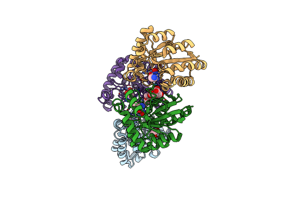
Crystal Structure Of The Polysaccharide Deacetylase Bc1974 From Bacillus Cereus In Complex With (E)-N-Hydroxy-3-(Naphthalen-1-Yl)Prop-2-Enamide
Organism: Bacillus cereus
Method: X-RAY DIFFRACTION Resolution:2.80 Å Release Date: 2018-02-21 Classification: HYDROLASE Ligands: ACT, ZN, EDO, 8SQ, PGE |
Organism: Bacillus cereus
Method: X-RAY DIFFRACTION
Release Date: 2018-02-21
Ligands: ACT, ZN, EDO, 8SQ, PGE
 |
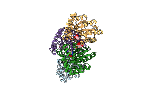
Crystal Structure Of The Polysaccharide Deacetylase Bc1974 From Bacillus Cereus In Complex With (2S)-2,6-Diamino-N-Hydroxyhexanamide
Organism: Bacillus cereus atcc 14579, Bacillus cereus
Method: X-RAY DIFFRACTION Resolution:2.44 Å Release Date: 2018-02-21 Classification: HYDROLASE Ligands: 8SZ, ZN, CIT, EDO, PGE |
Organism: Bacillus cereus atcc 14579, Bacillus cereus
Method: X-RAY DIFFRACTION
Release Date: 2018-02-21
Ligands: 8SZ, ZN, CIT, EDO, PGE
 |
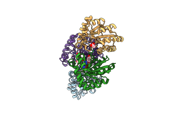
Crystal Structure Of The Polysaccharide Deacetylase Bc1974 From Bacillus Cereus In Complex With (2S)-2-Amino-5-(Diaminomethylideneamino)-N-Hydroxypentanamide
Organism: Bacillus cereus
Method: X-RAY DIFFRACTION Resolution:2.45 Å Release Date: 2018-02-21 Classification: HYDROLASE Ligands: ACT, ZN, AHL, CIT |
Organism: Bacillus cereus
Method: X-RAY DIFFRACTION
Release Date: 2018-02-21
Ligands: ACT, ZN, AHL, CIT
 |
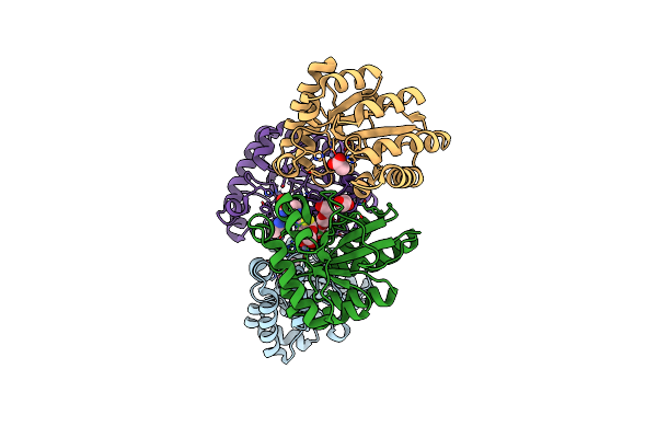
Crystal Structure Of The Polysaccharide Deacetylase Bc1974 From Bacillus Cereus In Complex With Acetazolamide
Organism: Bacillus cereus
Method: X-RAY DIFFRACTION Resolution:3.06 Å Release Date: 2018-02-21 Classification: HYDROLASE Ligands: ZN, ACT, AZM, PGE |
Organism: Bacillus cereus
Method: X-RAY DIFFRACTION
Release Date: 2018-02-21
Ligands: ZN, ACT, AZM, PGE
 |
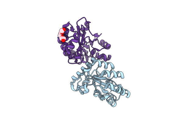
Crystal Structure Of The Polysaccharide Deacetylase Bc1974 From Bacillus Cereus In Complex With Thiametg
Organism: Bacillus cereus
Method: X-RAY DIFFRACTION Resolution:2.73 Å Release Date: 2018-02-21 Classification: HYDROLASE Ligands: ZN, ACT, NHT, PGE, EDO, CIT |
Organism: Bacillus cereus
Method: X-RAY DIFFRACTION
Release Date: 2018-02-21
Ligands: ZN, ACT, NHT, PGE, EDO, CIT
 |
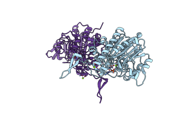
Structural Determination Of Ba0150, A Polysaccharide Deacetylase From Bacillus Anthracis
Organism: Bacillus anthracis
Method: X-RAY DIFFRACTION Resolution:1.99 Å Release Date: 2014-02-12 Classification: HYDROLASE Ligands: PGE |
Organism: Bacillus anthracis
Method: X-RAY DIFFRACTION
Release Date: 2014-02-12
Ligands: PGE
 |
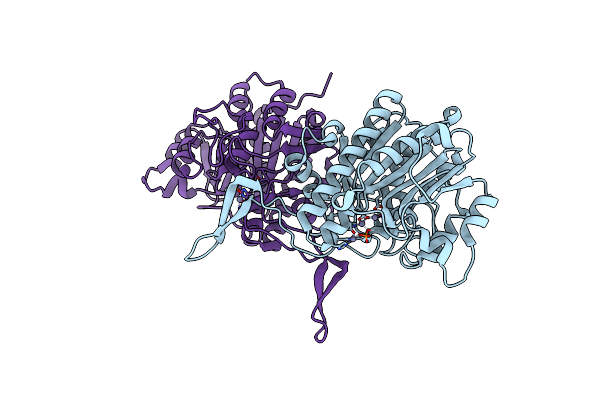
Structure Of Tab5 Alkaline Phosphatase Mutant His 135 Asp With Mg Bound In The M3 Site.
Organism: Antarctic bacterium tab5
Method: X-RAY DIFFRACTION Resolution:1.78 Å Release Date: 2009-11-24 Classification: HYDROLASE Ligands: ZN, MG |
Organism: Antarctic bacterium tab5
Method: X-RAY DIFFRACTION
Release Date: 2009-11-24
Ligands: ZN, MG
 |
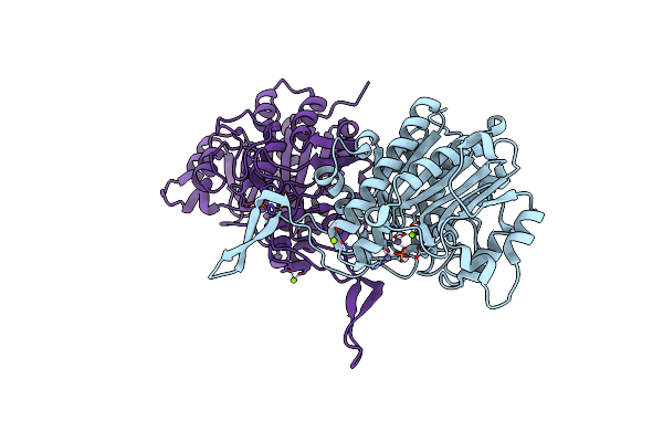
Structure Of Tab5 Alkaline Phosphatase Mutant His 135 Asp With Zn Bound In The M3 Site.
Organism: Antarctic bacterium tab5
Method: X-RAY DIFFRACTION Resolution:1.79 Å Release Date: 2009-11-24 Classification: HYDROLASE Ligands: ZN |
Organism: Antarctic bacterium tab5
Method: X-RAY DIFFRACTION
Release Date: 2009-11-24
Ligands: ZN
 |
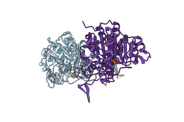
Structure Of Tab5 Alkaline Phosphatase Mutant His 135 Glu With Mg Bound In The M3 Site.
Organism: Antarctic bacterium tab5
Method: X-RAY DIFFRACTION Resolution:1.99 Å Release Date: 2009-11-24 Classification: HYDROLASE Ligands: ZN, MG |
Organism: Antarctic bacterium tab5
Method: X-RAY DIFFRACTION
Release Date: 2009-11-24
Ligands: ZN, MG
 |
Structure Of Alkaline Phosphatase From The Antarctic Bacterium Tab5
Organism: Antarctic bacterium tab5
Method: X-RAY DIFFRACTION Resolution:1.95 Å Release Date: 2006-11-28 Classification: HYDROLASE Ligands: ZN, MG, PO4, CAC |
Organism: Antarctic bacterium tab5
Method: X-RAY DIFFRACTION
Release Date: 2006-11-28
Ligands: ZN, MG, PO4, CAC