

Search Count: 14
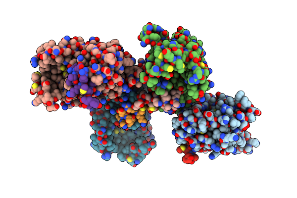 |
X-Ray Structure Of Human Pparalpha Ligand Binding Domain-Ncor2 Corepressor Peptide Co-Crystals Obtained By Co-Crystallization
Organism: Homo sapiens
Method: X-RAY DIFFRACTION Resolution:2.48 Å Release Date: 2025-05-07 Classification: TRANSCRIPTION |
Organism: Homo sapiens
Method: X-RAY DIFFRACTION
Release Date: 2025-05-07
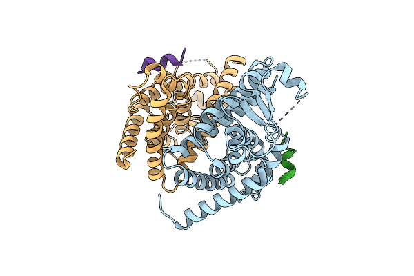 |
X-Ray Structure Of Human Ppargamma Ligand Binding Domain-Ncor2 Corepressor Peptide Co-Crystals Obtained By Co-Crystallization
Organism: Homo sapiens
Method: X-RAY DIFFRACTION Resolution:2.43 Å Release Date: 2025-05-07 Classification: TRANSCRIPTION |
Organism: Homo sapiens
Method: X-RAY DIFFRACTION
Release Date: 2025-05-07
 |
X-Ray Structure Of Human Pparalpha Ligand Binding Domain-Intrinsic Fatty Acid (E. Coli Origin)-Cbp Coactivator Peptide Co-Crystals Obtained By Cross-Seeding
Organism: Homo sapiens
Method: X-RAY DIFFRACTION Resolution:2.09 Å Release Date: 2025-05-07 Classification: TRANSCRIPTION Ligands: PLM, GOL |
Organism: Homo sapiens
Method: X-RAY DIFFRACTION
Release Date: 2025-05-07
Ligands: PLM, GOL
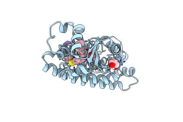 |
X-Ray Structure Of Human Pparalpha Ligand Binding Domain-Gw7647-Trap220 Coactivator Peptide Co-Crystals Obtained By Cross-Seeding
Organism: Homo sapiens
Method: X-RAY DIFFRACTION Resolution:1.39 Å Release Date: 2025-05-07 Classification: TRANSCRIPTION Ligands: 2VN, GOL |
Organism: Homo sapiens
Method: X-RAY DIFFRACTION
Release Date: 2025-05-07
Ligands: 2VN, GOL
 |
X-Ray Structure Of Human Pparalpha Ligand Binding Domain-Intrinsic Fatty Acid (E. Coli Origin)-Pgc1Alpha Coactivator Peptide Co-Crystals Obtained By Cross-Seeding
Organism: Homo sapiens
Method: X-RAY DIFFRACTION Resolution:1.59 Å Release Date: 2025-05-07 Classification: TRANSCRIPTION Ligands: PLM, GOL |
Organism: Homo sapiens
Method: X-RAY DIFFRACTION
Release Date: 2025-05-07
Ligands: PLM, GOL
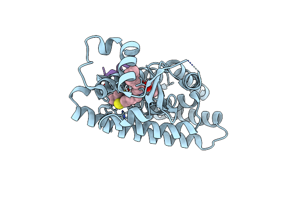 |
X-Ray Structure Of Human Pparalpha Ligand Binding Domain-Gw7647-Pgc1Alpha Coactivator Peptide Co-Crystals Obtained By Cross-Seeding
Organism: Homo sapiens
Method: X-RAY DIFFRACTION Resolution:1.49 Å Release Date: 2025-05-07 Classification: TRANSCRIPTION Ligands: 2VN |
Organism: Homo sapiens
Method: X-RAY DIFFRACTION
Release Date: 2025-05-07
Ligands: 2VN
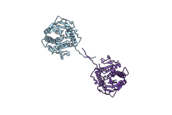 |
Crystal Structure Of Nucleotide-Free Human Kinesin-1 Motor Domain (G234V Mutant)
Organism: Homo sapiens
Method: X-RAY DIFFRACTION Resolution:2.80 Å Release Date: 2025-04-23 Classification: MOTOR PROTEIN |
Organism: Homo sapiens
Method: X-RAY DIFFRACTION
Release Date: 2025-04-23
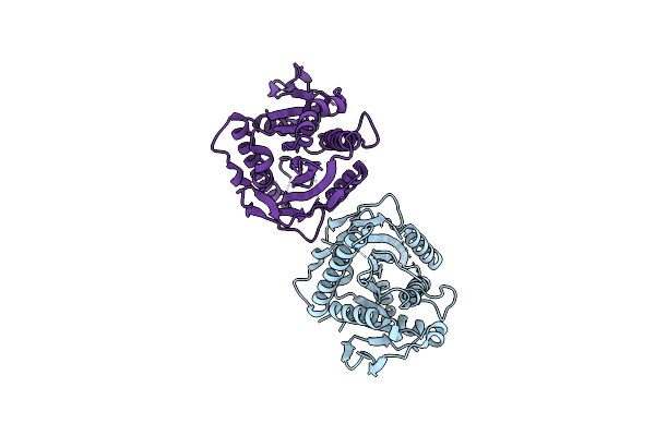 |
Crystal Structure Of Nucleotide-Free Human Kinesin-1 Motor Domain (G234A Mutant)
Organism: Homo sapiens
Method: X-RAY DIFFRACTION Resolution:2.82 Å Release Date: 2025-04-23 Classification: MOTOR PROTEIN Ligands: HOH |
Organism: Homo sapiens
Method: X-RAY DIFFRACTION
Release Date: 2025-04-23
Ligands: HOH
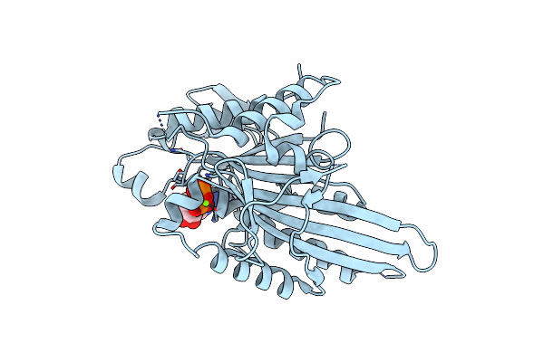 |
Crystal Structure Of Human Kinesin-1 Motor Domain (G234A Mutant) In Complex With Adp
Organism: Homo sapiens
Method: X-RAY DIFFRACTION Resolution:2.40 Å Release Date: 2025-04-23 Classification: MOTOR PROTEIN Ligands: MG, ADP |
Organism: Homo sapiens
Method: X-RAY DIFFRACTION
Release Date: 2025-04-23
Ligands: MG, ADP
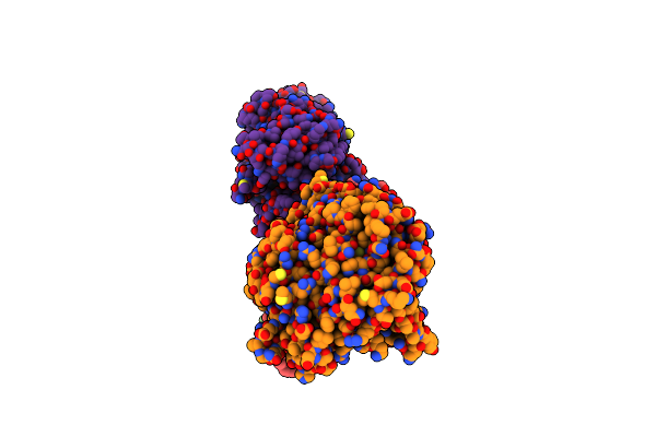 |
Nucleotide-Free Kinesin-1 Motor Domain Bound To The Microtubule
Organism: Homo sapiens, Sus scrofa
Method: ELECTRON MICROSCOPY Release Date: 2025-04-23 Classification: MOTOR PROTEIN Ligands: GTP, MG |
Organism: Homo sapiens, Sus scrofa
Method: ELECTRON MICROSCOPY
Release Date: 2025-04-23
Ligands: GTP, MG
 |
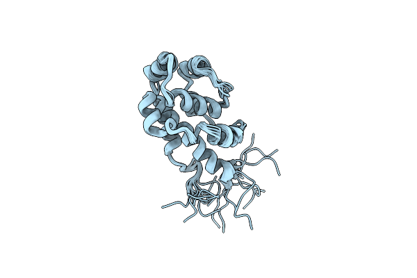
The Microtubule-Binding Domains Of Yeast Cytoplasmic Dynein In The High Affinity State
Organism: Saccharomyces cerevisiae (strain atcc 204508 / s288c)
Method: SOLUTION NMR Release Date: 2020-03-18 Classification: MOTOR PROTEIN |
Organism: Saccharomyces cerevisiae (strain atcc 204508 / s288c)
Method: SOLUTION NMR
Release Date: 2020-03-18
 |
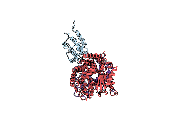
The Microtubule-Binding Domains Of Yeast Cytoplasmic Dynein In The Low Affinity State
Organism: Saccharomyces cerevisiae (strain atcc 204508 / s288c)
Method: SOLUTION NMR Release Date: 2020-03-18 Classification: MOTOR PROTEIN |
Organism: Saccharomyces cerevisiae (strain atcc 204508 / s288c)
Method: SOLUTION NMR
Release Date: 2020-03-18
 |
Complex Of Yeast Cytoplasmic Dynein Mtbd-High And Mt Without Dtt
Organism: Saccharomyces cerevisiae s288c, Sus scrofa
Method: ELECTRON MICROSCOPY Release Date: 2020-03-04 Classification: MOTOR PROTEIN/STRUCTURAL PROTEIN |
Organism: Saccharomyces cerevisiae s288c, Sus scrofa
Method: ELECTRON MICROSCOPY
Release Date: 2020-03-04
 |
Complex Of Yeast Cytoplasmic Dynein Mtbd-High And Mt With Dtt
Organism: Saccharomyces cerevisiae s288c, Sus scrofa
Method: ELECTRON MICROSCOPY Release Date: 2020-03-04 Classification: MOTOR PROTEIN/STRUCTURAL PROTEIN |
Organism: Saccharomyces cerevisiae s288c, Sus scrofa
Method: ELECTRON MICROSCOPY
Release Date: 2020-03-04