

Search Count: 24
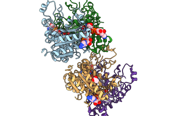 |
Xfel Structure Of Hnqo1 Mixed With Nadh In A Folded Orientation At 300 Ms
Organism: Homo sapiens
Method: X-RAY DIFFRACTION Resolution:2.51 Å Release Date: 2026-02-11 Classification: OXIDOREDUCTASE Ligands: NAD, FAD, ACT |
Organism: Homo sapiens
Method: X-RAY DIFFRACTION
Release Date: 2026-02-11
Ligands: NAD, FAD, ACT
 |
Xfel Structure Of Hnqo1 Mixed With Nadh In An Extended Orientation At 0.3 S
Organism: Homo sapiens
Method: X-RAY DIFFRACTION Resolution:2.51 Å Release Date: 2025-12-10 Classification: OXIDOREDUCTASE Ligands: NAD, FAD, ACT |
Organism: Homo sapiens
Method: X-RAY DIFFRACTION
Release Date: 2025-12-10
Ligands: NAD, FAD, ACT
 |
Room Temperature Structure Of Glycine Max Phya In Pfr/Pr Ensemble
Organism: Glycine max
Method: X-RAY DIFFRACTION Resolution:2.20 Å Release Date: 2025-06-04 Classification: PLANT PROTEIN Ligands: CYC |
Organism: Glycine max
Method: X-RAY DIFFRACTION
Release Date: 2025-06-04
Ligands: CYC
 |
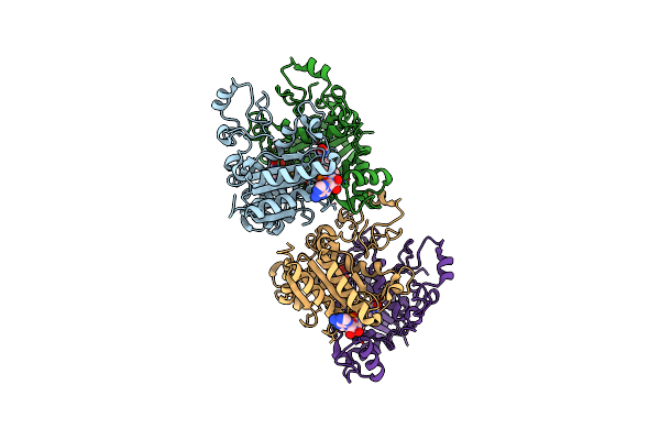
Xfel Structure Of The Free Hnqo1 Unmixed (P3083)
Organism: Homo sapiens
Method: X-RAY DIFFRACTION Resolution:2.50 Å Release Date: 2025-04-23 Classification: OXIDOREDUCTASE Ligands: FAD |
Organism: Homo sapiens
Method: X-RAY DIFFRACTION
Release Date: 2025-04-23
Ligands: FAD
 |
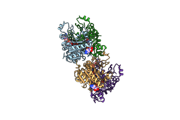
Xfel Structure Of Free Hnqo1 Unmixed (P4502)
Organism: Homo sapiens
Method: X-RAY DIFFRACTION Resolution:2.50 Å Release Date: 2025-04-23 Classification: OXIDOREDUCTASE Ligands: FAD |
Organism: Homo sapiens
Method: X-RAY DIFFRACTION
Release Date: 2025-04-23
Ligands: FAD
 |
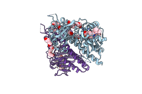
Xfel Structure Of Hnqo1 Mixed With Nadh For 1.2 S
Organism: Homo sapiens
Method: X-RAY DIFFRACTION Resolution:2.50 Å Release Date: 2025-04-23 Classification: OXIDOREDUCTASE Ligands: FAD, NAD |
Organism: Homo sapiens
Method: X-RAY DIFFRACTION
Release Date: 2025-04-23
Ligands: FAD, NAD
 |
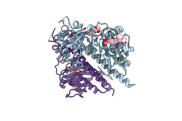
Pas-Gaf Bidomain Of Glycine Max Phytochrome A
Organism: Glycine max
Method: X-RAY DIFFRACTION Resolution:1.58 Å Release Date: 2025-01-08 Classification: PLANT PROTEIN Ligands: CYC, 1PE, PEG, PGE |
Organism: Glycine max
Method: X-RAY DIFFRACTION
Release Date: 2025-01-08
Ligands: CYC, 1PE, PEG, PGE
 |
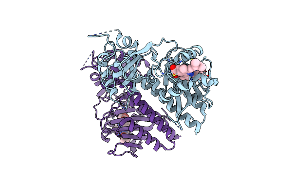
Phytochromobilin-Adducted Pas-Gaf Bidomain Of Glycine Max Phytochrome A
Organism: Glycine max
Method: X-RAY DIFFRACTION Resolution:1.86 Å Release Date: 2025-01-08 Classification: PLANT PROTEIN Ligands: O6E, PG4 |
Organism: Glycine max
Method: X-RAY DIFFRACTION
Release Date: 2025-01-08
Ligands: O6E, PG4
 |
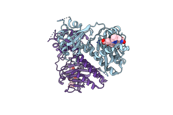
Room Temperature Structure Of Glycine Max Phya In Pr
Organism: Glycine max
Method: X-RAY DIFFRACTION Resolution:2.20 Å Release Date: 2025-01-08 Classification: PLANT PROTEIN Ligands: CYC |
Organism: Glycine max
Method: X-RAY DIFFRACTION
Release Date: 2025-01-08
Ligands: CYC
 |
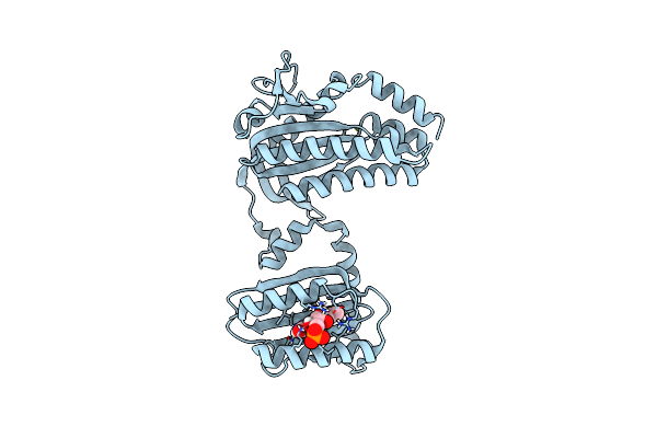
Room Temperature Structure Of Glycine Max Phya In Pfr
Organism: Glycine max
Method: X-RAY DIFFRACTION Resolution:2.20 Å Release Date: 2025-01-08 Classification: PLANT PROTEIN Ligands: CYC |
Organism: Glycine max
Method: X-RAY DIFFRACTION
Release Date: 2025-01-08
Ligands: CYC
 |
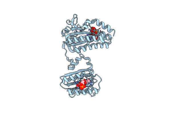
Cryogenic Crystal Structure Of The Photoactivated Adenylate Cyclase Oapac
Organism: Oscillatoria acuminata pcc 6304
Method: X-RAY DIFFRACTION Resolution:1.50 Å Release Date: 2023-11-01 Classification: LYASE Ligands: FMN, MG |
Organism: Oscillatoria acuminata pcc 6304
Method: X-RAY DIFFRACTION
Release Date: 2023-11-01
Ligands: FMN, MG
 |
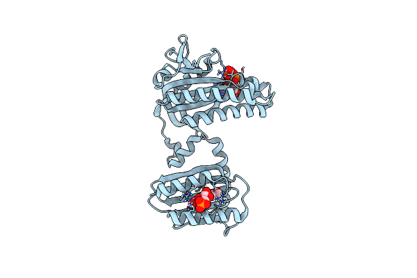
Cryogenic Crystal Structure Of The Photoactivated Adenylate Cyclase Oapac With Atp Bound
Organism: Oscillatoria acuminata pcc 6304
Method: X-RAY DIFFRACTION Resolution:2.10 Å Release Date: 2023-11-01 Classification: LYASE Ligands: FMN, ATP, MG |
Organism: Oscillatoria acuminata pcc 6304
Method: X-RAY DIFFRACTION
Release Date: 2023-11-01
Ligands: FMN, ATP, MG
 |
Cryogenic Crystal Structure Of The Photoactivated Adenylate Cyclase Oapac After 5 Seconds Of Blue Light Illumination
Organism: Oscillatoria acuminata pcc 6304
Method: X-RAY DIFFRACTION Resolution:1.70 Å Release Date: 2023-11-01 Classification: LYASE Ligands: FMN, ATP, MG |
Organism: Oscillatoria acuminata pcc 6304
Method: X-RAY DIFFRACTION
Release Date: 2023-11-01
Ligands: FMN, ATP, MG
 |
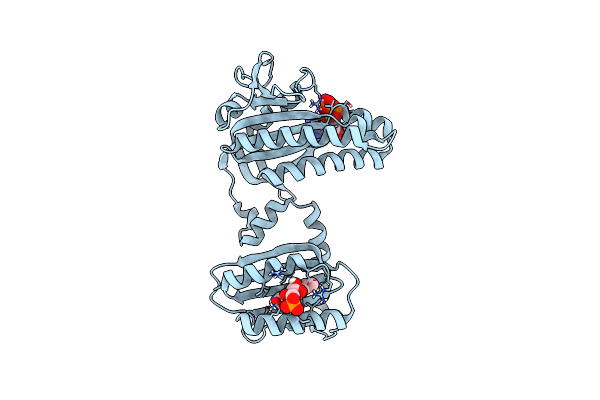
Room Temperature Crystal Structure Of The Photoactivated Adenylate Cyclase Oapac With Atp Bound
Organism: Oscillatoria acuminata pcc 6304
Method: X-RAY DIFFRACTION Resolution:1.80 Å Release Date: 2023-11-01 Classification: LYASE Ligands: FMN, ATP, MG |
Organism: Oscillatoria acuminata pcc 6304
Method: X-RAY DIFFRACTION
Release Date: 2023-11-01
Ligands: FMN, ATP, MG
 |
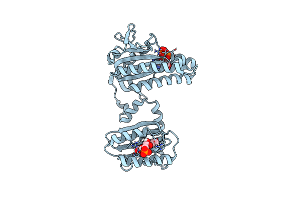
Room Temperature Crystal Structure Of The Photoactivated Adenylate Cyclase Oapac After Blue Light Excitation At 1.8 Us Delay
Organism: Oscillatoria acuminata pcc 6304
Method: X-RAY DIFFRACTION Resolution:1.90 Å Release Date: 2023-11-01 Classification: LYASE Ligands: FMN, ATP, MG |
Organism: Oscillatoria acuminata pcc 6304
Method: X-RAY DIFFRACTION
Release Date: 2023-11-01
Ligands: FMN, ATP, MG
 |
Room Temperature Crystal Structure Of The Photoactivated Adenylate Cyclase Oapac After Blue Light Excitation At 2.3 Us Delay
Organism: Oscillatoria acuminata pcc 6304
Method: X-RAY DIFFRACTION Resolution:1.90 Å Release Date: 2023-11-01 Classification: LYASE Ligands: FMN, ATP, MG |
Organism: Oscillatoria acuminata pcc 6304
Method: X-RAY DIFFRACTION
Release Date: 2023-11-01
Ligands: FMN, ATP, MG
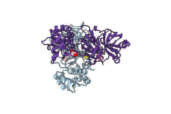 |
Crystal Structure Of Sars-Cov-2 Main Protease In Orthorhombic Space Group P212121
Organism: Severe acute respiratory syndrome coronavirus 2
Method: X-RAY DIFFRACTION Resolution:1.65 Å Release Date: 2023-03-22 Classification: HYDROLASE Ligands: CL, DMS, MLI |
Organism: Severe acute respiratory syndrome coronavirus 2
Method: X-RAY DIFFRACTION
Release Date: 2023-03-22
Ligands: CL, DMS, MLI
 |
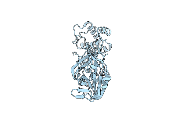
Oxidized Form Of Sars-Cov-2 Main Protease Determined By Xfel Radiation
Organism: Severe acute respiratory syndrome coronavirus 2
Method: X-RAY DIFFRACTION Resolution:2.25 Å Release Date: 2023-01-25 Classification: VIRAL PROTEIN Ligands: DMS |
Organism: Severe acute respiratory syndrome coronavirus 2
Method: X-RAY DIFFRACTION
Release Date: 2023-01-25
Ligands: DMS
 |
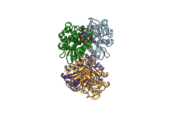
Reduced Form Of Sars-Cov-2 Main Protease Determined By Xfel Radiation
Organism: Severe acute respiratory syndrome coronavirus 2
Method: X-RAY DIFFRACTION Resolution:1.75 Å Release Date: 2023-01-18 Classification: VIRAL PROTEIN Ligands: CL |
Organism: Severe acute respiratory syndrome coronavirus 2
Method: X-RAY DIFFRACTION
Release Date: 2023-01-18
Ligands: CL
 |
Beta-Lactamase Mixed With Ceftriaxone, 5Ms
Organism: Mycobacterium tuberculosis
Method: X-RAY DIFFRACTION Resolution:2.40 Å Release Date: 2021-09-22 Classification: STRUCTURAL PROTEIN Ligands: PO4, 9F2 |
Organism: Mycobacterium tuberculosis
Method: X-RAY DIFFRACTION
Release Date: 2021-09-22
Ligands: PO4, 9F2