

Search Count: 20
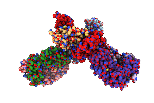 |
Tcr In Complex With Hla-E*01:03 Bound To Hbv Envelope 371-379 Index Peptide
Organism: Homo sapiens, Hepatitis b virus
Method: X-RAY DIFFRACTION Resolution:2.25 Å Release Date: 2024-10-16 Classification: IMMUNE SYSTEM |
Organism: Homo sapiens, Hepatitis b virus
Method: X-RAY DIFFRACTION
Release Date: 2024-10-16
 |
Tcr In Complex With Hla-E*01:03 Bound To Hbv Envelope 371-379 S3N Peptide
Organism: Homo sapiens, Hepatitis b virus
Method: X-RAY DIFFRACTION Resolution:2.35 Å Release Date: 2024-10-16 Classification: IMMUNE SYSTEM |
Organism: Homo sapiens, Hepatitis b virus
Method: X-RAY DIFFRACTION
Release Date: 2024-10-16
 |
Tcr In Complex With Hla-E*01:03 Bound To Hbv Envelope 371-379 L6I Peptide
Organism: Homo sapiens, Hepatitis b virus
Method: X-RAY DIFFRACTION Resolution:2.61 Å Release Date: 2024-10-16 Classification: IMMUNE SYSTEM |
Organism: Homo sapiens, Hepatitis b virus
Method: X-RAY DIFFRACTION
Release Date: 2024-10-16
 |
Crystal Structure Of High Affinity Tcr In Complex With Phla Harbouring Bacterial Peptide
Organism: Homo sapiens, Mycobacterium tuberculosis
Method: X-RAY DIFFRACTION Resolution:2.33 Å Release Date: 2024-05-15 Classification: IMMUNE SYSTEM Ligands: ACT |
Organism: Homo sapiens, Mycobacterium tuberculosis
Method: X-RAY DIFFRACTION
Release Date: 2024-05-15
Ligands: ACT
 |
Crystal Structure Of Inha:01 Tcr In Complex With Hla-E Bound To Inha (53-61)
Organism: Homo sapiens, Mycobacterium tuberculosis h37rv
Method: X-RAY DIFFRACTION Resolution:2.26 Å Release Date: 2022-01-26 Classification: IMMUNE SYSTEM |
Organism: Homo sapiens, Mycobacterium tuberculosis h37rv
Method: X-RAY DIFFRACTION
Release Date: 2022-01-26
 |
Crystal Structure Of Inha:01 Tcr In Complex With Hla-E (Y84C) Bound To Inha (53-61 Gcg)
Organism: Homo sapiens, Mycobacterium tuberculosis h37rv
Method: X-RAY DIFFRACTION Resolution:2.17 Å Release Date: 2022-01-26 Classification: IMMUNE SYSTEM Ligands: EDO |
Organism: Homo sapiens, Mycobacterium tuberculosis h37rv
Method: X-RAY DIFFRACTION
Release Date: 2022-01-26
Ligands: EDO
 |
Crystal Structure Of Inha:01 Tcr In Complex With Hla-E (S147C) Bound To Inha (53-61 H3C)
Organism: Homo sapiens, Mycobacterium tuberculosis h37rv
Method: X-RAY DIFFRACTION Resolution:2.65 Å Release Date: 2022-01-26 Classification: IMMUNE SYSTEM |
Organism: Homo sapiens, Mycobacterium tuberculosis h37rv
Method: X-RAY DIFFRACTION
Release Date: 2022-01-26
 |
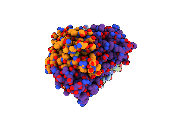
Crystal Structure Of Inha:01 Tcr In Complex With Hla-E (F116C) Bound To Inha (53-61 H4C)
Organism: Homo sapiens, Mycobacterium tuberculosis h37rv
Method: X-RAY DIFFRACTION Resolution:2.30 Å Release Date: 2022-01-26 Classification: IMMUNE SYSTEM Ligands: EDO |
Organism: Homo sapiens, Mycobacterium tuberculosis h37rv
Method: X-RAY DIFFRACTION
Release Date: 2022-01-26
Ligands: EDO
 |
Gag:02 Tcr In Complex With Hla-E.
Organism: Homo sapiens, Human immunodeficiency virus 1
Method: X-RAY DIFFRACTION Resolution:2.55 Å Release Date: 2022-01-26 Classification: IMMUNE SYSTEM |
Organism: Homo sapiens, Human immunodeficiency virus 1
Method: X-RAY DIFFRACTION
Release Date: 2022-01-26
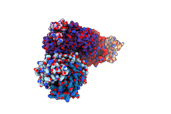 |
Ul40:01 Tcr In Complex With Hla-E With A Non-Natural Amino Acid
Organism: Homo sapiens
Method: X-RAY DIFFRACTION Resolution:3.00 Å Release Date: 2022-01-26 Classification: IMMUNE SYSTEM |
Organism: Homo sapiens
Method: X-RAY DIFFRACTION
Release Date: 2022-01-26
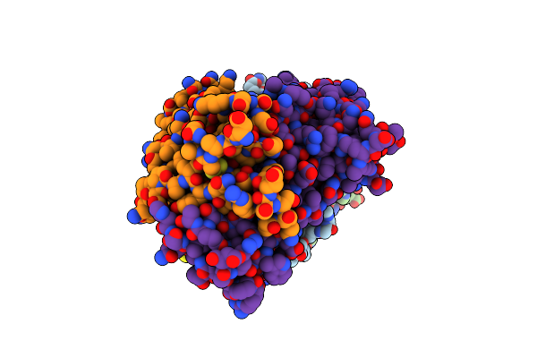 |
Gag:02 Tcr In Complex With Hla-E Featuring A Non-Natural Amino Acid
Organism: Homo sapiens, Human immunodeficiency virus 1
Method: X-RAY DIFFRACTION Resolution:2.90 Å Release Date: 2022-01-26 Classification: IMMUNE SYSTEM |
Organism: Homo sapiens, Human immunodeficiency virus 1
Method: X-RAY DIFFRACTION
Release Date: 2022-01-26
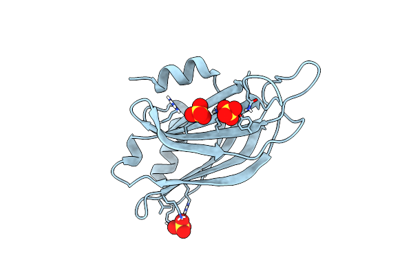 |
Crystal Structure Of Human Synaptotagmin 1 C2B Without Ca2+
Organism: Homo sapiens
Method: X-RAY DIFFRACTION Resolution:1.17 Å Release Date: 2020-05-13 Classification: EXOCYTOSIS Ligands: SO4 |
Organism: Homo sapiens
Method: X-RAY DIFFRACTION
Release Date: 2020-05-13
Ligands: SO4
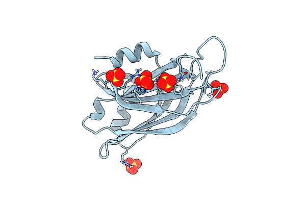 |
1.7 Angstrom Structure Of A Pathogenic Human Syt 1 C2B (D304G)
Organism: Homo sapiens
Method: X-RAY DIFFRACTION Resolution:1.70 Å Release Date: 2020-05-13 Classification: EXOCYTOSIS Ligands: SO4 |
Organism: Homo sapiens
Method: X-RAY DIFFRACTION
Release Date: 2020-05-13
Ligands: SO4
 |
1.3 A Structure Of A Pathogenic Human Syt 1 C2B (I368T)
Organism: Homo sapiens
Method: X-RAY DIFFRACTION Resolution:1.30 Å Release Date: 2020-05-13 Classification: EXOCYTOSIS Ligands: SO4 |
Organism: Homo sapiens
Method: X-RAY DIFFRACTION
Release Date: 2020-05-13
Ligands: SO4
 |
1.4 A Structure Of A Pathogenic Human Syt 1 C2B (D366E)
Organism: Homo sapiens
Method: X-RAY DIFFRACTION Resolution:1.40 Å Release Date: 2020-05-13 Classification: EXOCYTOSIS Ligands: SO4 |
Organism: Homo sapiens
Method: X-RAY DIFFRACTION
Release Date: 2020-05-13
Ligands: SO4
 |
Crystal Structure Of The T-Cell Receptor Nye_S3 Bound To Hla A2*01-Sllmwitqv
Organism: Homo sapiens
Method: X-RAY DIFFRACTION Resolution:3.12 Å Release Date: 2020-01-15 Classification: IMMUNE SYSTEM |
Organism: Homo sapiens
Method: X-RAY DIFFRACTION
Release Date: 2020-01-15
 |
Crystal Structure Of The T-Cell Receptor Nye_S2 Bound To Hla A2*01-Sllmwitqv
Organism: Homo sapiens
Method: X-RAY DIFFRACTION Resolution:2.56 Å Release Date: 2020-01-15 Classification: IMMUNE SYSTEM |
Organism: Homo sapiens
Method: X-RAY DIFFRACTION
Release Date: 2020-01-15
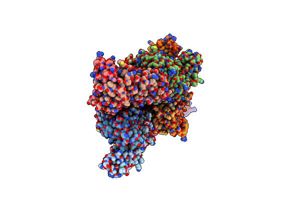 |
Crystal Structure Of The T-Cell Receptor Nye_S1 Bound To Hla A2*01-Sllmwitqv
Organism: Homo sapiens
Method: X-RAY DIFFRACTION Resolution:2.50 Å Release Date: 2020-01-15 Classification: IMMUNE SYSTEM |
Organism: Homo sapiens
Method: X-RAY DIFFRACTION
Release Date: 2020-01-15
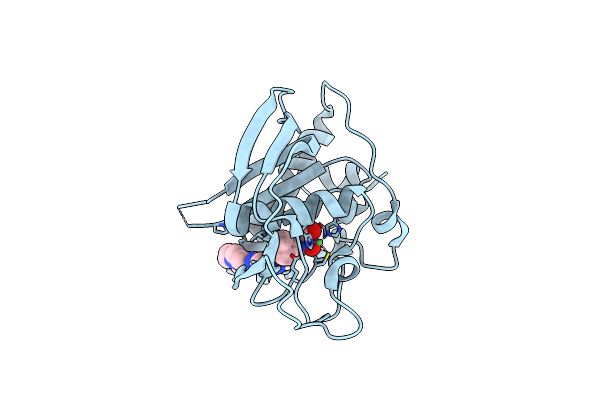 |
X-Ray Structure Of Polypeptide Deformylase
Organism: Streptococcus pneumoniae
Method: X-RAY DIFFRACTION Resolution:1.70 Å Release Date: 2019-06-26 Classification: HYDROLASE/HYDROLASE INHIBITOR Ligands: NI, NB4 |
Organism: Streptococcus pneumoniae
Method: X-RAY DIFFRACTION
Release Date: 2019-06-26
Ligands: NI, NB4
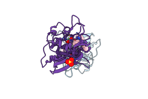 |
X-Ray Structure Of Polypeptide Deformylase With A Piperazic Acid
Organism: Streptococcus pneumoniae
Method: X-RAY DIFFRACTION Resolution:1.45 Å Release Date: 2019-06-26 Classification: HYDROLASE/HYDROLASE INHIBITOR Ligands: NI, SO4, N9Y |
Organism: Streptococcus pneumoniae
Method: X-RAY DIFFRACTION
Release Date: 2019-06-26
Ligands: NI, SO4, N9Y