

Search Count: 87
 |
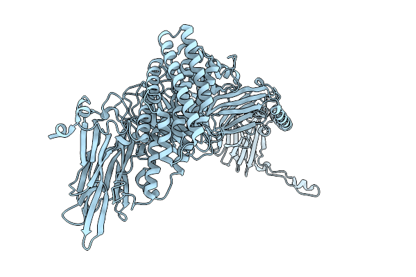
Native Structure Of Full-Length Pesticidal Protein Cry1Ca18 At Ph9, From Crystals Formed In Vivo
Organism: Bacillus thuringiensis
Method: X-RAY DIFFRACTION Resolution:1.65 Å Release Date: 2026-02-11 Classification: TOXIN Ligands: CA, ZN |
Organism: Bacillus thuringiensis
Method: X-RAY DIFFRACTION
Release Date: 2026-02-11
Ligands: CA, ZN
 |
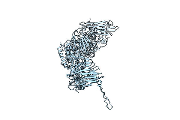
Native Structure Of The Full-Length Pesticidal Protein Cry8Ba2, From Crystals Formed In Vivo (Form 1)
Organism: Bacillus thuringiensis
Method: X-RAY DIFFRACTION Resolution:2.27 Å Release Date: 2025-12-03 Classification: TOXIN |
Organism: Bacillus thuringiensis
Method: X-RAY DIFFRACTION
Release Date: 2025-12-03
 |
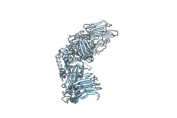
Native Structure Of The Full-Length Pesticidal Protein Cry8Ba2, From Crystals Formed In Vivo (Form 2)
Organism: Bacillus thuringiensis
Method: X-RAY DIFFRACTION Resolution:2.27 Å Release Date: 2025-12-03 Classification: TOXIN |
Organism: Bacillus thuringiensis
Method: X-RAY DIFFRACTION
Release Date: 2025-12-03
 |
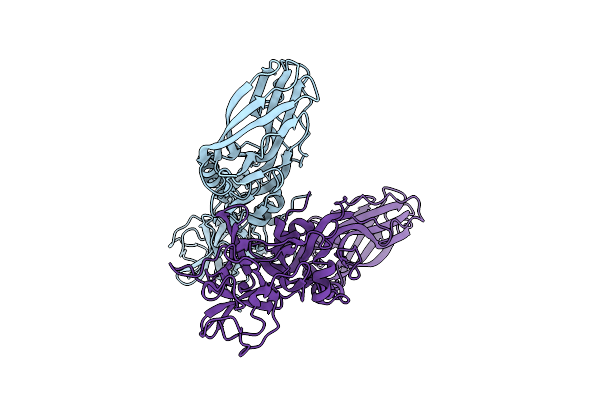
Native Structure Of The Full-Length Pesticidal Protein Cry1Ca18 At Ph7, From Crystals Formed In Vivo
Organism: Bacillus thuringiensis
Method: X-RAY DIFFRACTION Resolution:1.80 Å Release Date: 2025-11-12 Classification: TOXIN Ligands: ZN, CA |
Organism: Bacillus thuringiensis
Method: X-RAY DIFFRACTION
Release Date: 2025-11-12
Ligands: ZN, CA
 |
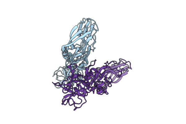
Structure Of The Lysinibacillus Sphaericus Tpp49Aa1 Pesticidal Protein At Ph 3
Organism: Lysinibacillus sphaericus
Method: X-RAY DIFFRACTION Resolution:1.78 Å Release Date: 2023-11-01 Classification: TOXIN |
Organism: Lysinibacillus sphaericus
Method: X-RAY DIFFRACTION
Release Date: 2023-11-01
 |
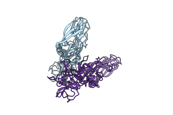
Structure Of The Lysinibacillus Sphaericus Tpp49Aa1 Pesticidal Protein At Ph 7
Organism: Lysinibacillus sphaericus
Method: X-RAY DIFFRACTION Resolution:1.62 Å Release Date: 2023-11-01 Classification: TOXIN |
Organism: Lysinibacillus sphaericus
Method: X-RAY DIFFRACTION
Release Date: 2023-11-01
 |
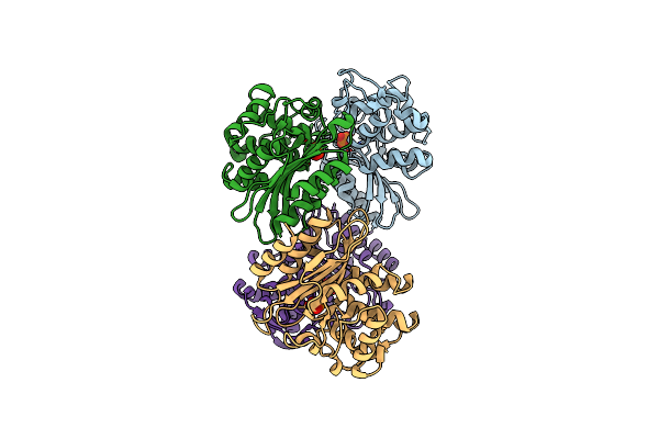
Structure Of The Lysinibacillus Sphaericus Tpp49Aa1 Pesticidal Protein At Ph 11
Organism: Lysinibacillus sphaericus
Method: X-RAY DIFFRACTION Resolution:1.75 Å Release Date: 2023-11-01 Classification: TOXIN |
Organism: Lysinibacillus sphaericus
Method: X-RAY DIFFRACTION
Release Date: 2023-11-01
 |
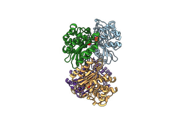
Xfel Structure Of Mycobacterium Tuberculosis Beta Lactamase Microcrystals Mixed With Sulbactam For 3 Ms
Organism: Mycobacterium tuberculosis str. beijing/w bt1
Method: X-RAY DIFFRACTION Resolution:2.62 Å Release Date: 2023-09-20 Classification: HYDROLASE/Inhibitor Ligands: PO4 |
Organism: Mycobacterium tuberculosis str. beijing/w bt1
Method: X-RAY DIFFRACTION
Release Date: 2023-09-20
Ligands: PO4
 |
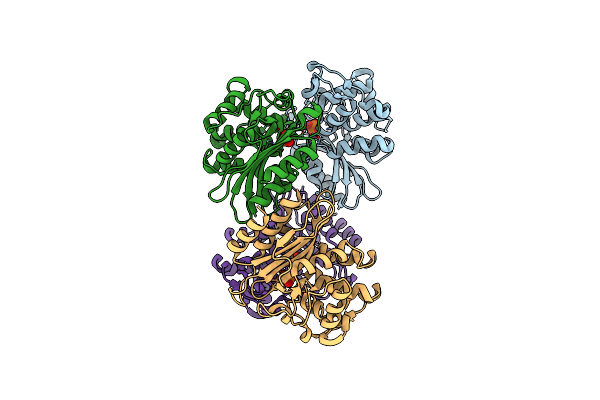
Xfel Structure Of Mycobacterium Tuberculosis Beta Lactamase Microcrystals Mixed With Sulbactam For 6 Ms
Organism: Mycobacterium tuberculosis
Method: X-RAY DIFFRACTION Resolution:2.75 Å Release Date: 2023-09-20 Classification: HYDROLASE/Inhibitor Ligands: PO4 |
Organism: Mycobacterium tuberculosis
Method: X-RAY DIFFRACTION
Release Date: 2023-09-20
Ligands: PO4
 |
Xfel Structure Of Mycobacterium Tuberculosis Beta Lactamase Microcrystals
Organism: Mycobacterium tuberculosis
Method: X-RAY DIFFRACTION Resolution:2.20 Å Release Date: 2023-09-20 Classification: HYDROLASE/Inhibitor Ligands: PO4 |
Organism: Mycobacterium tuberculosis
Method: X-RAY DIFFRACTION
Release Date: 2023-09-20
Ligands: PO4
 |
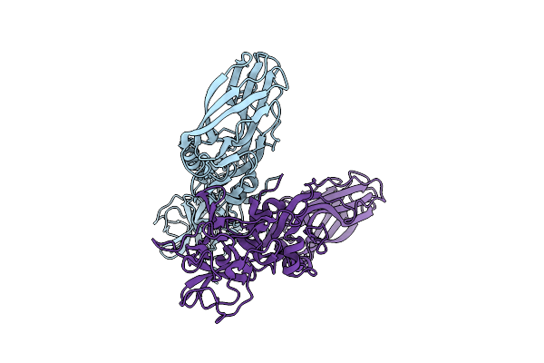
Xfel Structure Of Mycobacterium Tuberculosis Beta Lactamase Microcrystals Mixed With Sulbactam For 700 Ms
Organism: Mycobacterium tuberculosis
Method: X-RAY DIFFRACTION Resolution:3.20 Å Release Date: 2023-09-20 Classification: HYDROLASE/Inhibitor Ligands: PO4, TSL |
Organism: Mycobacterium tuberculosis
Method: X-RAY DIFFRACTION
Release Date: 2023-09-20
Ligands: PO4, TSL
 |
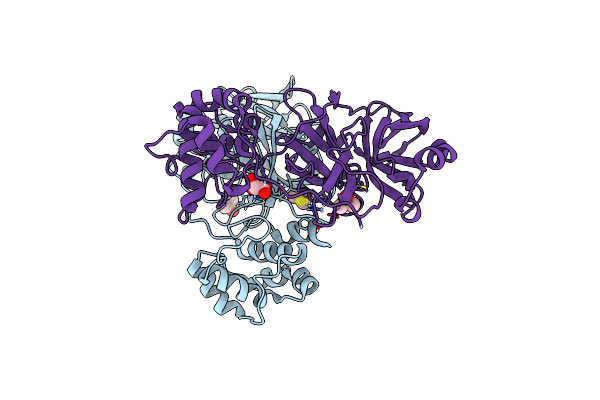
The Structure Of Natural Crystals Of The Lysinibacillus Sphaericus Tpp49Aa1 Pesticidal Protein Elucidated Using Serial Femtosecond Crystallography At An X-Ray Free Electron Laser
Organism: Lysinibacillus sphaericus
Method: X-RAY DIFFRACTION Resolution:2.20 Å Release Date: 2023-05-17 Classification: TOXIN |
Organism: Lysinibacillus sphaericus
Method: X-RAY DIFFRACTION
Release Date: 2023-05-17
 |
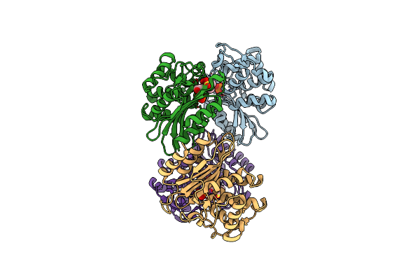
Crystal Structure Of Sars-Cov-2 Main Protease In Orthorhombic Space Group P212121
Organism: Severe acute respiratory syndrome coronavirus 2
Method: X-RAY DIFFRACTION Resolution:1.65 Å Release Date: 2023-03-22 Classification: HYDROLASE Ligands: CL, DMS, MLI |
Organism: Severe acute respiratory syndrome coronavirus 2
Method: X-RAY DIFFRACTION
Release Date: 2023-03-22
Ligands: CL, DMS, MLI
 |
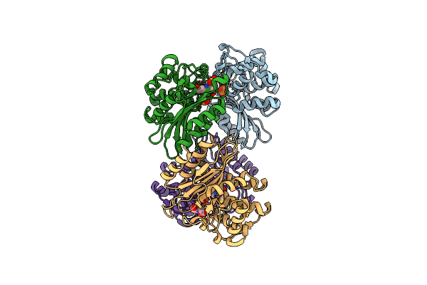
Xfel Structure Of Beta Lactamase Microcrystals Mixed With Sulbactam Solution For 15Ms
Organism: Mycobacterium tuberculosis
Method: X-RAY DIFFRACTION Resolution:2.70 Å Release Date: 2023-03-08 Classification: HYDROLASE Ligands: PO4, TSL |
Organism: Mycobacterium tuberculosis
Method: X-RAY DIFFRACTION
Release Date: 2023-03-08
Ligands: PO4, TSL
 |
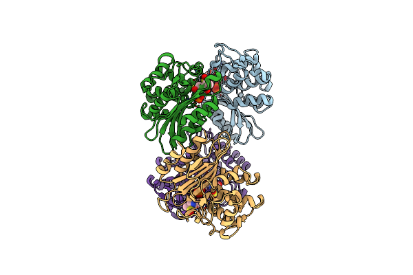
Xfel Structure Of Mycobacterium Tuberculosis Beta Lactamase Microcrystals Mixed With Sulbactam For 30Ms
Organism: Mycobacterium tuberculosis
Method: X-RAY DIFFRACTION Resolution:2.80 Å Release Date: 2023-03-08 Classification: HYDROLASE/Inhibitor Ligands: PO4, 0RN, TSL |
Organism: Mycobacterium tuberculosis
Method: X-RAY DIFFRACTION
Release Date: 2023-03-08
Ligands: PO4, 0RN, TSL
 |
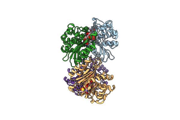
Xfel Structure Of Mycobacterium Tuberculosis Beta Lactamase Microcrystals Mixed With Sulbactam For 240Ms
Organism: Mycobacterium tuberculosis
Method: X-RAY DIFFRACTION Resolution:2.35 Å Release Date: 2023-03-08 Classification: HYDROLASE/Inhibitor Ligands: PO4, TSL |
Organism: Mycobacterium tuberculosis
Method: X-RAY DIFFRACTION
Release Date: 2023-03-08
Ligands: PO4, TSL
 |
Cryo Structure Of Mycobacterium Tuberculosis Beta Lactamase Microcrystals Mixed With Sulbactam For 3 Hours
Organism: Mycobacterium tuberculosis
Method: X-RAY DIFFRACTION Resolution:2.74 Å Release Date: 2023-03-08 Classification: HYDROLASE/Inhibitor Ligands: PO4, TSL |
Organism: Mycobacterium tuberculosis
Method: X-RAY DIFFRACTION
Release Date: 2023-03-08
Ligands: PO4, TSL
 |
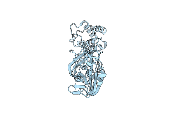
Oxidized Form Of Sars-Cov-2 Main Protease Determined By Xfel Radiation
Organism: Severe acute respiratory syndrome coronavirus 2
Method: X-RAY DIFFRACTION Resolution:2.25 Å Release Date: 2023-01-25 Classification: VIRAL PROTEIN Ligands: DMS |
Organism: Severe acute respiratory syndrome coronavirus 2
Method: X-RAY DIFFRACTION
Release Date: 2023-01-25
Ligands: DMS
 |
Reduced Form Of Sars-Cov-2 Main Protease Determined By Xfel Radiation
Organism: Severe acute respiratory syndrome coronavirus 2
Method: X-RAY DIFFRACTION Resolution:1.75 Å Release Date: 2023-01-18 Classification: VIRAL PROTEIN Ligands: CL |
Organism: Severe acute respiratory syndrome coronavirus 2
Method: X-RAY DIFFRACTION
Release Date: 2023-01-18
Ligands: CL
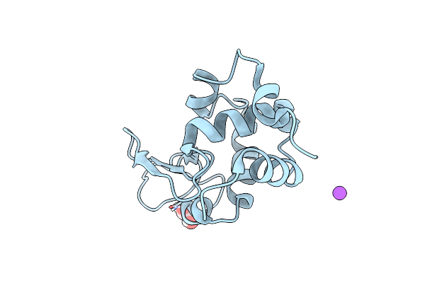 |
Multi-Hit Sfx Using Mhz Xfel Sources- First Hit
Organism: Gallus gallus
Method: X-RAY DIFFRACTION Resolution:3.20 Å Release Date: 2022-07-20 Classification: HYDROLASE Ligands: EDO, NA |
Organism: Gallus gallus
Method: X-RAY DIFFRACTION
Release Date: 2022-07-20
Ligands: EDO, NA