

Search Count: 13
 |
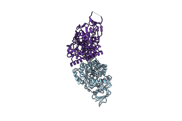
2.8 Angstrom Resolution Crystal Structure Of D-Hydantoinase From Bacillus Sp. Ar9 In C2221 Space Group
Organism: Bacillus sp. ar9
Method: X-RAY DIFFRACTION Resolution:2.80 Å Release Date: 2014-05-21 Classification: HYDROLASE Ligands: MN |
Organism: Bacillus sp. ar9
Method: X-RAY DIFFRACTION
Release Date: 2014-05-21
Ligands: MN
 |
Crystal Structure Of D-Hydantoinase From Bacillus Sp. Ar9 In C2221 Space Group
Organism: Bacillus sp.
Method: X-RAY DIFFRACTION Resolution:2.80 Å Release Date: 2014-05-07 Classification: HYDROLASE Ligands: MN |
Organism: Bacillus sp.
Method: X-RAY DIFFRACTION
Release Date: 2014-05-07
Ligands: MN
 |
Crystal Structure Of Proteolyzed Lysozyme
Organism: Gallus gallus
Method: X-RAY DIFFRACTION Resolution:2.00 Å Release Date: 2011-10-19 Classification: HYDROLASE |
Organism: Gallus gallus
Method: X-RAY DIFFRACTION
Release Date: 2011-10-19
 |
Molecular Structure Of D-Hydantoinase From A Bacillus Sp. Ar9: Evidence For Mercury Inhibition
Organism: Bacillus sp.
Method: X-RAY DIFFRACTION Resolution:2.30 Å Release Date: 2005-03-01 Classification: HYDROLASE Ligands: MN |
Organism: Bacillus sp.
Method: X-RAY DIFFRACTION
Release Date: 2005-03-01
Ligands: MN
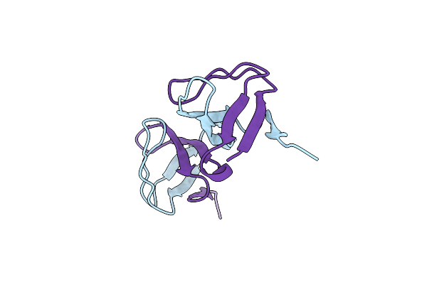 |
Eps8 Sh3 Domain Intertwined Dimer
Organism: Mus musculus
Method: X-RAY DIFFRACTION Resolution:1.80 Å Release Date: 2001-05-09 Classification: HORMONE/GROWTH FACTOR |
Organism: Mus musculus
Method: X-RAY DIFFRACTION
Release Date: 2001-05-09
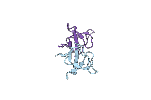 |
Eps8 Sh3 Closed Monomer
Organism: Mus musculus
Method: X-RAY DIFFRACTION Resolution:2.00 Å Release Date: 2001-05-09 Classification: HORMONE/GROWTH FACTOR |
Organism: Mus musculus
Method: X-RAY DIFFRACTION
Release Date: 2001-05-09
 |
The Sh3 Domain Of Eps8 Exists As A Novel Intertwined Dimer
Organism: Mus musculus
Method: X-RAY DIFFRACTION Resolution:2.50 Å Release Date: 1998-07-08 Classification: SIGNAL TRANSDUCTION |
Organism: Mus musculus
Method: X-RAY DIFFRACTION
Release Date: 1998-07-08
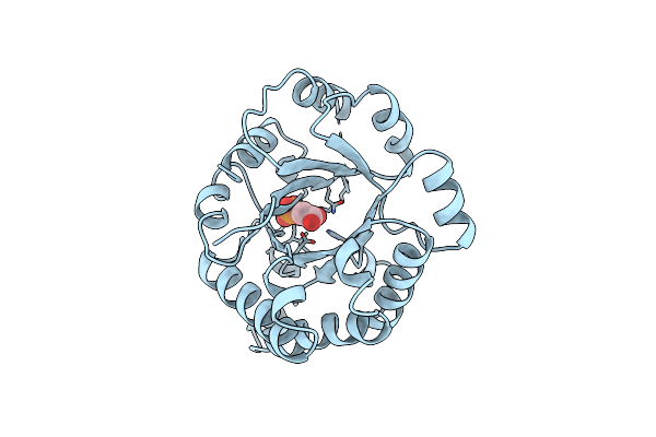 |
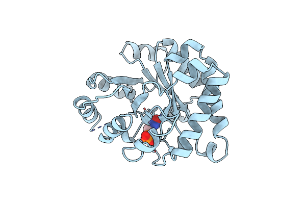
Three New Crystal Structures Of Point Mutation Variants Of Monotim: Conformational Flexibility Of Loop-1,Loop-4 And Loop-8
Organism: Trypanosoma brucei brucei
Method: X-RAY DIFFRACTION Resolution:2.40 Å Release Date: 1995-10-15 Classification: ISOMERASE(INTRAMOLECULAR OXIDOREDUCTASE) Ligands: PGA |
Organism: Trypanosoma brucei brucei
Method: X-RAY DIFFRACTION
Release Date: 1995-10-15
Ligands: PGA
 |
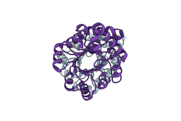
Three New Crystal Structures Of Point Mutation Variants Of Monotim: Conformational Flexibility Of Loop-1,Loop-4 And Loop-8
Organism: Trypanosoma brucei brucei
Method: X-RAY DIFFRACTION Resolution:2.40 Å Release Date: 1995-09-15 Classification: ISOMERASE(INTRAMOLECULAR OXIDOREDUCTASE) Ligands: PGH |
Organism: Trypanosoma brucei brucei
Method: X-RAY DIFFRACTION
Release Date: 1995-09-15
Ligands: PGH
 |
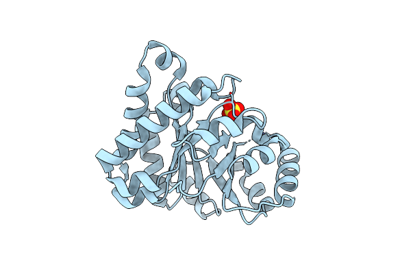
Large Scale Structural Rearrangements Of The Front Loops In Monomerised Triosephosphate Isomerase, As Deduced From The Comparison Of The Structural Properties Of Monotim And Its Point Mutation Variant Monoss
Organism: Trypanosoma brucei brucei
Method: X-RAY DIFFRACTION Resolution:2.40 Å Release Date: 1994-09-30 Classification: ISOMERASE(INTRAMOLECULAR OXIDOREDUCTASE) |
Organism: Trypanosoma brucei brucei
Method: X-RAY DIFFRACTION
Release Date: 1994-09-30
 |
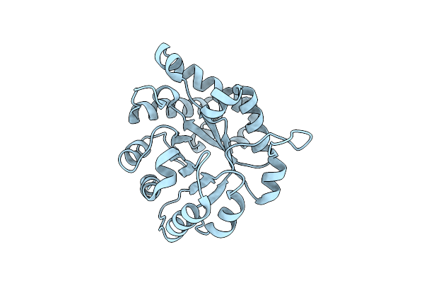
The Crystal Structure Of An Engineered Monomeric Triosephosphate Isomerase, Monotim: The Correct Modelling Of An Eight-Residue Loop
Organism: Trypanosoma brucei brucei
Method: X-RAY DIFFRACTION Resolution:2.40 Å Release Date: 1994-07-31 Classification: ISOMERASE(INTRAMOLECULAR OXIDOREDUCTASE) Ligands: SO4 |
Organism: Trypanosoma brucei brucei
Method: X-RAY DIFFRACTION
Release Date: 1994-07-31
Ligands: SO4
 |
Comparison Of The Structures And The Crystal Contacts Of Trypanosomal Triosephosphate Isomerase In Four Different Crystal Forms
Organism: Trypanosoma brucei brucei
Method: X-RAY DIFFRACTION Resolution:2.10 Å Release Date: 1994-05-31 Classification: ISOMERASE(INTRAMOLECULAR OXIDOREDUCTASE) |
Organism: Trypanosoma brucei brucei
Method: X-RAY DIFFRACTION
Release Date: 1994-05-31
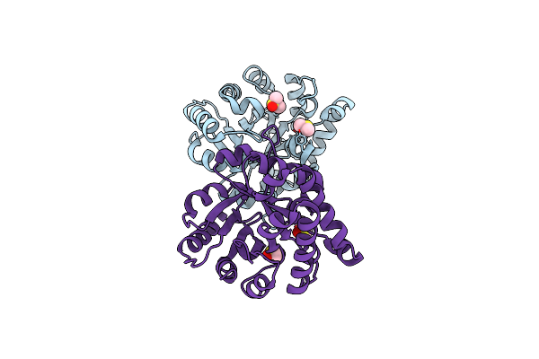 |
Comparison Of The Structures And The Crystal Contacts Of Trypanosomal Triosephosphate Isomerase In Four Different Crystal Forms
Organism: Trypanosoma brucei brucei
Method: X-RAY DIFFRACTION Resolution:1.80 Å Release Date: 1994-05-31 Classification: ISOMERASE(INTRAMOLECULAR OXIDOREDUCTASE) Ligands: DMS |
Organism: Trypanosoma brucei brucei
Method: X-RAY DIFFRACTION
Release Date: 1994-05-31
Ligands: DMS