

Search Count: 43
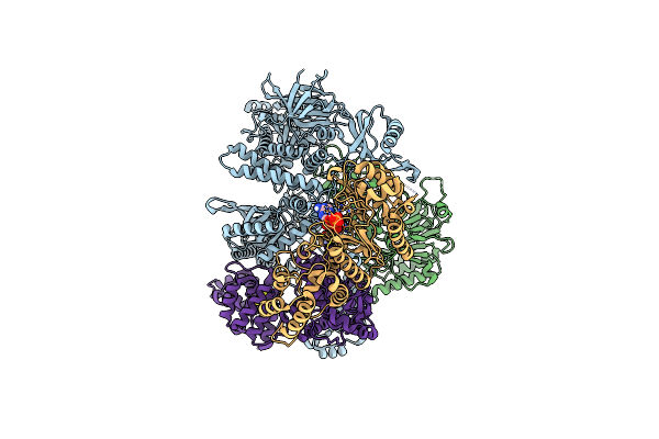 |
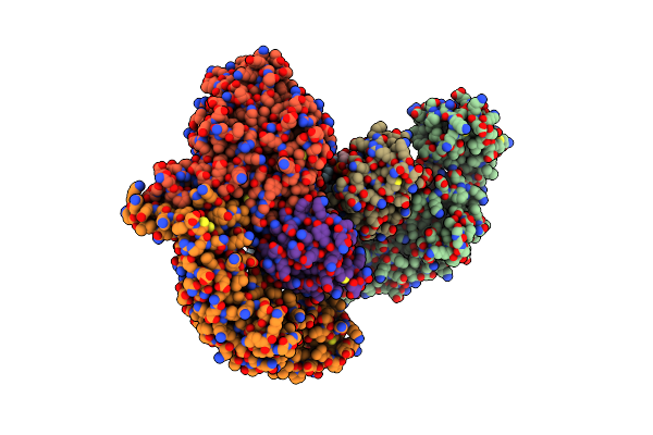
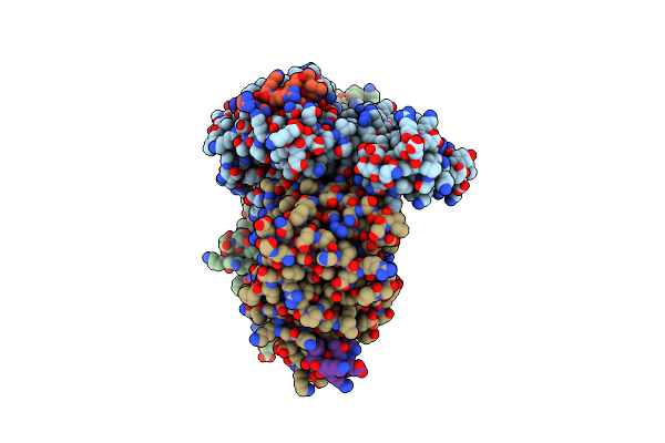
Cryo-Em Structure Of Human Dna Polymerase Alpha-Primase In Pre-Initiation Stage 1
Organism: Homo sapiens
Method: ELECTRON MICROSCOPY Release Date: 2024-02-21 Classification: DNA BINDING PROTEIN Ligands: ZN, MN, ATP, SF4 |
Organism: Homo sapiens
Method: ELECTRON MICROSCOPY
Release Date: 2024-02-21
Ligands: ZN, MN, ATP, SF4
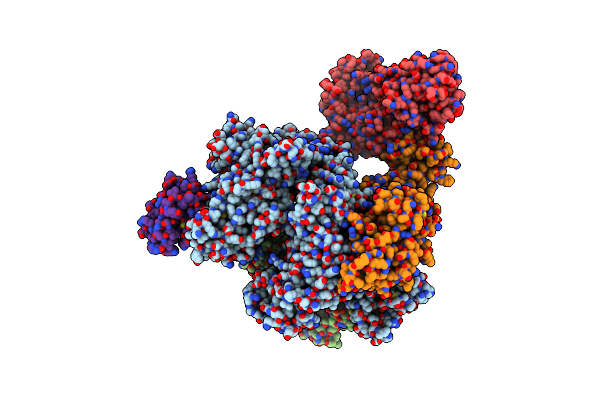 |
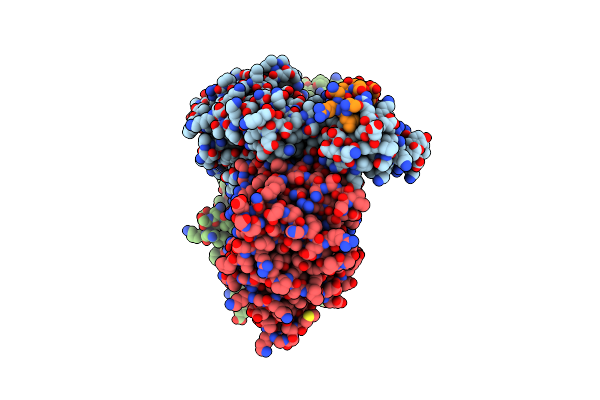
Cryoem Structure Of Dna Polymerase Alpha - Primase Bound To Sars Cov Nsp1
Organism: Homo sapiens, Severe acute respiratory syndrome coronavirus
Method: ELECTRON MICROSCOPY Release Date: 2021-11-10 Classification: DNA BINDING PROTEIN Ligands: ZN, SF4 |
Organism: Homo sapiens, Severe acute respiratory syndrome coronavirus
Method: ELECTRON MICROSCOPY
Release Date: 2021-11-10
Ligands: ZN, SF4
 |
Crystal Structure Of The Second, C-Terminal Repeat Of The Dna-Binding Domain Of Human Timeless
Organism: Homo sapiens
Method: X-RAY DIFFRACTION Resolution:1.15 Å Release Date: 2020-07-01 Classification: REPLICATION |
Organism: Homo sapiens
Method: X-RAY DIFFRACTION
Release Date: 2020-07-01
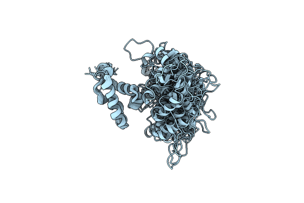 |
Timeless Couples G Quadruplex Detection With Processing By Ddx11 During Dna Replication
Organism: Homo sapiens
Method: SOLUTION NMR Release Date: 2020-07-01 Classification: DNA BINDING PROTEIN |
Organism: Homo sapiens
Method: SOLUTION NMR
Release Date: 2020-07-01
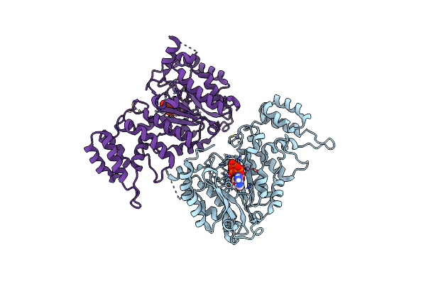 |
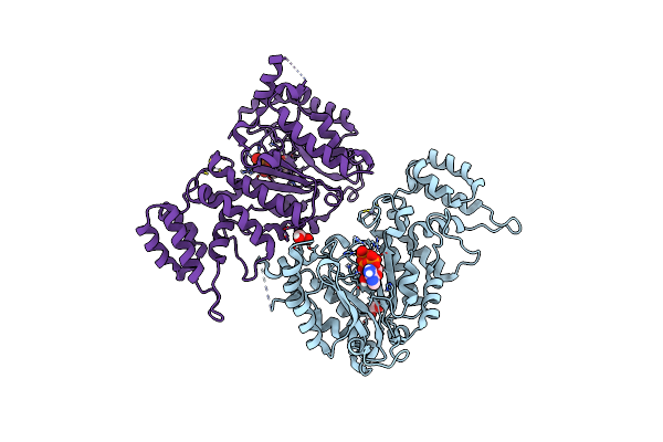
Crystal Structure Of The Pri1 Subunit Of Human Primase Bound To Atp
Organism: Homo sapiens
Method: X-RAY DIFFRACTION Resolution:2.75 Å Release Date: 2019-09-11 Classification: REPLICATION Ligands: ZN, MN, ATP |
Organism: Homo sapiens
Method: X-RAY DIFFRACTION
Release Date: 2019-09-11
Ligands: ZN, MN, ATP
 |
Crystal Structure Of The Pri1 Subunit Of Human Primase Bound To Vidarabine Triphosphate
Organism: Homo sapiens
Method: X-RAY DIFFRACTION Resolution:2.35 Å Release Date: 2019-09-11 Classification: REPLICATION Ligands: ZN, MN, HEJ, EDO |
Organism: Homo sapiens
Method: X-RAY DIFFRACTION
Release Date: 2019-09-11
Ligands: ZN, MN, HEJ, EDO
 |
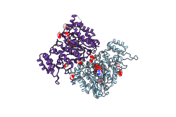
Crystal Structure Of The Pri1 Subunit Of Human Primase Bound To Fludarabine Triphosphate
Organism: Homo sapiens
Method: X-RAY DIFFRACTION Resolution:2.20 Å Release Date: 2019-09-11 Classification: REPLICATION Ligands: ZN, MN, HFD, EDO |
Organism: Homo sapiens
Method: X-RAY DIFFRACTION
Release Date: 2019-09-11
Ligands: ZN, MN, HFD, EDO
 |
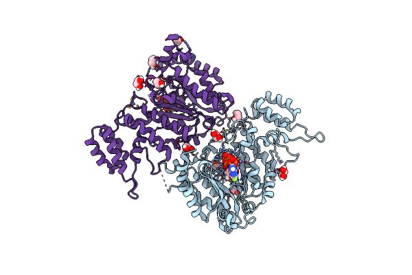
Crystal Structure Of The Pri1 Subunit Of Human Primase Bound To Datp
Organism: Homo sapiens
Method: X-RAY DIFFRACTION Resolution:1.95 Å Release Date: 2019-09-11 Classification: REPLICATION Ligands: ZN, MN, DTP, EDO |
Organism: Homo sapiens
Method: X-RAY DIFFRACTION
Release Date: 2019-09-11
Ligands: ZN, MN, DTP, EDO
 |
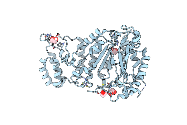
Crystal Structure Of The Pri1 Subunit Of Human Primase Bound To 2F-Atp
Organism: Homo sapiens
Method: X-RAY DIFFRACTION Resolution:1.85 Å Release Date: 2019-09-11 Classification: REPLICATION Ligands: ZN, MN, JSQ, EDO |
Organism: Homo sapiens
Method: X-RAY DIFFRACTION
Release Date: 2019-09-11
Ligands: ZN, MN, JSQ, EDO
 |
Crystal Structure Of The Pri1 Subunit Of Human Primase
Organism: Homo sapiens
Method: X-RAY DIFFRACTION Resolution:1.50 Å Release Date: 2019-09-11 Classification: REPLICATION Ligands: EDO, ZN |
Organism: Homo sapiens
Method: X-RAY DIFFRACTION
Release Date: 2019-09-11
Ligands: EDO, ZN
 |
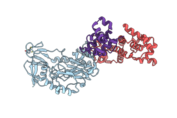
Crystal Structure Of The Heterotrimeric Prislx Primase From S. Solfataricus.
Organism: Sulfolobus solfataricus (strain atcc 35092 / dsm 1617 / jcm 11322 / p2)
Method: X-RAY DIFFRACTION Resolution:2.91 Å Release Date: 2017-11-29 Classification: REPLICATION Ligands: APC, MN, ZN |
Organism: Sulfolobus solfataricus (strain atcc 35092 / dsm 1617 / jcm 11322 / p2)
Method: X-RAY DIFFRACTION
Release Date: 2017-11-29
Ligands: APC, MN, ZN
 |
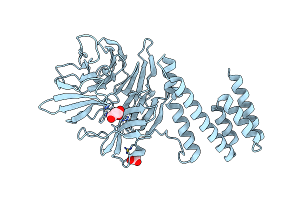
Crystal Structure Of The Heterotrimeric Prislx Primase From S. Solfataricus.
Organism: Sulfolobus solfataricus (strain atcc 35092 / dsm 1617 / jcm 11322 / p2), Sulfolobus solfataricus
Method: X-RAY DIFFRACTION Resolution:3.01 Å Release Date: 2017-11-29 Classification: REPLICATION Ligands: ZN |
Organism: Sulfolobus solfataricus (strain atcc 35092 / dsm 1617 / jcm 11322 / p2), Sulfolobus solfataricus
Method: X-RAY DIFFRACTION
Release Date: 2017-11-29
Ligands: ZN
 |
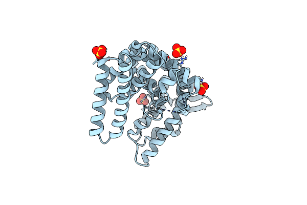
Crystal Structure Of Human And-1 Sepb Domain
Organism: Homo sapiens
Method: X-RAY DIFFRACTION Resolution:2.50 Å Release Date: 2017-11-29 Classification: REPLICATION Ligands: MLI |
Organism: Homo sapiens
Method: X-RAY DIFFRACTION
Release Date: 2017-11-29
Ligands: MLI
 |
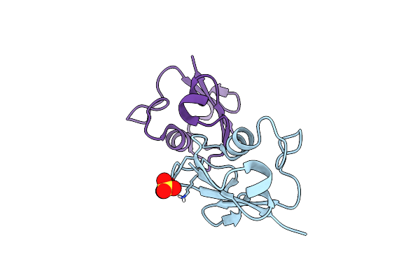
Crystal Structure Of The N-Terminal Domain Of Human Timeless
Organism: Homo sapiens
Method: X-RAY DIFFRACTION Resolution:1.85 Å Release Date: 2017-03-08 Classification: REPLICATION Ligands: SO4 |
Organism: Homo sapiens
Method: X-RAY DIFFRACTION
Release Date: 2017-03-08
Ligands: SO4
 |
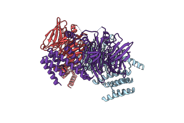
Crystal Structure Of The Carboxy-Terminal Domain Of Yeast Ctf4 Bound To Dna2.
Organism: Saccharomyces cerevisiae
Method: X-RAY DIFFRACTION Resolution:3.09 Å Release Date: 2016-06-22 Classification: REPLICATION |
Organism: Saccharomyces cerevisiae
Method: X-RAY DIFFRACTION
Release Date: 2016-06-22
 |
Crystal Structure Of The Carboxy-Terminal Domain Of Yeast Ctf4 Bound To Tof2.
Organism: Saccharomyces cerevisiae
Method: X-RAY DIFFRACTION Resolution:3.30 Å Release Date: 2016-06-22 Classification: REPLICATION |
Organism: Saccharomyces cerevisiae
Method: X-RAY DIFFRACTION
Release Date: 2016-06-22
 |
X-Ray Crystal Structure Of Sulfolobus Solfataricus Urm1
Organism: Sulfolobus solfataricus
Method: X-RAY DIFFRACTION Resolution:2.20 Å Release Date: 2015-09-23 Classification: SIGNALING PROTEIN Ligands: SO4 |
Organism: Sulfolobus solfataricus
Method: X-RAY DIFFRACTION
Release Date: 2015-09-23
Ligands: SO4
 |
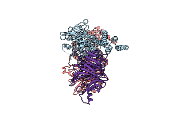
Crystal Structure Of The C-Terminal Region Of Yeast Ctf4, Selenomethionine Protein.
Organism: Saccharomyces cerevisiae
Method: X-RAY DIFFRACTION Resolution:2.69 Å Release Date: 2014-04-30 Classification: DNA REPLICATION |
Organism: Saccharomyces cerevisiae
Method: X-RAY DIFFRACTION
Release Date: 2014-04-30
 |
Crystal Structure Of The C-Terminal Region Of Yeast Ctf4
Organism: Saccharomyces cerevisiae
Method: X-RAY DIFFRACTION Resolution:3.00 Å Release Date: 2014-04-30 Classification: DNA REPLICATION |
Organism: Saccharomyces cerevisiae
Method: X-RAY DIFFRACTION
Release Date: 2014-04-30
 |
Crystal Structure Of The Carboxy-Terminal Domain Of Yeast Ctf4 Bound To Pol Alpha.
Organism: Saccharomyces cerevisiae
Method: X-RAY DIFFRACTION Resolution:2.69 Å Release Date: 2014-04-30 Classification: DNA REPLICATION |
Organism: Saccharomyces cerevisiae
Method: X-RAY DIFFRACTION
Release Date: 2014-04-30