

Search Count: 54
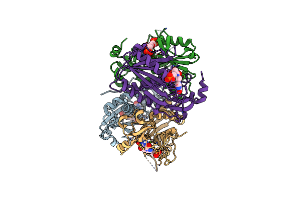 |
Structure Of Human Hypoxanthine Guanine Phosphoribzosyltransferase In Complex With [2S,4R]-4-Guanin-9-Yl-2-(2-Phosphonoethoxymethyl)-1-N-(3-Phosphonopropionyl)Pyrrolidine
Organism: Homo sapiens
Method: X-RAY DIFFRACTION Resolution:2.27 Å Release Date: 2024-05-08 Classification: TRANSFERASE Ligands: JEI, MG |
Organism: Homo sapiens
Method: X-RAY DIFFRACTION
Release Date: 2024-05-08
Ligands: JEI, MG
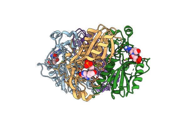 |
Structure Of Human Hypoxanthine Guanine Phosphoribzosyltransferase In Complex With [2S,4R] 4-Guanin-9-Yl-2-Hydroxymethyl-1-N-(3-Phosphonopropionyl)Pyrrolidine
Organism: Homo sapiens
Method: X-RAY DIFFRACTION Resolution:2.50 Å Release Date: 2024-05-08 Classification: TRANSFERASE Ligands: JG6, MG |
Organism: Homo sapiens
Method: X-RAY DIFFRACTION
Release Date: 2024-05-08
Ligands: JG6, MG
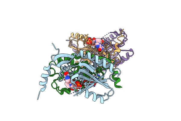 |
Crystal Structure Of Trypanosome Brucei Hypoxanthine Guanine Phosphopribosyltransferase In Complex With [2S,4R]-4-Guanin-9-Yl-2-(2- Phosphonoethoxymethyl)-1-N-(3-Phosphonopropionyl)Pyrrolidine
Organism: Trypanosoma brucei
Method: X-RAY DIFFRACTION Resolution:2.46 Å Release Date: 2024-05-08 Classification: TRANSFERASE Ligands: JEI |
Organism: Trypanosoma brucei
Method: X-RAY DIFFRACTION
Release Date: 2024-05-08
Ligands: JEI
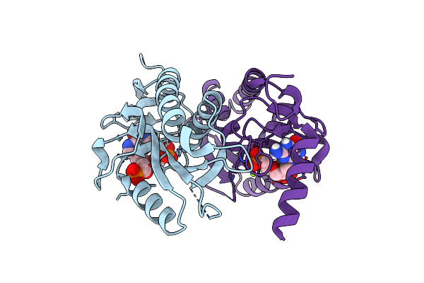 |
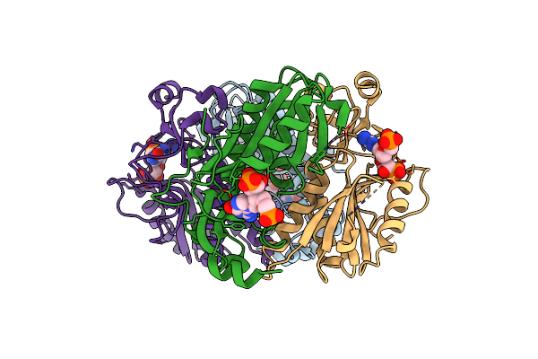
Crystal Structure Of T. Brucei Hypoxanthine Guanine Phosphoribosyltransferase In Complex With [2S,4S]-4-Guanin-9-Yl-2-(2-Phosphonoethoxymethyl)-1-N-(3-Phosphonopropionyl)Pyrrolidine
Organism: Trypanosoma brucei
Method: X-RAY DIFFRACTION Resolution:2.20 Å Release Date: 2024-05-08 Classification: TRANSFERASE Ligands: MG, KFF |
Organism: Trypanosoma brucei
Method: X-RAY DIFFRACTION
Release Date: 2024-05-08
Ligands: MG, KFF
 |
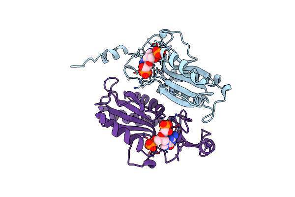
Crystal Structure Of Human Hypoxanthine Guanine Phosphoribzosyltransferase In Complex With (4S,7S)-7-Hydroxy-4-((Guanin-9-Yl)Methyl)-2,5-Dioxaheptan-1,7-Diphosphonate
Organism: Homo sapiens
Method: X-RAY DIFFRACTION Resolution:2.58 Å Release Date: 2022-05-18 Classification: TRANSFERASE/TRANSFERASE INHIBITOR Ligands: 8QI |
Organism: Homo sapiens
Method: X-RAY DIFFRACTION
Release Date: 2022-05-18
Ligands: 8QI
 |
Crystal Structure Of T. Brucei Hypoxanthine Guanine Phosphoribosyltransferase In Complex With (4S,7S)-7-Hydroxy-4-((Guanin-9-Yl)Methyl)-2,5-Dioxaheptan-1,7-Diphosphonate
Organism: Trypanosoma brucei brucei
Method: X-RAY DIFFRACTION Resolution:2.65 Å Release Date: 2022-03-02 Classification: TRANSFERASE Ligands: 8QI |
Organism: Trypanosoma brucei brucei
Method: X-RAY DIFFRACTION
Release Date: 2022-03-02
Ligands: 8QI
 |
Crystal Structure Of Trypanosome Brucei Hypoxanthine-Guanine-Xanthine Phosphoribzosyltransferase In Complex With (4S,7S)-7-Hydroxy-4-((Guanin-9-Yl)Methyl)-2,5-Dioxaheptan-1,7-Diphosphonate
Organism: Trypanosoma brucei brucei
Method: X-RAY DIFFRACTION Resolution:2.12 Å Release Date: 2022-03-02 Classification: TRANSFERASE/INHIBITOR Ligands: 8QI |
Organism: Trypanosoma brucei brucei
Method: X-RAY DIFFRACTION
Release Date: 2022-03-02
Ligands: 8QI
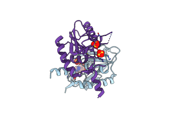 |
Helicobacter Pylori Xanthine-Guanine-Hypoxanthine Phosphoribosyltransferase
Organism: Helicobacter pylori
Method: X-RAY DIFFRACTION Resolution:1.97 Å Release Date: 2021-05-05 Classification: TRANSFERASE Ligands: WG7, SO4 |
Organism: Helicobacter pylori
Method: X-RAY DIFFRACTION
Release Date: 2021-05-05
Ligands: WG7, SO4
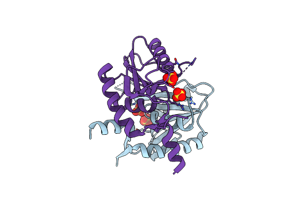 |
Helicobacter Pylori Xanthine-Guanine-Hypoxanthine Phosphoribosyltransferase
Organism: Helicobacter pylori
Method: X-RAY DIFFRACTION Resolution:1.47 Å Release Date: 2021-05-05 Classification: TRANSFERASE Ligands: SO4 |
Organism: Helicobacter pylori
Method: X-RAY DIFFRACTION
Release Date: 2021-05-05
Ligands: SO4
 |
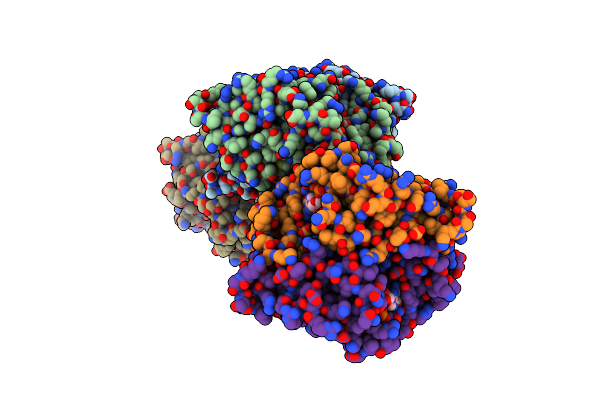
Crystal Structure Of Trypanosoma Brucei Hypoxanthine-Guanine-Xanthine Phosphoribosyltranferase In Complex With Xmp
Organism: Trypanosoma brucei brucei (strain 927/4 gutat10.1)
Method: X-RAY DIFFRACTION Resolution:2.19 Å Release Date: 2019-07-24 Classification: TRANSFERASE Ligands: XMP, MG |
Organism: Trypanosoma brucei brucei (strain 927/4 gutat10.1)
Method: X-RAY DIFFRACTION
Release Date: 2019-07-24
Ligands: XMP, MG
 |
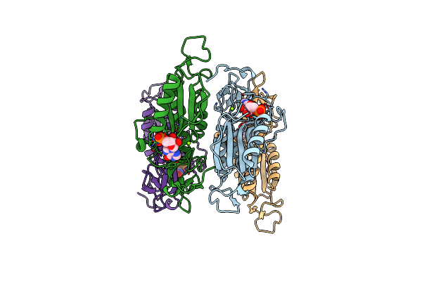
Crystal Structure Of Trypanosoma Brucei Hypoxanthine-Guanine-Xanthine Phosphoribosyltranferase In Complex With Gmp
Organism: Trypanosoma brucei brucei (strain 927/4 gutat10.1)
Method: X-RAY DIFFRACTION Resolution:1.99 Å Release Date: 2019-07-24 Classification: TRANSFERASE Ligands: 5GP, MG |
Organism: Trypanosoma brucei brucei (strain 927/4 gutat10.1)
Method: X-RAY DIFFRACTION
Release Date: 2019-07-24
Ligands: 5GP, MG
 |
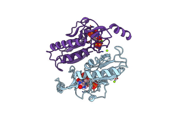
Crystal Structure Of Trypanosoma Brucei Hypoxanthine-Guanine-Xanthine Phosphoribosyltranferase In Complex With Imp
Organism: Trypanosoma brucei brucei (strain 927/4 gutat10.1)
Method: X-RAY DIFFRACTION Resolution:2.96 Å Release Date: 2019-07-24 Classification: TRANSFERASE Ligands: IMP, MG |
Organism: Trypanosoma brucei brucei (strain 927/4 gutat10.1)
Method: X-RAY DIFFRACTION
Release Date: 2019-07-24
Ligands: IMP, MG
 |
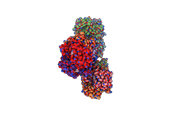
Crystal Structure Of Trypanosoma Brucei Hypoxanthine-Guanine Phosphoribosyltranferase In Complex With Xmp
Organism: Trypanosoma brucei brucei
Method: X-RAY DIFFRACTION Resolution:2.39 Å Release Date: 2019-07-24 Classification: TRANSFERASE Ligands: XMP, MG, SO4 |
Organism: Trypanosoma brucei brucei
Method: X-RAY DIFFRACTION
Release Date: 2019-07-24
Ligands: XMP, MG, SO4
 |
Crystal Structure Of Plasmodium Vivax Hypoxanthine Guanine Phosphoribosyltransferase In Complex With [3R,4R]-4-Guanin-9-Yl-3-((S)-2-Hydroxy-2-Phosphonoethyl)Oxy-1-N-(Phosphonopropionyl)Pyrrolidine
Organism: Plasmodium vivax
Method: X-RAY DIFFRACTION Resolution:2.86 Å Release Date: 2017-12-13 Classification: TRANSFERASE/INHIBITOR Ligands: MG, YPG |
Organism: Plasmodium vivax
Method: X-RAY DIFFRACTION
Release Date: 2017-12-13
Ligands: MG, YPG
 |
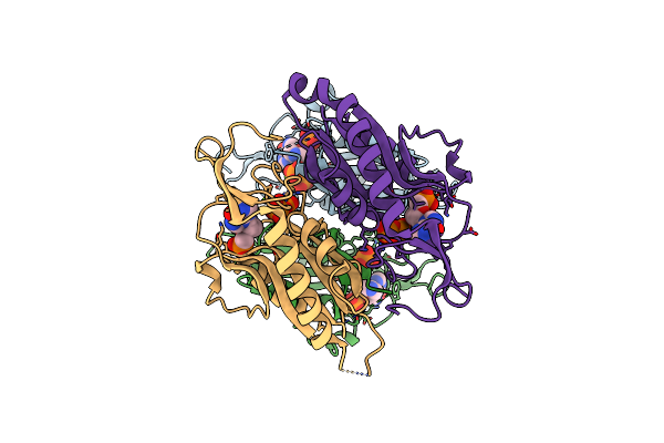
Human Hypoxanthine Guanine Phosphoribosyltransferase In Complex With [3R,4R]-4-Guanin-9-Yl-3-((R)-2-Hydroxy-2-Phosphonoethyl)Oxy-1-N-(Phosphonopropionyl)Pyrrolidine
Organism: Homo sapiens
Method: X-RAY DIFFRACTION Resolution:1.91 Å Release Date: 2017-12-06 Classification: TRANSFERASE/INHIBITOR Ligands: WPG, MG |
Organism: Homo sapiens
Method: X-RAY DIFFRACTION
Release Date: 2017-12-06
Ligands: WPG, MG
 |
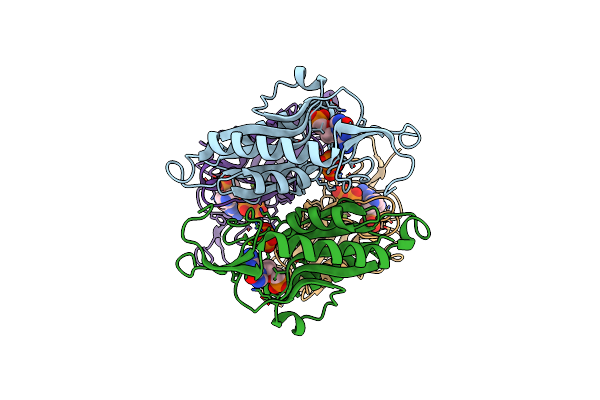
Crystal Structure Of Mycobacterium Tuberculosis Hypoxanthine Guanine Phosphoribosyltransferase In Complex With [3S,4R]-(4-(Hypoxanthin-9-Yl)Pyrrolidin-3-Yl)-Oxymethanephosphonic Acid
Organism: Mycobacterium tuberculosis (strain atcc 25618 / h37rv)
Method: X-RAY DIFFRACTION Resolution:2.45 Å Release Date: 2017-09-20 Classification: TRANSFERASE/INHIBITOR Ligands: 6W7, MG, POP |
Organism: Mycobacterium tuberculosis (strain atcc 25618 / h37rv)
Method: X-RAY DIFFRACTION
Release Date: 2017-09-20
Ligands: 6W7, MG, POP
 |
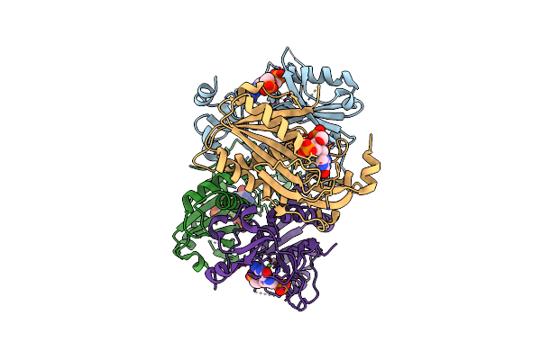
Crystal Structure Of Mycobacterium Tuberculosis Hypoxanthine Guanine Phosphoribosyltransferase In Complex With [3S,4R]-(4-(Guanin-9-Yl)Pyrrolidin-3-Yl)Oxymethanephosphonic Acid And Pyrophosphate
Organism: Mycobacterium tuberculosis (strain atcc 25618 / h37rv)
Method: X-RAY DIFFRACTION Resolution:2.55 Å Release Date: 2017-09-20 Classification: TRANSFERASE/INHIBITOR Ligands: 6W8, MG, POP |
Organism: Mycobacterium tuberculosis (strain atcc 25618 / h37rv)
Method: X-RAY DIFFRACTION
Release Date: 2017-09-20
Ligands: 6W8, MG, POP
 |
Crystal Structure Of Mycobacterium Tuberculosis Hypoxanthine Guanine Phosphoribosyltransferase In Complex With (3-((3R,4R)-3-(Guanin-9-Yl)-4-((S)-2-Hydroxy-2-Phosphonoethoxy)Pyrrolidin-1-Yl)-3-Oxopropyl)Phosphonic Acid
Organism: Mycobacterium tuberculosis (strain atcc 25618 / h37rv)
Method: X-RAY DIFFRACTION Resolution:2.91 Å Release Date: 2017-09-20 Classification: TRANSFERASE/INHIBITOR Ligands: YPG, MG |
Organism: Mycobacterium tuberculosis (strain atcc 25618 / h37rv)
Method: X-RAY DIFFRACTION
Release Date: 2017-09-20
Ligands: YPG, MG
 |
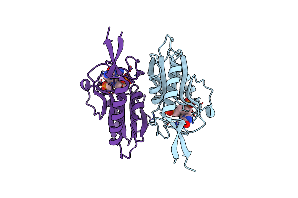
Human Hgprt In Complex With [(2-[(Guanin-9-Yl)Methyl]Propane-1,3-Diyl)Bis(Oxy)]Bis(Methylene)Diphosphonic Acid
Organism: Homo sapiens
Method: X-RAY DIFFRACTION Resolution:2.35 Å Release Date: 2017-08-30 Classification: TRANSFERASE Ligands: 9YP, MG |
Organism: Homo sapiens
Method: X-RAY DIFFRACTION
Release Date: 2017-08-30
Ligands: 9YP, MG
 |
E. Coli Hprt In Complexed With 9-[(N-Phosphonoethyl-N-Phosphonoethoxyethyl)-2-Aminoethyl]-Guanine
Organism: Escherichia coli
Method: X-RAY DIFFRACTION Resolution:2.86 Å Release Date: 2017-07-19 Classification: TRANSFERASE/INHIBITOR Ligands: 3L5, MG |
Organism: Escherichia coli
Method: X-RAY DIFFRACTION
Release Date: 2017-07-19
Ligands: 3L5, MG