

Search Count: 84
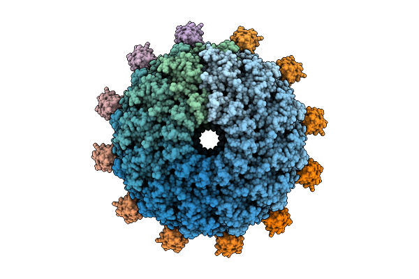 |
C3 Reconstruction Of The Thermophilic Bacteriophage P74-26 Neck
Organism: Oshimavirus p7426
Method: ELECTRON MICROSCOPY Release Date: 2026-01-28 Classification: VIRUS |
Organism: Oshimavirus p7426
Method: ELECTRON MICROSCOPY
Release Date: 2026-01-28
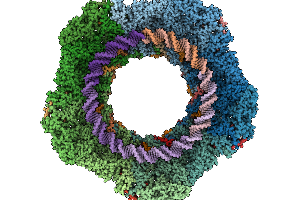 |
C5 Reconstruction Of The Thermophilic Bacteriophage P74-26 Portal Vertex
Organism: Oshimavirus p7426
Method: ELECTRON MICROSCOPY Release Date: 2026-01-28 Classification: VIRUS |
Organism: Oshimavirus p7426
Method: ELECTRON MICROSCOPY
Release Date: 2026-01-28
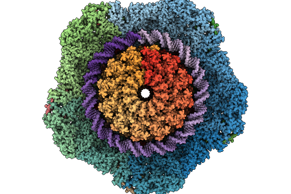 |
C1 Reconstruction Of The Thermophilic Bacteriophage P74-26 Portal And Portal Vertex
Organism: Oshimavirus p7426
Method: ELECTRON MICROSCOPY Release Date: 2026-01-28 Classification: VIRUS |
Organism: Oshimavirus p7426
Method: ELECTRON MICROSCOPY
Release Date: 2026-01-28
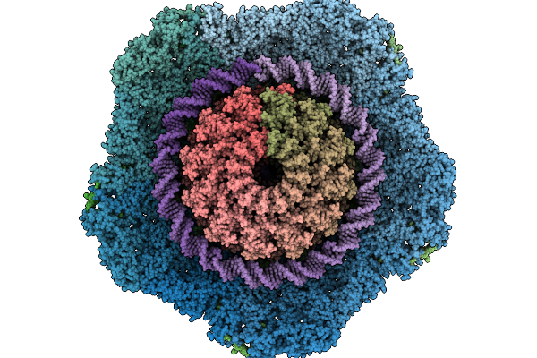 |
Composite Reconstruction Of The Thermophilic Bacteriophage P74-26 Neck And Portal Vertex
Organism: Oshimavirus p7426
Method: ELECTRON MICROSCOPY Release Date: 2026-01-28 Classification: VIRUS/DNA |
Organism: Oshimavirus p7426
Method: ELECTRON MICROSCOPY
Release Date: 2026-01-28
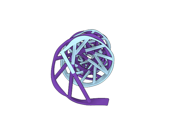 |
Dsdna In The Central Channel Of The Bacteriophage P74-26 Neck
Organism: Oshimavirus p7426
Method: ELECTRON MICROSCOPY Release Date: 2026-01-28 Classification: VIRUS/DNA |
Organism: Oshimavirus p7426
Method: ELECTRON MICROSCOPY
Release Date: 2026-01-28
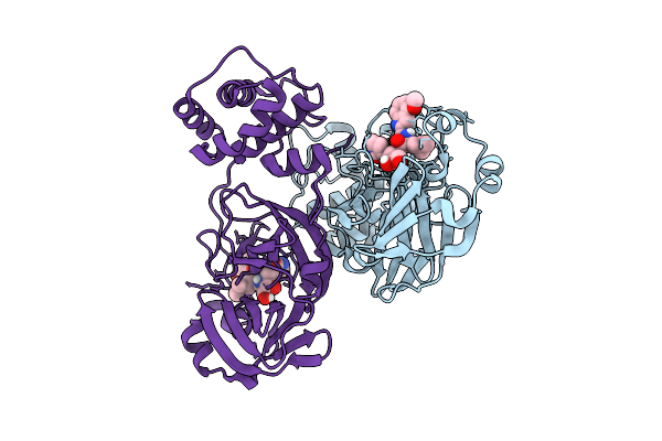 |
Crystal Structure Of Sars-Cov-2 Mpro S10A In Complex With Pfizer Intravenous Inhibitor Pf-00835231
Organism: Severe acute respiratory syndrome coronavirus 2
Method: X-RAY DIFFRACTION Resolution:1.90 Å Release Date: 2025-12-10 Classification: VIRAL PROTEIN Ligands: V2M |
Organism: Severe acute respiratory syndrome coronavirus 2
Method: X-RAY DIFFRACTION
Release Date: 2025-12-10
Ligands: V2M
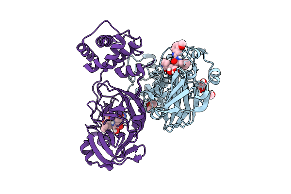 |
Crystal Structure Of Sars-Cov-2 Mpro S10C In Complex With Pfizer Intravenous Inhibitor Pf-00835231
Organism: Severe acute respiratory syndrome coronavirus 2
Method: X-RAY DIFFRACTION Resolution:1.99 Å Release Date: 2025-12-10 Classification: VIRAL PROTEIN Ligands: DMS, EDO, V2M |
Organism: Severe acute respiratory syndrome coronavirus 2
Method: X-RAY DIFFRACTION
Release Date: 2025-12-10
Ligands: DMS, EDO, V2M
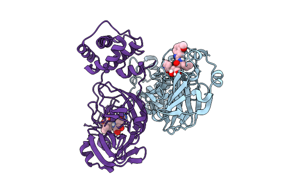 |
Crystal Structure Of Sars-Cov-2 Mpro S113A In Complex With Pfizer Intravenous Inhibitor Pf-00835231
Organism: Severe acute respiratory syndrome coronavirus 2
Method: X-RAY DIFFRACTION Resolution:1.97 Å Release Date: 2025-12-10 Classification: VIRAL PROTEIN Ligands: V2M |
Organism: Severe acute respiratory syndrome coronavirus 2
Method: X-RAY DIFFRACTION
Release Date: 2025-12-10
Ligands: V2M
 |
Crystal Structure Of Sars-Cov-2 Mpro S113C In Complex With Pfizer Intravenous Inhibitor Pf-00835231
Organism: Severe acute respiratory syndrome coronavirus 2
Method: X-RAY DIFFRACTION Resolution:2.28 Å Release Date: 2025-12-10 Classification: VIRAL PROTEIN Ligands: V2M |
Organism: Severe acute respiratory syndrome coronavirus 2
Method: X-RAY DIFFRACTION
Release Date: 2025-12-10
Ligands: V2M
 |
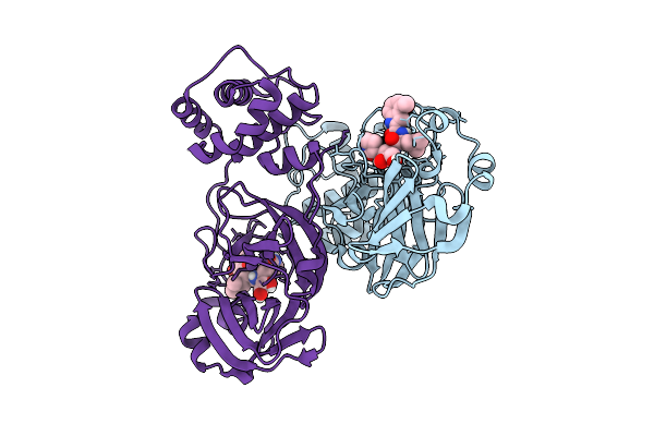
Crystal Structure Of Sars-Cov-2 Mpro L115A In Complex With Pfizer Intravenous Inhibitor Pf-00835231
Organism: Severe acute respiratory syndrome coronavirus 2
Method: X-RAY DIFFRACTION Resolution:2.21 Å Release Date: 2025-12-10 Classification: VIRAL PROTEIN Ligands: V2M |
Organism: Severe acute respiratory syndrome coronavirus 2
Method: X-RAY DIFFRACTION
Release Date: 2025-12-10
Ligands: V2M
 |
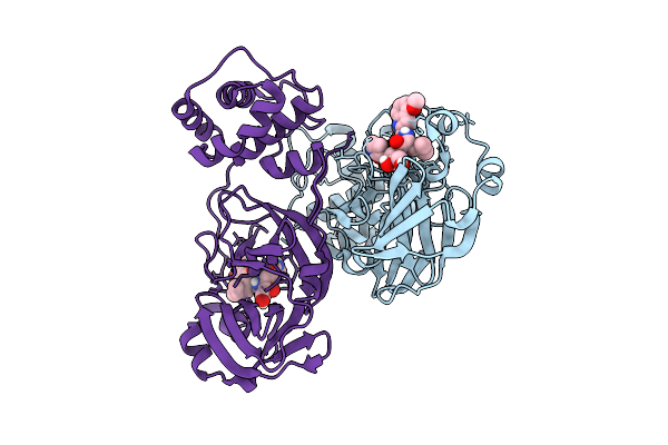
Crystal Structure Of Sars-Cov-2 Mpro L115M In Complex With Pfizer Intravenous Inhibitor Pf-00835231
Organism: Severe acute respiratory syndrome coronavirus 2
Method: X-RAY DIFFRACTION Resolution:2.19 Å Release Date: 2025-12-10 Classification: VIRAL PROTEIN Ligands: V2M |
Organism: Severe acute respiratory syndrome coronavirus 2
Method: X-RAY DIFFRACTION
Release Date: 2025-12-10
Ligands: V2M
 |
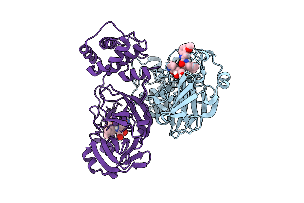
Crystal Structure Of Sars-Cov-2 Mpro S147A In Complex With Pfizer Intravenous Inhibitor Pf-00835231
Organism: Severe acute respiratory syndrome coronavirus 2
Method: X-RAY DIFFRACTION Resolution:2.13 Å Release Date: 2025-12-10 Classification: VIRAL PROTEIN Ligands: V2M |
Organism: Severe acute respiratory syndrome coronavirus 2
Method: X-RAY DIFFRACTION
Release Date: 2025-12-10
Ligands: V2M
 |
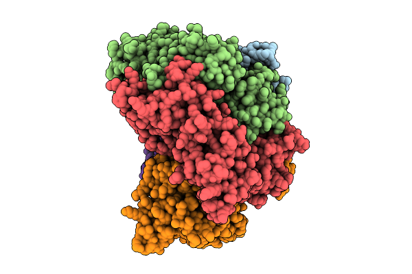
Crystal Structure Of Sars-Cov-2 Mpro S147N In Complex With Pfizer Intravenous Inhibitor Pf-00835231
Organism: Severe acute respiratory syndrome coronavirus 2
Method: X-RAY DIFFRACTION Resolution:1.77 Å Release Date: 2025-12-10 Classification: VIRAL PROTEIN Ligands: V2M |
Organism: Severe acute respiratory syndrome coronavirus 2
Method: X-RAY DIFFRACTION
Release Date: 2025-12-10
Ligands: V2M
 |
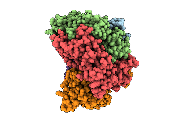
Structure Of The S. Cerevisiae Clamp Loader Replication Factor C (Rfc) With Mixed Nucleotide Occupancy
Organism: Saccharomyces cerevisiae
Method: ELECTRON MICROSCOPY Release Date: 2025-11-12 Classification: REPLICATION Ligands: ACT, ADP, GDP |
Organism: Saccharomyces cerevisiae
Method: ELECTRON MICROSCOPY
Release Date: 2025-11-12
Ligands: ACT, ADP, GDP
 |
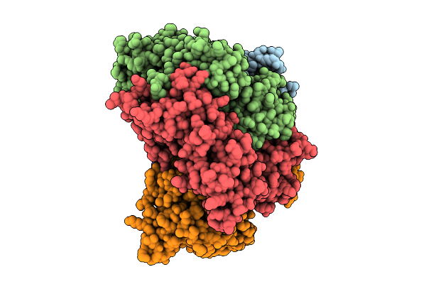
Structure Of The S. Cerevisiae Clamp Loader Replication Factor C (Rfc) With Mixed Nucleotide Occupancy
Organism: Saccharomyces cerevisiae
Method: ELECTRON MICROSCOPY Release Date: 2025-11-12 Classification: REPLICATION Ligands: ACT, ADP, GDP |
Organism: Saccharomyces cerevisiae
Method: ELECTRON MICROSCOPY
Release Date: 2025-11-12
Ligands: ACT, ADP, GDP
 |
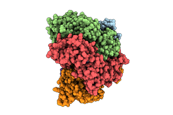
Structure Of The S. Cerevisiae Clamp Loader Replication Factor C (Rfc) With Mixed Nucleotide Occupancy
Organism: Saccharomyces cerevisiae
Method: ELECTRON MICROSCOPY Release Date: 2025-11-12 Classification: REPLICATION Ligands: ACT, ADP, GDP |
Organism: Saccharomyces cerevisiae
Method: ELECTRON MICROSCOPY
Release Date: 2025-11-12
Ligands: ACT, ADP, GDP
 |
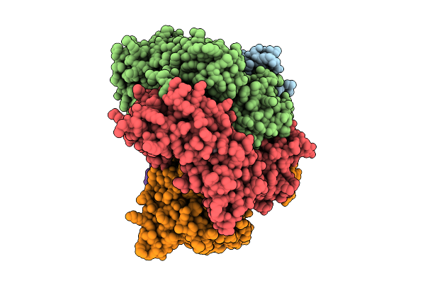
Structure Of The S. Cerevisiae Clamp Loader Replication Factor C (Rfc) With Mixed Nucleotide Occupancy
Organism: Saccharomyces cerevisiae
Method: ELECTRON MICROSCOPY Release Date: 2025-11-12 Classification: REPLICATION Ligands: ACT, ADP, GDP |
Organism: Saccharomyces cerevisiae
Method: ELECTRON MICROSCOPY
Release Date: 2025-11-12
Ligands: ACT, ADP, GDP
 |
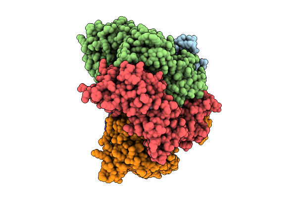
Structure Of The S. Cerevisiae Clamp Loader Replication Factor C (Rfc) With Mixed Nucleotide Occupancy
Organism: Saccharomyces cerevisiae
Method: ELECTRON MICROSCOPY Release Date: 2025-11-12 Classification: REPLICATION Ligands: ACT, ADP, GDP |
Organism: Saccharomyces cerevisiae
Method: ELECTRON MICROSCOPY
Release Date: 2025-11-12
Ligands: ACT, ADP, GDP
 |
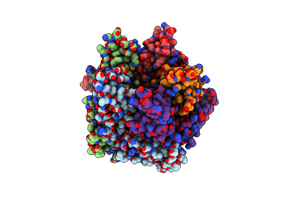
Structure Of The S. Cerevisiae Clamp Loader Replication Factor C (Rfc) With Mixed Nucleotide Occupancy
Organism: Saccharomyces cerevisiae
Method: ELECTRON MICROSCOPY Release Date: 2025-11-12 Classification: REPLICATION Ligands: ACT, ADP, GDP |
Organism: Saccharomyces cerevisiae
Method: ELECTRON MICROSCOPY
Release Date: 2025-11-12
Ligands: ACT, ADP, GDP
 |
Structure Of The Five-Fold Capsomer Of The Drosophila Retrotransposon Copia Capsid
Organism: Drosophila
Method: ELECTRON MICROSCOPY Release Date: 2025-02-12 Classification: VIRUS LIKE PARTICLE |
Organism: Drosophila
Method: ELECTRON MICROSCOPY
Release Date: 2025-02-12