
Search Count: 60
All
Selected
 |
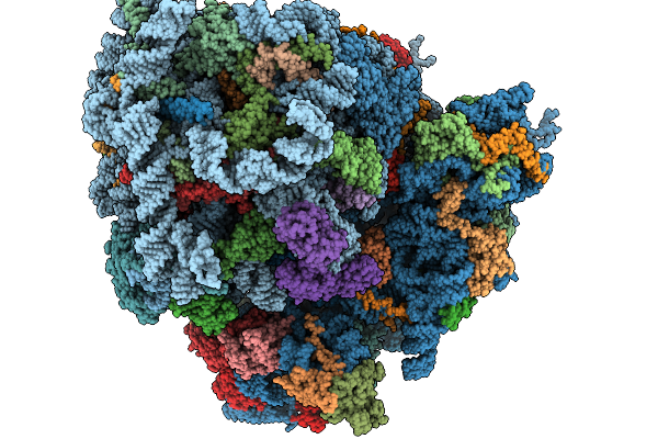
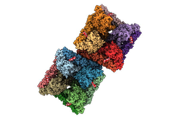
Organism: Heterocephalus glaber
Method: ELECTRON MICROSCOPY Resolution:5.00 Å Release Date: 2026-02-18 Classification: RIBOSOME Ligands: MG, ZN |
 |
Organism: Heterocephalus glaber
Method: ELECTRON MICROSCOPY Release Date: 2026-02-18 Classification: RIBOSOME Ligands: MG, ZN |
 |
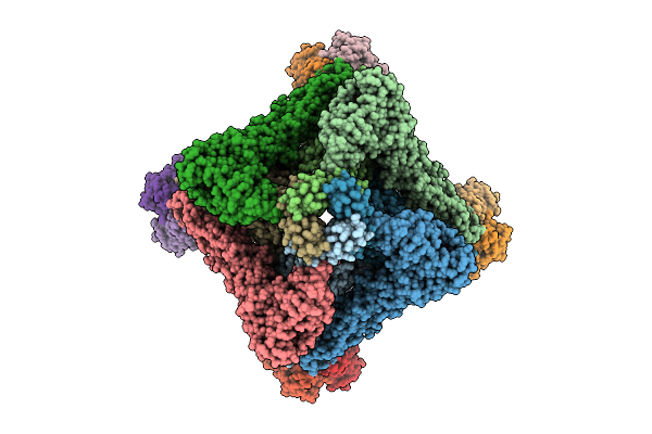
Organism: Ctenomyidae
Method: ELECTRON MICROSCOPY Resolution:3.40 Å Release Date: 2026-02-18 Classification: RIBOSOME |
 |
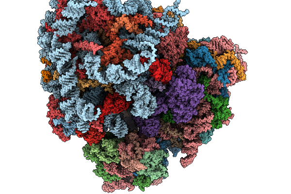
Organism: Ctenomyidae
Method: ELECTRON MICROSCOPY Resolution:3.30 Å Release Date: 2026-02-18 Classification: RIBOSOME Ligands: MG |
 |
Organism: Cavia porcellus
Method: ELECTRON MICROSCOPY Release Date: 2026-02-18 Classification: RIBOSOME |
 |
Organism: Heterocephalus glaber
Method: ELECTRON MICROSCOPY Release Date: 2026-02-18 Classification: RIBOSOME |
 |
Organism: Orthohantavirus andesense
Method: ELECTRON MICROSCOPY Release Date: 2025-07-30 Classification: VIRAL PROTEIN |
 |
Organism: Orthohantavirus andesense
Method: ELECTRON MICROSCOPY Release Date: 2025-07-30 Classification: VIRAL PROTEIN |
 |
Organism: Orthohantavirus andesense
Method: ELECTRON MICROSCOPY Release Date: 2025-07-30 Classification: VIRAL PROTEIN |
 |
Organism: Orthohantavirus andesense
Method: ELECTRON MICROSCOPY Release Date: 2025-07-30 Classification: VIRAL PROTEIN |
 |
Organism: Homo sapiens, Orthohantavirus andesense
Method: ELECTRON MICROSCOPY Release Date: 2025-07-30 Classification: VIRAL PROTEIN/IMMUNE SYSTEM Ligands: NAG |
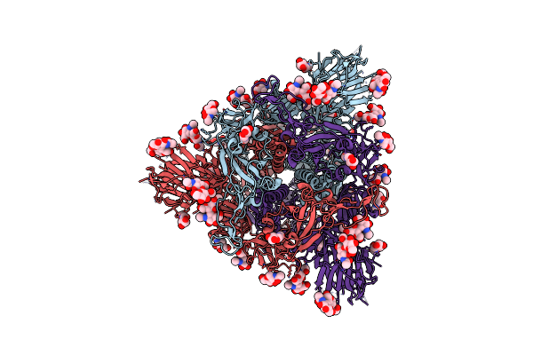 |
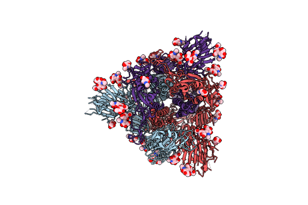
Cryo-Em Structure Of Sars-Cov-2 Spike Proteins On Intact Virions: B.1 Variant 3 Closed Rbds
Organism: Severe acute respiratory syndrome coronavirus 2
Method: ELECTRON MICROSCOPY Release Date: 2024-11-27 Classification: VIRAL PROTEIN Ligands: NAG |
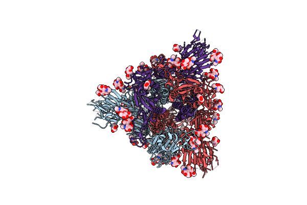 |
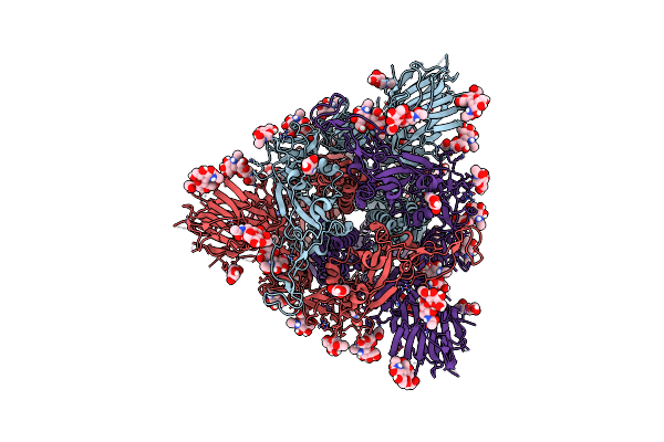
Cryo-Em Structure Of Sars-Cov-2 Spike Proteins On Intact Virions: B.1 Variant 1 Open Rbd
Organism: Severe acute respiratory syndrome coronavirus 2
Method: ELECTRON MICROSCOPY Release Date: 2024-11-27 Classification: VIRAL PROTEIN Ligands: NAG |
 |
Cryo-Em Structure Of Sars-Cov-2 Spike Proteins On Intact Virions: Alpha (B.1.1.7) Variant 3 Closed Rbds
Organism: Severe acute respiratory syndrome coronavirus 2
Method: ELECTRON MICROSCOPY Release Date: 2024-11-27 Classification: VIRAL PROTEIN Ligands: NAG, ZN |
 |
Cryo-Em Structure Of Sars-Cov-2 Spike Proteins On Intact Virions: Alpha (B.1.1.7) Variant 1 Open Rbd
Organism: Severe acute respiratory syndrome coronavirus 2
Method: ELECTRON MICROSCOPY Release Date: 2024-11-27 Classification: VIRAL PROTEIN Ligands: NAG |
 |
Cryo-Em Structure Of Sars-Cov-2 Spike Proteins On Intact Virions: Gamma (P.1) Variant 3 Closed Rbds
Organism: Severe acute respiratory syndrome coronavirus 2
Method: ELECTRON MICROSCOPY Release Date: 2024-11-27 Classification: VIRUS Ligands: NAG |
 |
Cryo-Em Structure Of Sars-Cov-2 Spike Proteins On Intact Virions: Delta (B.1.617.2) Variant 3 Closed Rbds
Organism: Severe acute respiratory syndrome coronavirus 2
Method: ELECTRON MICROSCOPY Release Date: 2024-11-27 Classification: VIRAL PROTEIN Ligands: NAG |
 |
Cryo-Em Structure Of Sars-Cov-2 Spike Proteins On Intact Virions: Mu (B.1.621) Variant 3 Closed Rbds
Organism: Severe acute respiratory syndrome coronavirus 2
Method: ELECTRON MICROSCOPY Release Date: 2024-11-27 Classification: VIRAL PROTEIN Ligands: NAG |
 |
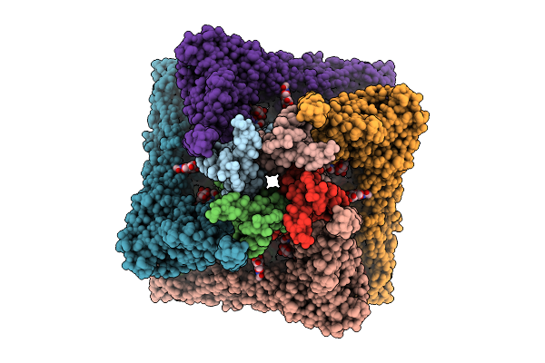
Organism: Mycobacterium tuberculosis
Method: X-RAY DIFFRACTION Resolution:2.70 Å Release Date: 2024-11-06 Classification: DNA BINDING PROTEIN |
 |
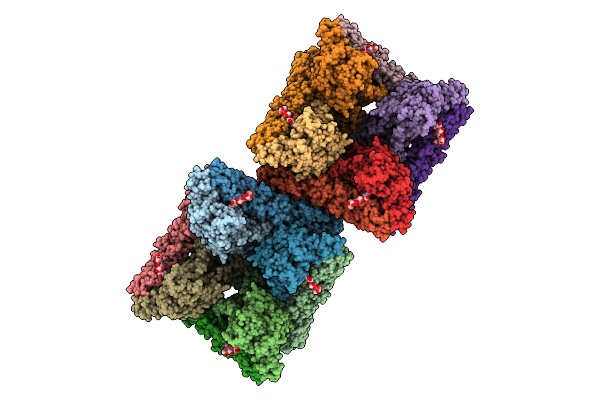
Identification Of Small-Molecule Binding Sites Of A Ubiquitin-Conjugating Enzyme-Ube2T Through Fragment-Based Screening
Organism: Homo sapiens
Method: X-RAY DIFFRACTION Resolution:1.54 Å Release Date: 2024-02-28 Classification: LIGASE Ligands: V23, EDO |