

Search Count: 27
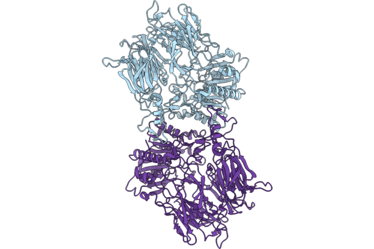 |
Chloroplast Glutamyl Peptidase S781R In Closed-Closed Conformation
Organism: Arabidopsis thaliana
Method: ELECTRON MICROSCOPY Resolution:3.20 Å Release Date: 2026-05-27 Classification: PLANT PROTEIN |
Organism: Arabidopsis thaliana
Method: ELECTRON MICROSCOPY
Release Date: 2026-05-27
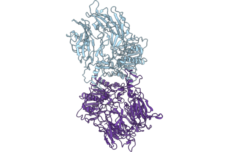 |
Chloroplast Glutamyl Peptidase S781R In Open-Closed Conformation
Organism: Arabidopsis thaliana
Method: ELECTRON MICROSCOPY Resolution:3.20 Å Release Date: 2026-05-27 Classification: PLANT PROTEIN |
Organism: Arabidopsis thaliana
Method: ELECTRON MICROSCOPY
Release Date: 2026-05-27
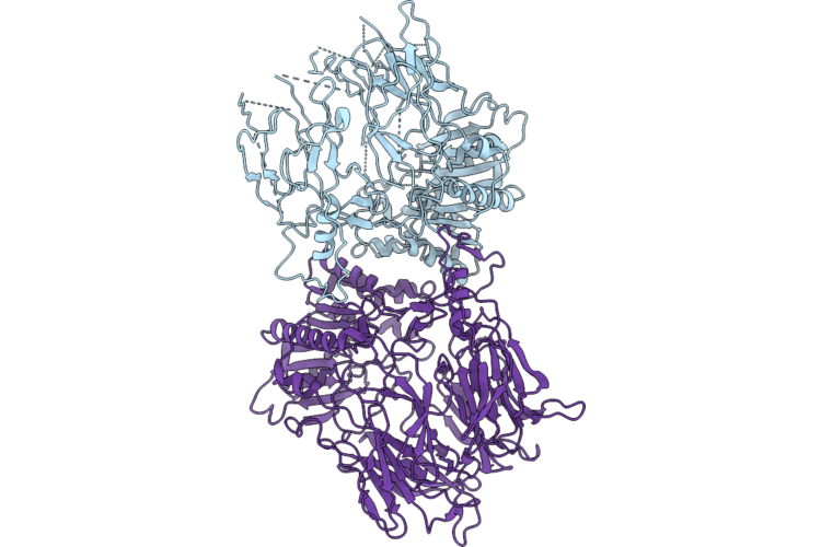 |
Chloroplast Glutamyl Peptidase Wt In Open-Closed Conformation
Organism: Arabidopsis thaliana
Method: ELECTRON MICROSCOPY Resolution:3.30 Å Release Date: 2026-05-27 Classification: PLANT PROTEIN |
Organism: Arabidopsis thaliana
Method: ELECTRON MICROSCOPY
Release Date: 2026-05-27
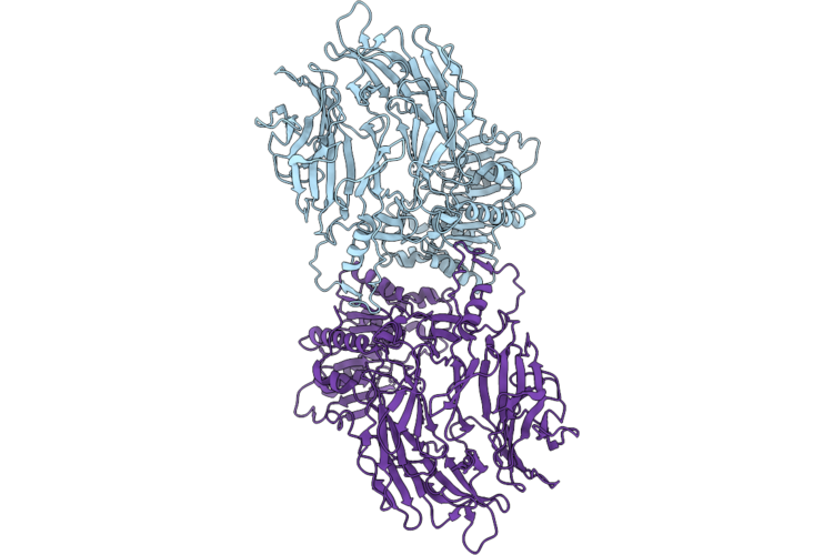 |
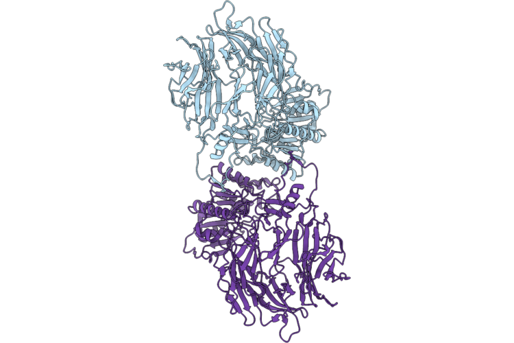
Chloroplast Glutamyl Peptidase S781R In Open-Open Conformation
Organism: Arabidopsis thaliana
Method: ELECTRON MICROSCOPY Resolution:3.20 Å Release Date: 2026-05-27 Classification: PLANT PROTEIN |
Organism: Arabidopsis thaliana
Method: ELECTRON MICROSCOPY
Release Date: 2026-05-27
 |
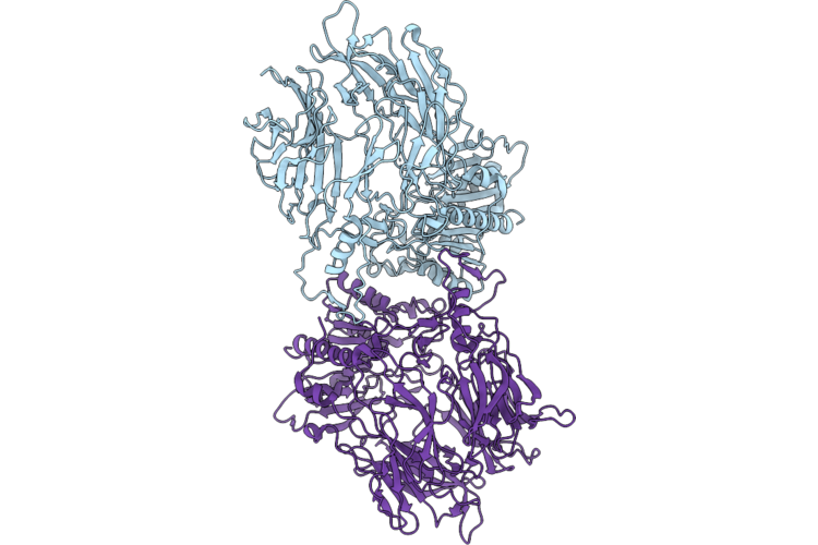
Chloroplast Glutamyl Peptidase Wt In Open-Open Conformation
Organism: Arabidopsis thaliana
Method: ELECTRON MICROSCOPY Resolution:3.00 Å Release Date: 2026-05-27 Classification: PLANT PROTEIN |
Organism: Arabidopsis thaliana
Method: ELECTRON MICROSCOPY
Release Date: 2026-05-27
 |
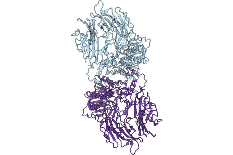
Chloroplast Glutamyl Peptidase D855N In Open-Closed Conformation
Organism: Arabidopsis thaliana
Method: ELECTRON MICROSCOPY Resolution:3.00 Å Release Date: 2026-05-27 Classification: PLANT PROTEIN |
Organism: Arabidopsis thaliana
Method: ELECTRON MICROSCOPY
Release Date: 2026-05-27
 |
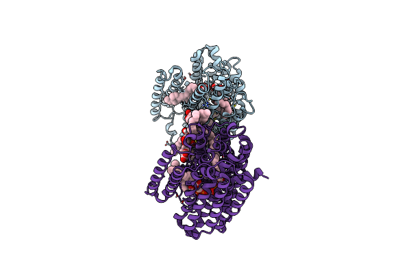
Chloroplast Glutamyl Peptidase D855N In Open-Open Conformation
Organism: Arabidopsis thaliana
Method: ELECTRON MICROSCOPY Resolution:3.00 Å Release Date: 2026-05-27 Classification: PLANT PROTEIN |
Organism: Arabidopsis thaliana
Method: ELECTRON MICROSCOPY
Release Date: 2026-05-27
 |
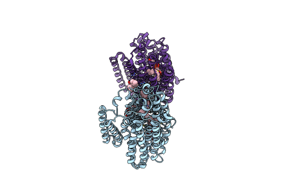
Mycobacterium Tuberculosis Efpa Antiparallel Dimer
Organism: Mycobacterium tuberculosis h37rv
Method: ELECTRON MICROSCOPY Release Date: 2025-02-05 Classification: MEMBRANE PROTEIN Ligands: A1H2V, Y01 |
Organism: Mycobacterium tuberculosis h37rv
Method: ELECTRON MICROSCOPY
Release Date: 2025-02-05
Ligands: A1H2V, Y01
 |
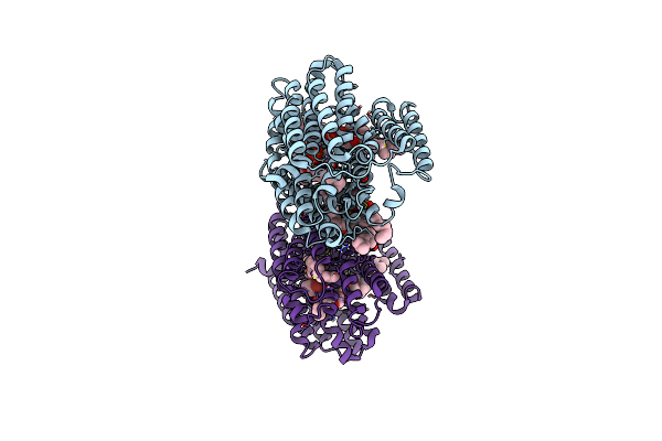
Brd-8000.3 Bound Efpa Transporter Of Mycobacterium Tuberculosis
Organism: Mycobacterium tuberculosis h37rv
Method: ELECTRON MICROSCOPY Release Date: 2025-02-05 Classification: MEMBRANE PROTEIN Ligands: A1AQR, A1H2V, Y01 |
Organism: Mycobacterium tuberculosis h37rv
Method: ELECTRON MICROSCOPY
Release Date: 2025-02-05
Ligands: A1AQR, A1H2V, Y01
 |
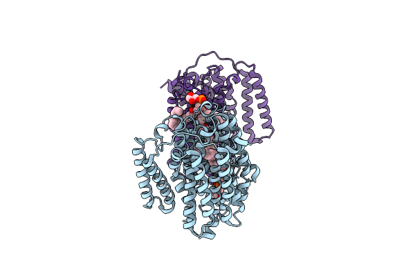
Brd-9327 Bound Efpa Transporter Of Mycobacterium Tuberculosis
Organism: Mycobacterium tuberculosis h37rv
Method: ELECTRON MICROSCOPY Release Date: 2025-02-05 Classification: MEMBRANE PROTEIN Ligands: A1APN, A1H2V, Y01 |
Organism: Mycobacterium tuberculosis h37rv
Method: ELECTRON MICROSCOPY
Release Date: 2025-02-05
Ligands: A1APN, A1H2V, Y01
 |
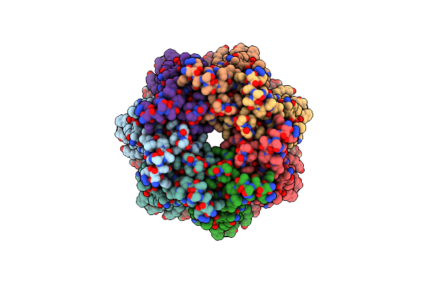
Mycobacterium Tuberculosis Efpa Parallel Dimer
Organism: Mycobacterium tuberculosis h37rv
Method: ELECTRON MICROSCOPY Release Date: 2025-02-05 Classification: MEMBRANE PROTEIN Ligands: A1ATG |
Organism: Mycobacterium tuberculosis h37rv
Method: ELECTRON MICROSCOPY
Release Date: 2025-02-05
Ligands: A1ATG
 |
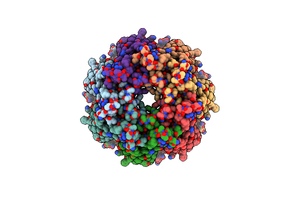
Pannexin 1 Lacking C-Terminal Activating Domain
Organism: Xenopus tropicalis
Method: ELECTRON MICROSCOPY Release Date: 2024-12-11 Classification: MEMBRANE PROTEIN |
Organism: Xenopus tropicalis
Method: ELECTRON MICROSCOPY
Release Date: 2024-12-11
 |
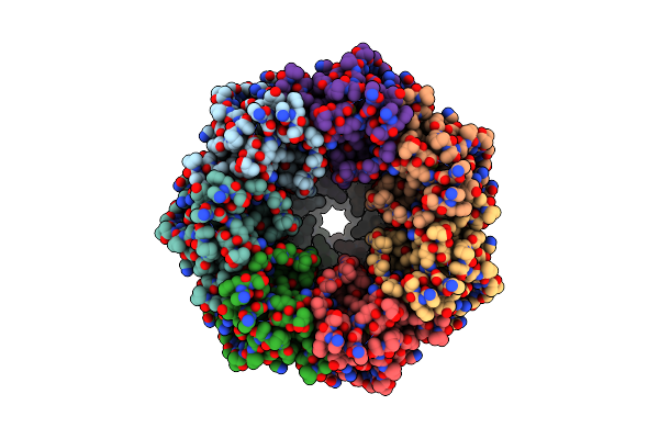
Pannexin 1 Containing C-Terminal Activating Domain
Organism: Xenopus tropicalis
Method: ELECTRON MICROSCOPY Release Date: 2024-12-04 Classification: MEMBRANE PROTEIN |
Organism: Xenopus tropicalis
Method: ELECTRON MICROSCOPY
Release Date: 2024-12-04
 |
Cryo-Em Structure Of Xenopus Tropicalis Pannexin 1 Channel
Organism: Xenopus tropicalis
Method: ELECTRON MICROSCOPY Release Date: 2020-02-26 Classification: TRANSPORT PROTEIN |
Organism: Xenopus tropicalis
Method: ELECTRON MICROSCOPY
Release Date: 2020-02-26
 |
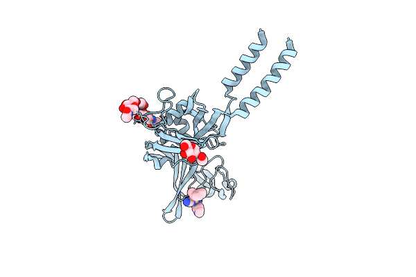
Crystal Structure Of The Atp-Gated P2X7 Ion Channel Bound To Allosteric Antagonist A740003
Organism: Ailuropoda melanoleuca
Method: X-RAY DIFFRACTION Resolution:3.60 Å Release Date: 2017-01-11 Classification: MEMBRANE PROTEIN Ligands: NAG, 7S4 |
Organism: Ailuropoda melanoleuca
Method: X-RAY DIFFRACTION
Release Date: 2017-01-11
Ligands: NAG, 7S4
 |
Crystal Structure Of The Atp-Gated P2X7 Ion Channel Bound To Allosteric Antagonist A804598
Organism: Ailuropoda melanoleuca
Method: X-RAY DIFFRACTION Resolution:3.40 Å Release Date: 2017-01-11 Classification: MEMBRANE PROTEIN Ligands: NAG, 7S1 |
Organism: Ailuropoda melanoleuca
Method: X-RAY DIFFRACTION
Release Date: 2017-01-11
Ligands: NAG, 7S1
 |
Crystal Structure Of The Atp-Gated P2X7 Ion Channel In The Closed, Apo State
Organism: Ailuropoda melanoleuca
Method: X-RAY DIFFRACTION Resolution:3.40 Å Release Date: 2017-01-04 Classification: MEMBRANE PROTEIN Ligands: NAG |
Organism: Ailuropoda melanoleuca
Method: X-RAY DIFFRACTION
Release Date: 2017-01-04
Ligands: NAG
 |
Crystal Structure Of The Atp-Gated P2X7 Ion Channel Bound To Allosteric Antagonist Az10606120
Organism: Ailuropoda melanoleuca
Method: X-RAY DIFFRACTION Resolution:3.50 Å Release Date: 2017-01-04 Classification: MEMBRANE PROTEIN Ligands: NAG, 7RY |
Organism: Ailuropoda melanoleuca
Method: X-RAY DIFFRACTION
Release Date: 2017-01-04
Ligands: NAG, 7RY
 |
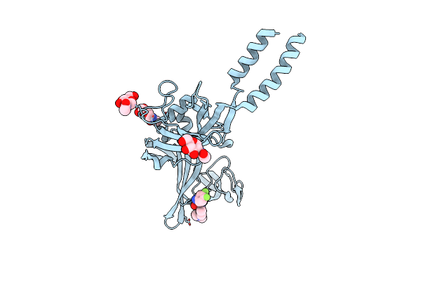
Crystal Structure Of The Atp-Gated P2X7 Ion Channel Bound To Allosteric Antagonist Jnj47965567
Organism: Ailuropoda melanoleuca
Method: X-RAY DIFFRACTION Resolution:3.20 Å Release Date: 2017-01-04 Classification: MEMBRANE PROTEIN Ligands: NAG, 7RV |
Organism: Ailuropoda melanoleuca
Method: X-RAY DIFFRACTION
Release Date: 2017-01-04
Ligands: NAG, 7RV
 |
Crystal Structure Of The Atp-Gated P2X7 Ion Channel Bound To Allosteric Antagonist Gw791343
Organism: Ailuropoda melanoleuca
Method: X-RAY DIFFRACTION Resolution:3.30 Å Release Date: 2017-01-04 Classification: MEMBRANE PROTEIN Ligands: NAG, 7RS |
Organism: Ailuropoda melanoleuca
Method: X-RAY DIFFRACTION
Release Date: 2017-01-04
Ligands: NAG, 7RS