

Search Count: 68
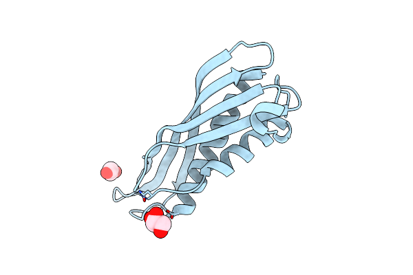 |
Crystal Structure Of Cj1090C
Organism: Campylobacter jejuni
Method: X-RAY DIFFRACTION Resolution:2.40 Å Release Date: 2022-08-31 Classification: TRANSPORT PROTEIN Ligands: GOL, EDO |
Organism: Campylobacter jejuni
Method: X-RAY DIFFRACTION
Release Date: 2022-08-31
Ligands: GOL, EDO
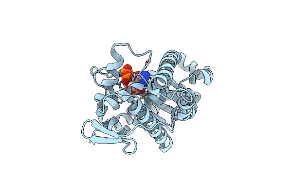 |
Crystal Structure Of Tam41 From Firmicutes Bacterium, Complex With Ctp-Mg
Organism: Firmicutes bacterium cag:884
Method: X-RAY DIFFRACTION Resolution:2.60 Å Release Date: 2022-01-19 Classification: TRANSFERASE Ligands: CTP, MG, BR |
Organism: Firmicutes bacterium cag:884
Method: X-RAY DIFFRACTION
Release Date: 2022-01-19
Ligands: CTP, MG, BR
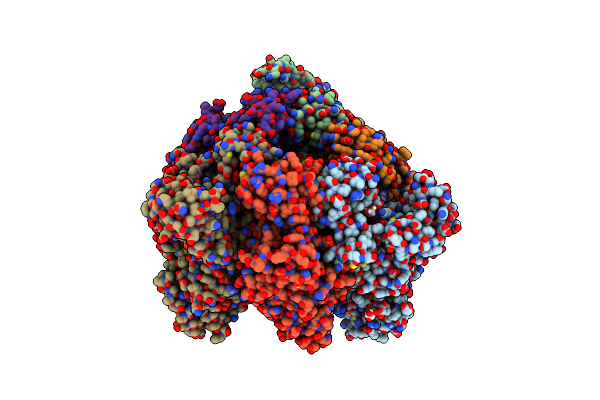 |
Crystal Structure Of Nucleotide-Free Mutant A(S23C)3B(N64C)3 Complex From Enterococcus Hirae V-Atpase
Organism: Enterococcus hirae (strain atcc 9790 / dsm 20160 / jcm 8729 / lmg 6399 / nbrc 3181 / ncimb 6459 / ncdo 1258)
Method: X-RAY DIFFRACTION Resolution:2.71 Å Release Date: 2021-12-29 Classification: HYDROLASE Ligands: GOL |
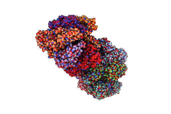 |
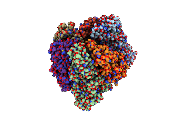
Crystal Structure Of The Amp-Pnp-Bound Mutant A(S23C)3B(N64C)3 Complex From Enterococcus Hirae V-Atpase
Organism: Enterococcus hirae (strain atcc 9790 / dsm 20160 / jcm 8729 / lmg 6399 / nbrc 3181 / ncimb 6459 / ncdo 1258)
Method: X-RAY DIFFRACTION Resolution:3.38 Å Release Date: 2021-12-29 Classification: HYDROLASE Ligands: GOL, ANP, MG |
Organism: Enterococcus hirae (strain atcc 9790 / dsm 20160 / jcm 8729 / lmg 6399 / nbrc 3181 / ncimb 6459 / ncdo 1258)
Method: X-RAY DIFFRACTION
Release Date: 2021-12-29
Ligands: GOL, ANP, MG
 |
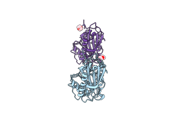
Crystal Structure Of The Adp-Bound Mutant A(S23C)3B(N64C)3 Complex From Enterococcus Hirae V-Atpase
Organism: Enterococcus hirae (strain atcc 9790 / dsm 20160 / jcm 8729 / lmg 6399 / nbrc 3181 / ncimb 6459 / ncdo 1258)
Method: X-RAY DIFFRACTION Resolution:2.69 Å Release Date: 2021-12-29 Classification: HYDROLASE Ligands: MG, ADP, SO4 |
Organism: Enterococcus hirae (strain atcc 9790 / dsm 20160 / jcm 8729 / lmg 6399 / nbrc 3181 / ncimb 6459 / ncdo 1258)
Method: X-RAY DIFFRACTION
Release Date: 2021-12-29
Ligands: MG, ADP, SO4
 |
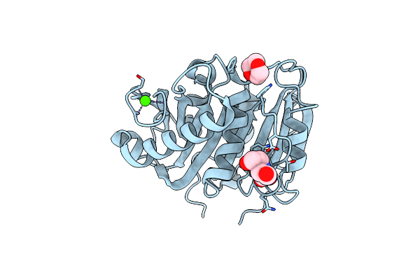
Closed Form Of Pet-Degrading Cutinase Cut190 With Thermostability-Improving Mutations Of S226P/R228S/Q138A/D250C-E296C/Q123H/N202H
Organism: Saccharomonospora viridis
Method: X-RAY DIFFRACTION Resolution:1.20 Å Release Date: 2021-02-03 Classification: HYDROLASE Ligands: DIO |
Organism: Saccharomonospora viridis
Method: X-RAY DIFFRACTION
Release Date: 2021-02-03
Ligands: DIO
 |
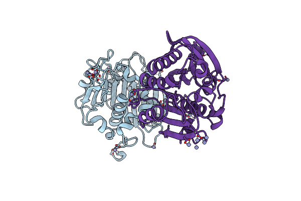
Open Form Of Pet-Degrading Cutinase Cut190 With Thermostability-Improving Mutations Of S226P/R228S/Q138A/D250C-E296C/Q123H/N202H And S176A Inactivation
Organism: Saccharomonospora viridis
Method: X-RAY DIFFRACTION Resolution:1.10 Å Release Date: 2021-02-03 Classification: HYDROLASE Ligands: DIO, BCN, CA |
Organism: Saccharomonospora viridis
Method: X-RAY DIFFRACTION
Release Date: 2021-02-03
Ligands: DIO, BCN, CA
 |
Crystal Structure Of Pet-Degrading Cutinase Cut190 /S226P/R228S/ Mutant With The C-Terminal Three Residues Deletion
Organism: Saccharomonospora viridis
Method: X-RAY DIFFRACTION Resolution:1.60 Å Release Date: 2020-08-26 Classification: HYDROLASE Ligands: CA, ZN |
Organism: Saccharomonospora viridis
Method: X-RAY DIFFRACTION
Release Date: 2020-08-26
Ligands: CA, ZN
 |
Crystal Structure Of Pet-Degrading Cutinase Cut190 S176A/S226P/R228S/ Mutant With The C-Terminal Three Residues Deletion In Ligand Ejecting Form
Organism: Saccharomonospora viridis
Method: X-RAY DIFFRACTION Resolution:1.09 Å Release Date: 2020-08-26 Classification: HYDROLASE Ligands: CA |
Organism: Saccharomonospora viridis
Method: X-RAY DIFFRACTION
Release Date: 2020-08-26
Ligands: CA
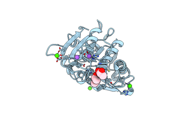 |
Structural Insights Into The Unique Polylactate Degrading Mechanism Of Thermobifida Alba Cutinase
Organism: Thermobifida alba
Method: X-RAY DIFFRACTION Resolution:1.30 Å Release Date: 2019-02-27 Classification: HYDROLASE Ligands: CA, 9YL, NA, LAC |
Organism: Thermobifida alba
Method: X-RAY DIFFRACTION
Release Date: 2019-02-27
Ligands: CA, 9YL, NA, LAC
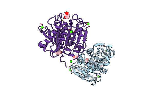 |
Crystal Structure Of Pet-Degrading Cutinase Cut190 S176A/S226P/R228S/ Mutant In Ca(2+)-Bound State
Organism: Saccharomonospora viridis
Method: X-RAY DIFFRACTION Resolution:1.60 Å Release Date: 2018-09-12 Classification: HYDROLASE Ligands: CA, GOL |
Organism: Saccharomonospora viridis
Method: X-RAY DIFFRACTION
Release Date: 2018-09-12
Ligands: CA, GOL
 |
Crystal Structure Of Pet-Degrading Cutinase Cut190 S176A/S226P/R228S Mutant In Zn(2+)-Bound State
Organism: Saccharomonospora viridis
Method: X-RAY DIFFRACTION Resolution:1.12 Å Release Date: 2018-09-12 Classification: HYDROLASE Ligands: ZN, CA, SO4, GOL |
Organism: Saccharomonospora viridis
Method: X-RAY DIFFRACTION
Release Date: 2018-09-12
Ligands: ZN, CA, SO4, GOL
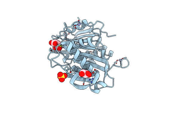 |
Crystal Structure Of Pet-Degrading Cutinase Cut190 S176A/S226P/R228S Mutant In Monoethyl Succinate Bound State
Organism: Saccharomonospora viridis
Method: X-RAY DIFFRACTION Resolution:1.34 Å Release Date: 2018-09-12 Classification: HYDROLASE Ligands: 9J3, ZN, GOL, SO4 |
Organism: Saccharomonospora viridis
Method: X-RAY DIFFRACTION
Release Date: 2018-09-12
Ligands: 9J3, ZN, GOL, SO4
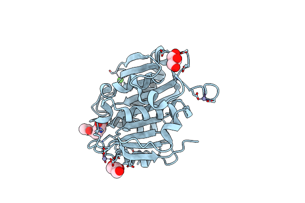 |
Crystal Structure Of Pet-Degrading Cutinase Cut190 S176A/S226P/R228S Mutant In Monoethyl Adipate Bound State
Organism: Saccharomonospora viridis
Method: X-RAY DIFFRACTION Resolution:1.40 Å Release Date: 2018-09-12 Classification: HYDROLASE Ligands: 9J6, ZN, CA, GOL |
Organism: Saccharomonospora viridis
Method: X-RAY DIFFRACTION
Release Date: 2018-09-12
Ligands: 9J6, ZN, CA, GOL
 |
Engineered Chitinase, Smchiab-Fysfv
Organism: Serratia marcescens
Method: X-RAY DIFFRACTION Resolution:2.60 Å Release Date: 2018-07-11 Classification: HYDROLASE Ligands: GOL |
Organism: Serratia marcescens
Method: X-RAY DIFFRACTION
Release Date: 2018-07-11
Ligands: GOL
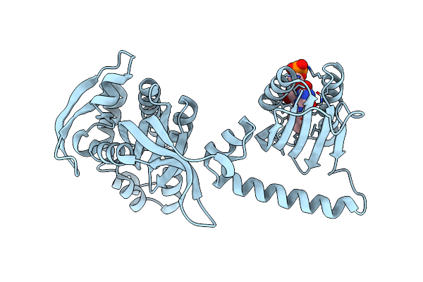 |
Crystal Structure Of Photoactivated Adenylyl Cyclase Of A Cyanobacteriaoscillatoria Acuminata In Hexagonal Form
Organism: Oscillatoria acuminata pcc 6304
Method: X-RAY DIFFRACTION Resolution:1.80 Å Release Date: 2016-06-01 Classification: LYASE Ligands: FMN |
Organism: Oscillatoria acuminata pcc 6304
Method: X-RAY DIFFRACTION
Release Date: 2016-06-01
Ligands: FMN
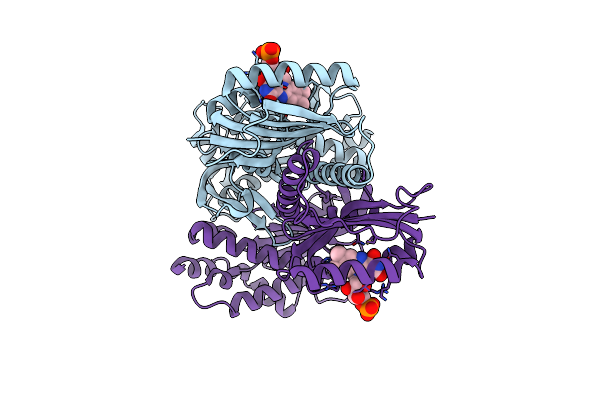 |
Crystal Structure Of Photoactivated Adenylyl Cyclase Of A Cyanobacteriaoscillatoria Acuminata In Orthorhombic Form
Organism: Oscillatoria acuminata pcc 6304
Method: X-RAY DIFFRACTION Resolution:2.90 Å Release Date: 2016-06-01 Classification: LYASE Ligands: FMN |
Organism: Oscillatoria acuminata pcc 6304
Method: X-RAY DIFFRACTION
Release Date: 2016-06-01
Ligands: FMN
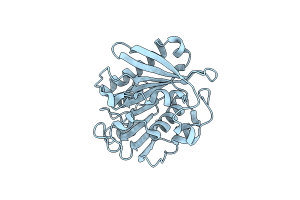 |
Crystal Structure Of Pet-Degrading Cutinase Cut190 S226P Mutant In Ca(2+)-Free State
Organism: Saccharomonospora viridis
Method: X-RAY DIFFRACTION Resolution:1.45 Å Release Date: 2014-12-24 Classification: HYDROLASE |
Organism: Saccharomonospora viridis
Method: X-RAY DIFFRACTION
Release Date: 2014-12-24
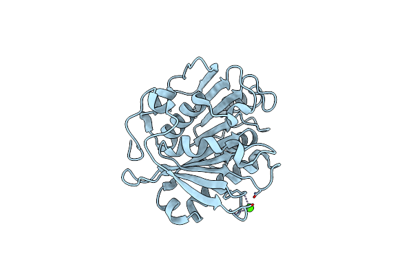 |
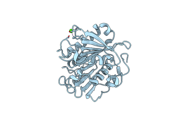
Crystal Structure Of Pet-Degrading Cutinase Cut190 S226P Mutant In Ca(2+)-Bound State At 1.75 Angstrom Resolution
Organism: Saccharomonospora viridis
Method: X-RAY DIFFRACTION Resolution:1.75 Å Release Date: 2014-12-24 Classification: HYDROLASE Ligands: CA, CL |
Organism: Saccharomonospora viridis
Method: X-RAY DIFFRACTION
Release Date: 2014-12-24
Ligands: CA, CL
 |
Crystal Structure Of Pet-Degrading Cutinase Cut190 S226P Mutant In Ca(2+)-Bound State At 2.35 Angstrom Resolution
Organism: Saccharomonospora viridis
Method: X-RAY DIFFRACTION Resolution:2.35 Å Release Date: 2014-12-24 Classification: HYDROLASE Ligands: CA, CL |
Organism: Saccharomonospora viridis
Method: X-RAY DIFFRACTION
Release Date: 2014-12-24
Ligands: CA, CL