

Search Count: 26
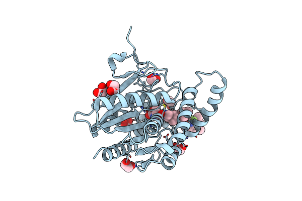 |
Crystal Structure Of Monoacylglycerol Lipase With Bodipy Labeled Probe
Organism: Homo sapiens
Method: X-RAY DIFFRACTION Resolution:1.41 Å Release Date: 2025-06-11 Classification: HYDROLASE Ligands: A1IIH, EDO |
Organism: Homo sapiens
Method: X-RAY DIFFRACTION
Release Date: 2025-06-11
Ligands: A1IIH, EDO
 |
Crystal Structure Of Human Monoglyceride Lipase In Complex With Compound 5
Organism: Homo sapiens
Method: X-RAY DIFFRACTION Resolution:1.43 Å Release Date: 2025-02-12 Classification: HYDROLASE Ligands: A1H35, EDO |
Organism: Homo sapiens
Method: X-RAY DIFFRACTION
Release Date: 2025-02-12
Ligands: A1H35, EDO
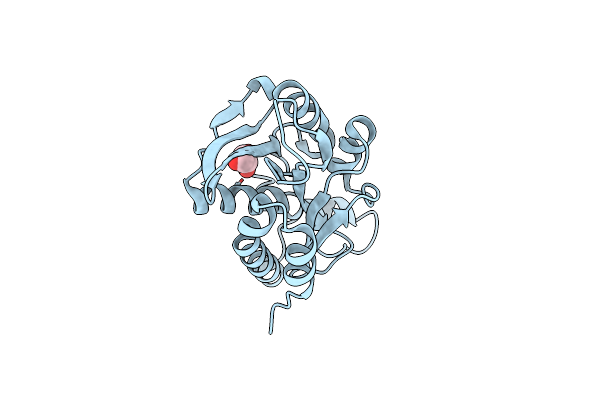 |
Crystal Structure Of Native Unbound Peptidyl Trna Hydrolase From Acinetobacter Baumannii At 1.36 A Resolution
Organism: Acinetobacter baumannii (strain atcc 19606 / dsm 30007 / cip 70.34 / jcm 6841 / nbrc 109757 / ncimb 12457 / nctc 12156 / 81)
Method: X-RAY DIFFRACTION Resolution:1.36 Å Release Date: 2017-09-13 Classification: HYDROLASE Ligands: GOL |
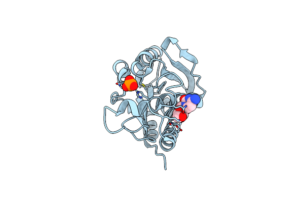 |
Crystal Structure Of The Complex Of Peptidyl Trna Hydrolase With A Phosphate Ion At The Substrate Binding Site And Cytarabine At A New Ligand Binding Site At 1.1 A Resolution
Organism: Acinetobacter baumannii (strain atcc 19606 / dsm 30007 / cip 70.34 / jcm 6841 / nbrc 109757 / ncimb 12457 / nctc 12156 / 81)
Method: X-RAY DIFFRACTION Resolution:1.10 Å Release Date: 2017-09-13 Classification: HYDROLASE Ligands: AR3, PO4 |
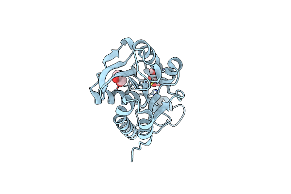 |
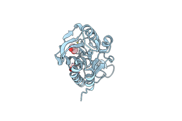
Crystal Structure Of Arg119Gln Mutant Of Peptidyl-Trna Hydrolase From Acinetobacter Baumannii At 1.49 A Resolution
Organism: Acinetobacter baumannii
Method: X-RAY DIFFRACTION Resolution:1.49 Å Release Date: 2014-02-12 Classification: HYDROLASE Ligands: GOL, TLA |
Organism: Acinetobacter baumannii
Method: X-RAY DIFFRACTION
Release Date: 2014-02-12
Ligands: GOL, TLA
 |
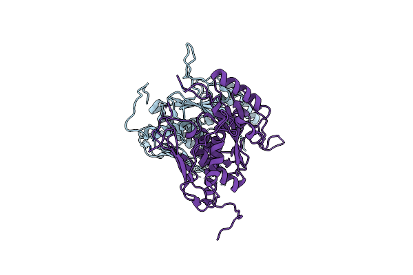
Crystal Structure Of Native Peptidyl T-Rna Hydrolase From Acinetobacter Baumannii At 1.38A Resolution
Organism: Acinetobacter baumannii
Method: X-RAY DIFFRACTION Resolution:1.38 Å Release Date: 2013-08-14 Classification: HYDROLASE Ligands: GOL |
Organism: Acinetobacter baumannii
Method: X-RAY DIFFRACTION
Release Date: 2013-08-14
Ligands: GOL
 |
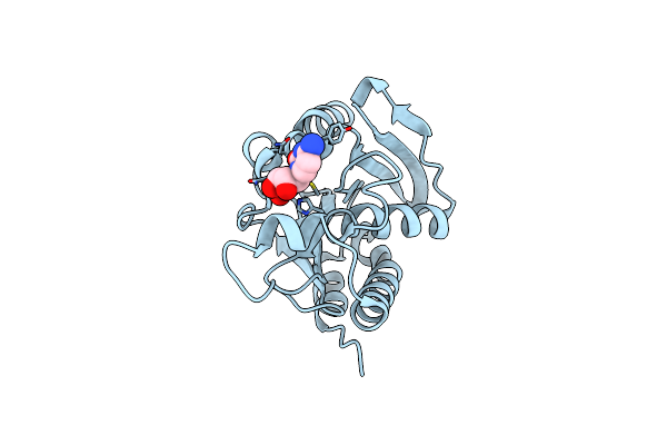
Crystal Structure Of Chorismate Synthase From Acinetobacter Baumannii At 3.15A Resolution
Organism: Acinetobacter baumannii
Method: X-RAY DIFFRACTION Resolution:3.15 Å Release Date: 2013-07-24 Classification: LYASE |
Organism: Acinetobacter baumannii
Method: X-RAY DIFFRACTION
Release Date: 2013-07-24
 |
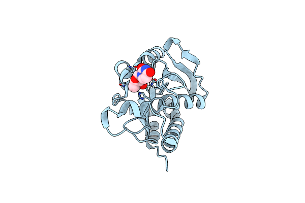
Crystal Structure Of The Complex Of Peptidyl-Trna Hydrolase From Acinetobacter Baumannii With Cytidine At 1.87 A Resolution
Organism: Acinetobacter baumannii atcc 19606 = cip 70.34
Method: X-RAY DIFFRACTION Resolution:1.87 Å Release Date: 2013-06-05 Classification: HYDROLASE Ligands: CTN |
Organism: Acinetobacter baumannii atcc 19606 = cip 70.34
Method: X-RAY DIFFRACTION
Release Date: 2013-06-05
Ligands: CTN
 |
Crystal Structure Of The Complex Of Peptidyl T-Rna Hydrolase From Acinetobacter Baumannii With Uridine At 1.4A Resolution
Organism: Acinetobacter baumannii atcc 19606 = cip 70.34
Method: X-RAY DIFFRACTION Resolution:1.40 Å Release Date: 2013-06-05 Classification: HYDROLASE Ligands: URI |
Organism: Acinetobacter baumannii atcc 19606 = cip 70.34
Method: X-RAY DIFFRACTION
Release Date: 2013-06-05
Ligands: URI
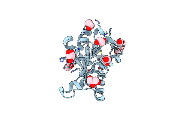 |
Crystal Structure Of Acinetobacter Baumannii Peptidyl-Trna Hydrolase
Organism: Acinetobacter baumannii
Method: X-RAY DIFFRACTION Resolution:1.90 Å Release Date: 2013-04-17 Classification: HYDROLASE Ligands: GOL, EDO, ACT, PEG |
Organism: Acinetobacter baumannii
Method: X-RAY DIFFRACTION
Release Date: 2013-04-17
Ligands: GOL, EDO, ACT, PEG
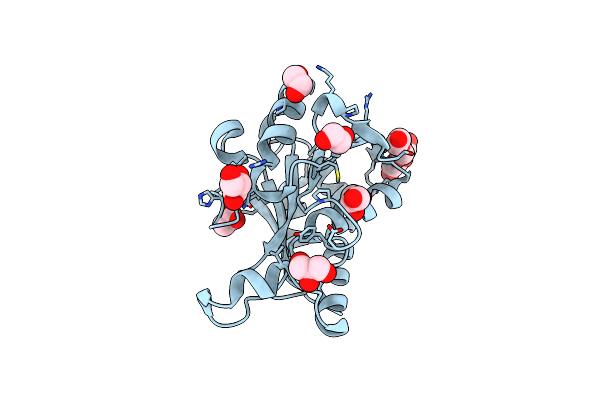 |
Structure Of Peptidyl- Trna Hydrolase From Acinetobacter Baumannii At 1.90 A Resolution
Organism: Acinetobacter baumannii
Method: X-RAY DIFFRACTION Resolution:1.90 Å Release Date: 2013-01-30 Classification: HYDROLASE Ligands: GOL, EDO, ACT, PEG |
Organism: Acinetobacter baumannii
Method: X-RAY DIFFRACTION
Release Date: 2013-01-30
Ligands: GOL, EDO, ACT, PEG
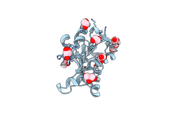 |
Crystal Structure Of Peptidyl- Trna Hydrolase From Acinetobacter Baumannii At 1.78 A Resolution
Organism: Acinetobacter baumannii
Method: X-RAY DIFFRACTION Resolution:1.78 Å Release Date: 2012-11-07 Classification: HYDROLASE Ligands: GOL, EDO, ACT, PEG |
Organism: Acinetobacter baumannii
Method: X-RAY DIFFRACTION
Release Date: 2012-11-07
Ligands: GOL, EDO, ACT, PEG
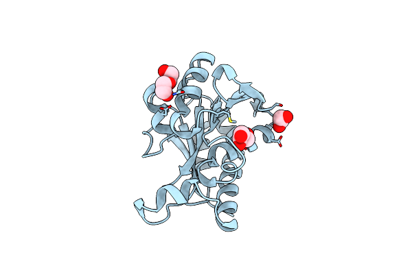 |
Crystal Structure Of Peptidyl- Trna Hydrolase From Acinetobacter Baumannii At 2.20 A Resolution
Organism: Acinetobacter baumannii
Method: X-RAY DIFFRACTION Resolution:2.20 Å Release Date: 2012-07-11 Classification: HYDROLASE Ligands: GOL, EDO, PEG |
Organism: Acinetobacter baumannii
Method: X-RAY DIFFRACTION
Release Date: 2012-07-11
Ligands: GOL, EDO, PEG
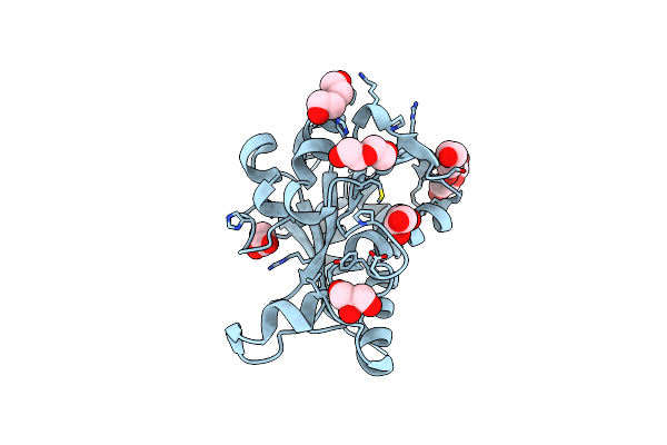 |
Crystal Structure Of Peptidyl-Trna Hydrolase From Acinetobacter Baumannii At 1.86 A Resolution
Organism: Acinetobacter baumannii
Method: X-RAY DIFFRACTION Resolution:1.86 Å Release Date: 2012-07-04 Classification: HYDROLASE Ligands: GOL, PEG, ACT |
Organism: Acinetobacter baumannii
Method: X-RAY DIFFRACTION
Release Date: 2012-07-04
Ligands: GOL, PEG, ACT
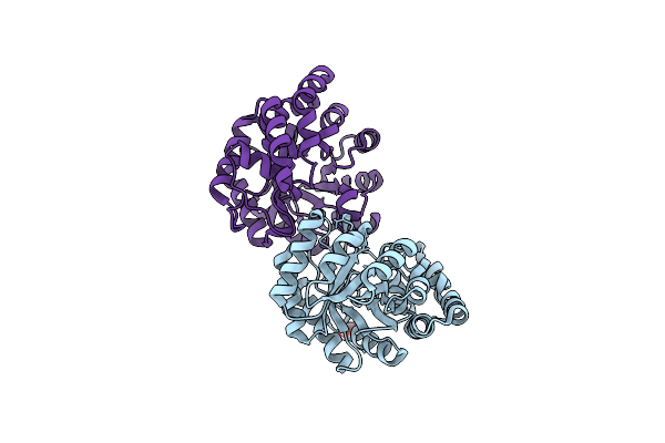 |
Crystal Structure Of Dihydrodipicolinate Synthase From Acinetobacter Baumannii Complexed With Mg And Cl Ions At 1.80 A Resolution
Organism: Acinetobacter baumannii
Method: X-RAY DIFFRACTION Resolution:1.80 Å Release Date: 2012-03-14 Classification: LYASE Ligands: CL, GOL, MG |
Organism: Acinetobacter baumannii
Method: X-RAY DIFFRACTION
Release Date: 2012-03-14
Ligands: CL, GOL, MG
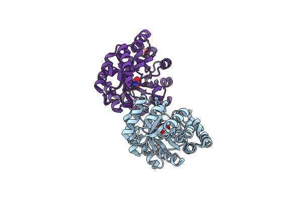 |
Crystal Structure Of Dihydrodipicolinate Synthase From Acinetobacter Baumannii Complexed With Oxamic Acid At 1.9 Angstrom Resolution
Organism: Acinetobacter baumannii
Method: X-RAY DIFFRACTION Resolution:1.94 Å Release Date: 2011-12-07 Classification: LYASE Ligands: GOL, OXM |
Organism: Acinetobacter baumannii
Method: X-RAY DIFFRACTION
Release Date: 2011-12-07
Ligands: GOL, OXM
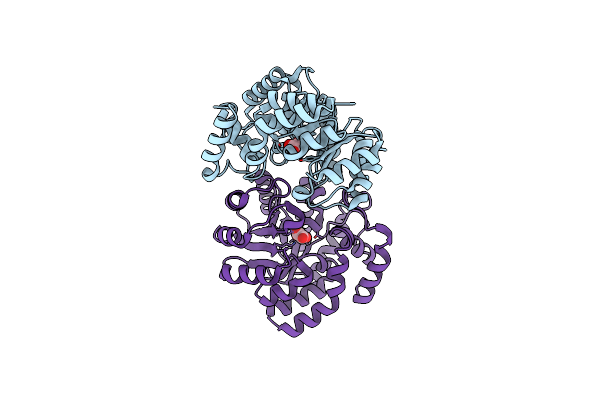 |
Crystal Structure Of The Complex Of Dihydrodipicolinate Synthase From Acinetobacter Baumannii With Oxalic Acid At 1.80 A Resolution
Organism: Acinetobacter baumannii
Method: X-RAY DIFFRACTION Resolution:1.80 Å Release Date: 2011-11-02 Classification: LYASE Ligands: OXL |
Organism: Acinetobacter baumannii
Method: X-RAY DIFFRACTION
Release Date: 2011-11-02
Ligands: OXL
 |
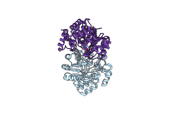
Crystal Structure Of C-Lobe Of Bovine Lactoferrin Complexed With Meta-Hydroxy Benzoic Acid At 2.5 A Resolution
Organism: Bos taurus
Method: X-RAY DIFFRACTION Resolution:2.50 Å Release Date: 2011-10-19 Classification: HYDROLASE Ligands: NAG, FE, ZN, CO3, SO4, 3HB |
Organism: Bos taurus
Method: X-RAY DIFFRACTION
Release Date: 2011-10-19
Ligands: NAG, FE, ZN, CO3, SO4, 3HB
 |
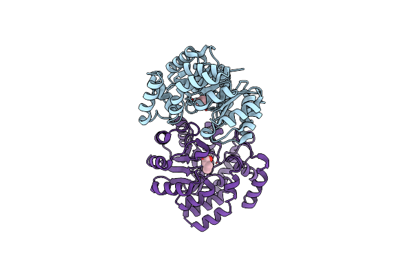
Crystal Structure Of The Complex Of Dihydrodipicolinate Synthase From Acinetobacter Baumannii With 5-Hydroxylysine At 2.6 A Resolution
Organism: Acinetobacter baumannii
Method: X-RAY DIFFRACTION Resolution:2.60 Å Release Date: 2011-08-31 Classification: LYASE Ligands: LYZ |
Organism: Acinetobacter baumannii
Method: X-RAY DIFFRACTION
Release Date: 2011-08-31
Ligands: LYZ
 |
Crystal Structure Of The Complex Of Dihydrodipicolinate Synthase From Acinetobacter Baumannii With 2-Ketobutanoic Acid At 1.99 A Resolution
Organism: Acinetobacter baumannii
Method: X-RAY DIFFRACTION Resolution:1.99 Å Release Date: 2011-08-31 Classification: LYASE Ligands: 2KT |
Organism: Acinetobacter baumannii
Method: X-RAY DIFFRACTION
Release Date: 2011-08-31
Ligands: 2KT