

Search Count: 58
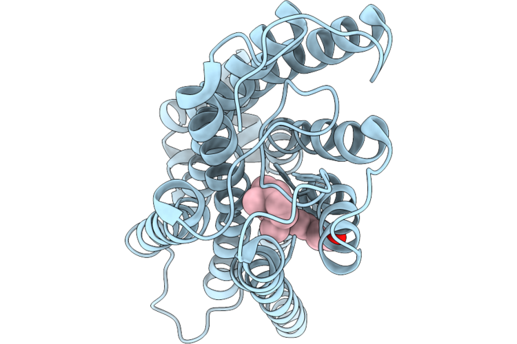 |
Cryo-Em Structure Of Macaque Red Cone Pigment With Q114N Mutation
Organism: Macaca fascicularis, Escherichia coli
Method: ELECTRON MICROSCOPY Resolution:3.21 Å Release Date: 2026-06-24 Classification: MEMBRANE PROTEIN Ligands: RET |
Organism: Macaca fascicularis, Escherichia coli
Method: ELECTRON MICROSCOPY
Release Date: 2026-06-24
Ligands: RET
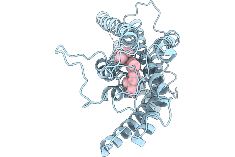 |
Cryo-Em Structure Of Macaque Green Cone Pigment With Q114N Mutation
Organism: Macaca fascicularis, Escherichia coli
Method: ELECTRON MICROSCOPY Resolution:3.37 Å Release Date: 2026-06-24 Classification: MEMBRANE PROTEIN Ligands: RET |
Organism: Macaca fascicularis, Escherichia coli
Method: ELECTRON MICROSCOPY
Release Date: 2026-06-24
Ligands: RET
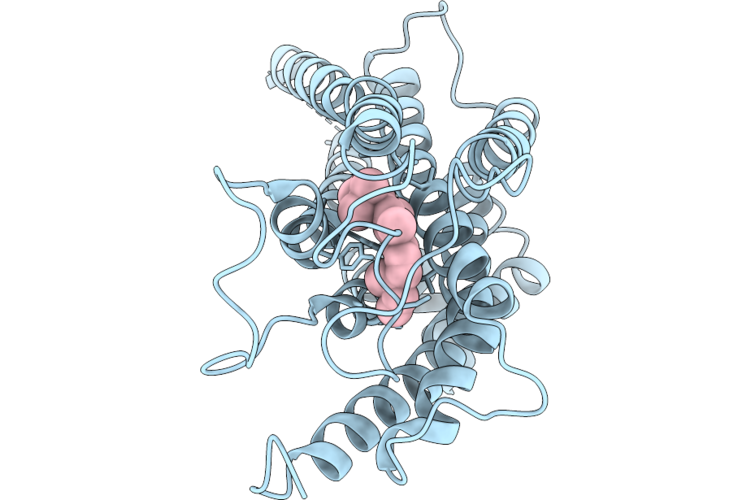 |
Cryo-Em Structure Of Macaque Green Cone Pigment Wild Type
Organism: Macaca fascicularis, Escherichia coli
Method: ELECTRON MICROSCOPY Resolution:3.71 Å Release Date: 2026-06-24 Classification: MEMBRANE PROTEIN Ligands: RET |
Organism: Macaca fascicularis, Escherichia coli
Method: ELECTRON MICROSCOPY
Release Date: 2026-06-24
Ligands: RET
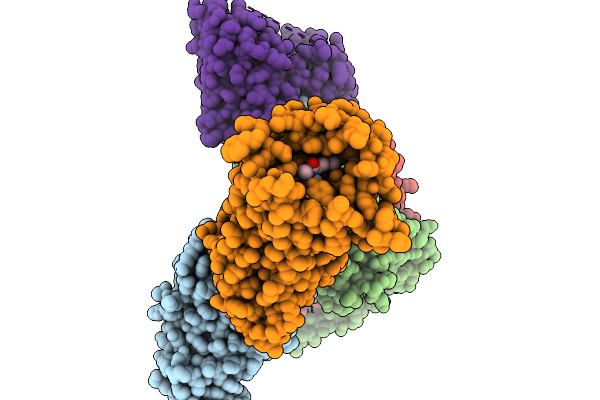 |
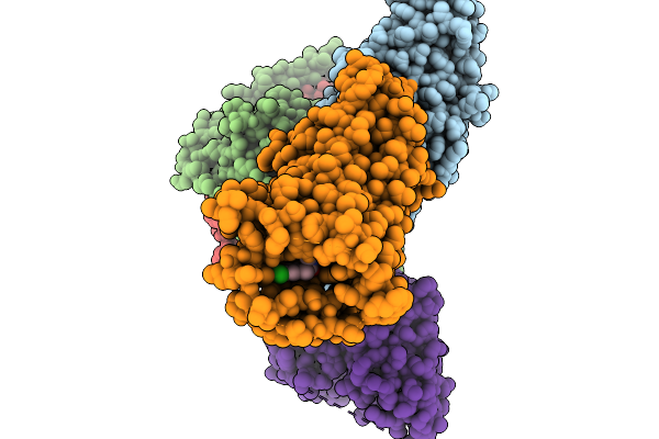
Cryo-Em Structure Of Human Kappa Opioid Receptor - G Protein Signaling Complex Bound With Nalfurafine.
Organism: Homo sapiens
Method: ELECTRON MICROSCOPY Resolution:2.76 Å Release Date: 2025-11-19 Classification: MEMBRANE PROTEIN Ligands: IVB |
Organism: Homo sapiens
Method: ELECTRON MICROSCOPY
Release Date: 2025-11-19
Ligands: IVB
 |
Cryo-Em Structure Of Human Kappa Opioid Receptor -G Protein Signaling Complex Bound With U-50488H
Organism: Homo sapiens, Mus musculus
Method: ELECTRON MICROSCOPY Resolution:2.90 Å Release Date: 2025-11-19 Classification: MEMBRANE PROTEIN Ligands: A1LWX |
Organism: Homo sapiens, Mus musculus
Method: ELECTRON MICROSCOPY
Release Date: 2025-11-19
Ligands: A1LWX
 |
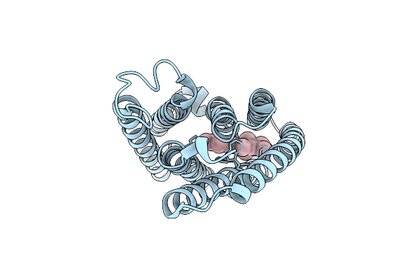
Crystal Structure Of The Light-Driven Proton Pump Heimdallarchaeial Rhodopsin Heimdallr1
Organism: Candidatus heimdallarchaeota archaeon
Method: X-RAY DIFFRACTION Resolution:2.00 Å Release Date: 2025-04-02 Classification: PROTON TRANSPORT Ligands: RET, OLC, NO3 |
Organism: Candidatus heimdallarchaeota archaeon
Method: X-RAY DIFFRACTION
Release Date: 2025-04-02
Ligands: RET, OLC, NO3
 |
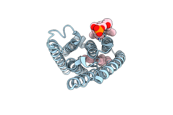
Cryo-Em Structure Of The Channelrhodopsin Gtccr2 Focused On The Monomer
Organism: Guillardia theta ccmp2712
Method: ELECTRON MICROSCOPY Resolution:2.86 Å Release Date: 2024-09-04 Classification: MEMBRANE PROTEIN Ligands: RET |
Organism: Guillardia theta ccmp2712
Method: ELECTRON MICROSCOPY
Release Date: 2024-09-04
Ligands: RET
 |
Cryo-Em Structure Of The Channelrhodopsin Gtccr2
Organism: Guillardia theta ccmp2712
Method: ELECTRON MICROSCOPY Resolution:2.73 Å Release Date: 2024-09-04 Classification: MEMBRANE PROTEIN Ligands: RET, PC1 |
Organism: Guillardia theta ccmp2712
Method: ELECTRON MICROSCOPY
Release Date: 2024-09-04
Ligands: RET, PC1
 |
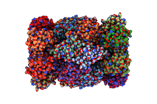
Cryo-Em Structure Of The Channelrhodopsin Gtccr4
Organism: Guillardia theta
Method: ELECTRON MICROSCOPY Resolution:2.71 Å Release Date: 2024-09-04 Classification: MEMBRANE PROTEIN Ligands: RET, PC1 |
Organism: Guillardia theta
Method: ELECTRON MICROSCOPY
Release Date: 2024-09-04
Ligands: RET, PC1
 |
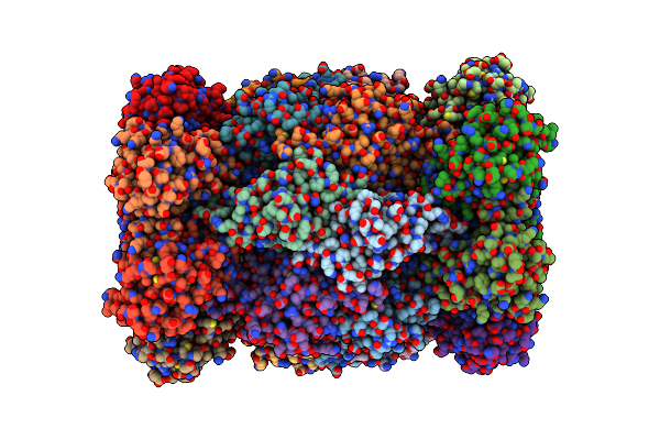
Mouse Proteasome 20S Subunit In Complex With Compound 1
Organism: Mus musculus
Method: ELECTRON MICROSCOPY Resolution:2.70 Å Release Date: 2024-07-31 Classification: HYDROLASE Ligands: A1L0C |
Organism: Mus musculus
Method: ELECTRON MICROSCOPY
Release Date: 2024-07-31
Ligands: A1L0C
 |
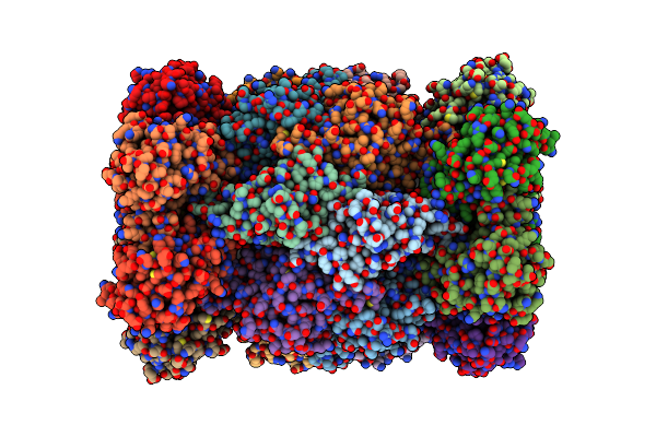
Canine Immunoproteasome 20S Subunit In Complex With Compound 2
Organism: Canis lupus familiaris
Method: ELECTRON MICROSCOPY Resolution:2.20 Å Release Date: 2024-07-31 Classification: HYDROLASE Ligands: A1L0D |
Organism: Canis lupus familiaris
Method: ELECTRON MICROSCOPY
Release Date: 2024-07-31
Ligands: A1L0D
 |
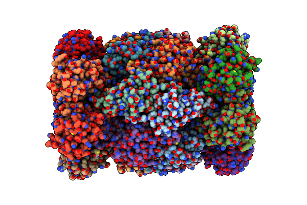
Canine Immunoproteasome 20S Subunit In Complex With Compound 1
Organism: Canis lupus familiaris
Method: ELECTRON MICROSCOPY Resolution:2.00 Å Release Date: 2024-07-31 Classification: HYDROLASE Ligands: A1L0C |
Organism: Canis lupus familiaris
Method: ELECTRON MICROSCOPY
Release Date: 2024-07-31
Ligands: A1L0C
 |
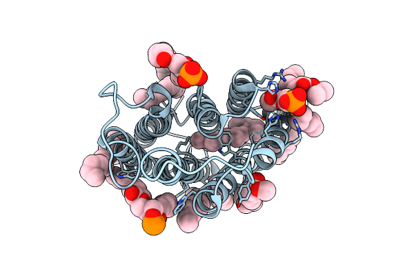
Canine Immunoproteasome 20S Subunit In Complex With Compound 1
Organism: Mus musculus
Method: ELECTRON MICROSCOPY Resolution:2.50 Å Release Date: 2024-07-31 Classification: HYDROLASE Ligands: A1L0D, THR |
Organism: Mus musculus
Method: ELECTRON MICROSCOPY
Release Date: 2024-07-31
Ligands: A1L0D, THR
 |
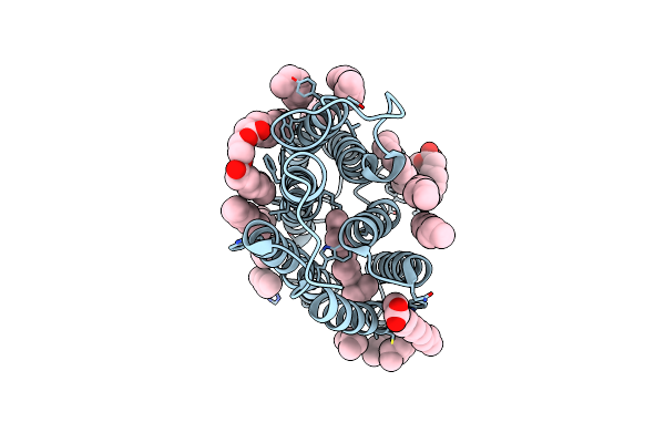
Cryo-Em Structure Of The Potassium-Selective Channelrhodopsin Hckcr1 In Lipid Nanodisc
Organism: Hyphochytrium catenoides
Method: ELECTRON MICROSCOPY Resolution:2.56 Å Release Date: 2023-09-06 Classification: MEMBRANE PROTEIN Ligands: RET, PSC, PLM |
Organism: Hyphochytrium catenoides
Method: ELECTRON MICROSCOPY
Release Date: 2023-09-06
Ligands: RET, PSC, PLM
 |
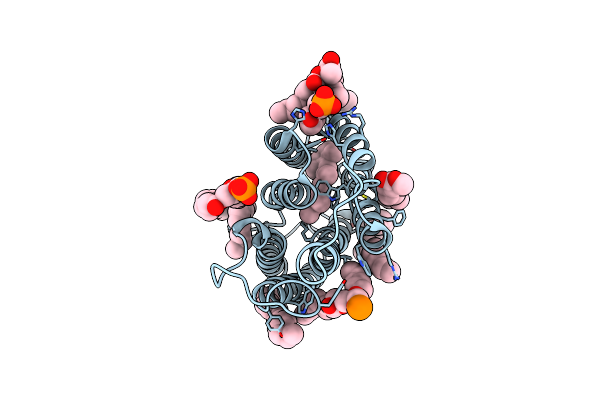
Cryo-Em Structure Of The Potassium-Selective Channelrhodopsin Hckcr2 In Lipid Nanodisc
Organism: Hyphochytrium catenoides
Method: ELECTRON MICROSCOPY Resolution:2.53 Å Release Date: 2023-09-06 Classification: MEMBRANE PROTEIN Ligands: RET, PSC, PLM |
Organism: Hyphochytrium catenoides
Method: ELECTRON MICROSCOPY
Release Date: 2023-09-06
Ligands: RET, PSC, PLM
 |
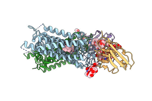
Cryo-Em Structure Of The Potassium-Selective Channelrhodopsin Hckcr1 H225F Mutant In Lipid Nanodisc
Organism: Hyphochytrium catenoides
Method: ELECTRON MICROSCOPY Resolution:2.66 Å Release Date: 2023-09-06 Classification: MEMBRANE PROTEIN Ligands: RET, PSC, PLM |
Organism: Hyphochytrium catenoides
Method: ELECTRON MICROSCOPY
Release Date: 2023-09-06
Ligands: RET, PSC, PLM
 |
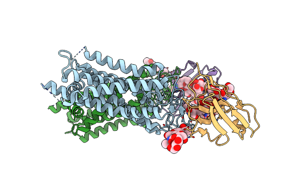
Crystal Structure Of Ground-State Rhodopsin In Complex With A Nanobody
Organism: Lama glama, Bos taurus
Method: X-RAY DIFFRACTION Resolution:3.70 Å Release Date: 2023-08-30 Classification: MEMBRANE PROTEIN Ligands: RET |
Organism: Lama glama, Bos taurus
Method: X-RAY DIFFRACTION
Release Date: 2023-08-30
Ligands: RET
 |
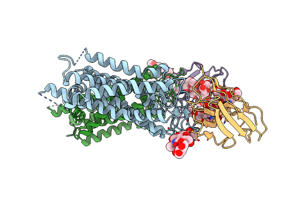
Crystal Structure Of Bovine Rod Opsin In Complex With A Nanobody
Organism: Lama glama, Bos taurus
Method: X-RAY DIFFRACTION Resolution:3.71 Å Release Date: 2023-08-30 Classification: MEMBRANE PROTEIN |
Organism: Lama glama, Bos taurus
Method: X-RAY DIFFRACTION
Release Date: 2023-08-30
 |
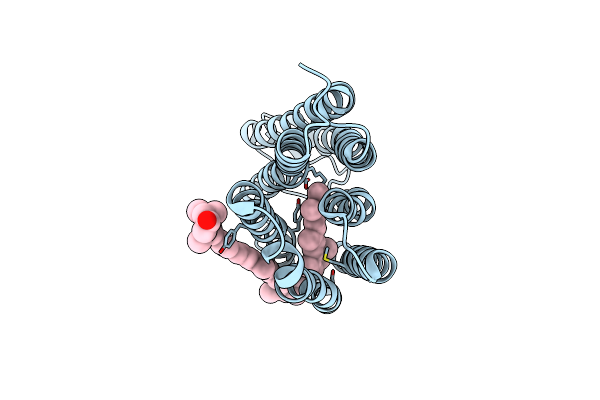
Crystal Structure Of Photoactivated Rhodopsin In Complex With A Nanobody
Organism: Lama glama, Bos taurus
Method: X-RAY DIFFRACTION Resolution:4.25 Å Release Date: 2023-08-30 Classification: MEMBRANE PROTEIN |
Organism: Lama glama, Bos taurus
Method: X-RAY DIFFRACTION
Release Date: 2023-08-30
 |
Cryo-Em Structure Of The Zeaxanthin-Bound Kin4B8
Organism: Uncultured bdellovibrionales bacterium
Method: ELECTRON MICROSCOPY Resolution:2.30 Å Release Date: 2023-03-29 Classification: PROTON TRANSPORT Ligands: RET, K3I |
Organism: Uncultured bdellovibrionales bacterium
Method: ELECTRON MICROSCOPY
Release Date: 2023-03-29
Ligands: RET, K3I